. 2022 Sep 3;13(1):5202.
doi: 10.1038/s41467-022-32788-x.

Spermidine-mediated hypusination of translation factor EIF5A improves mitochondrial fatty acid oxidation and prevents non-alcoholic steatohepatitis progression

Affiliations

Spermidine-mediated hypusination of translation factor EIF5A improves mitochondrial fatty acid oxidation and prevents non-alcoholic steatohepatitis progression

Jin Zhou et al. Nat Commun. .

Abstract

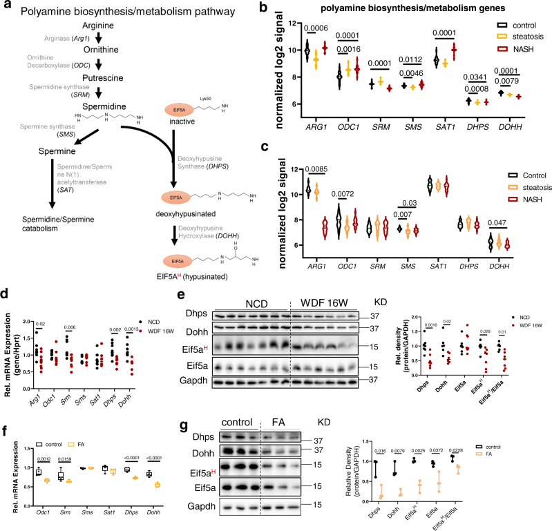
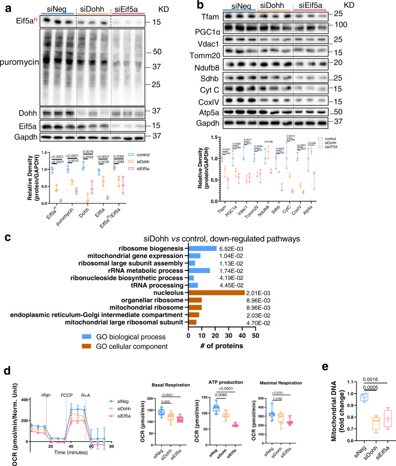
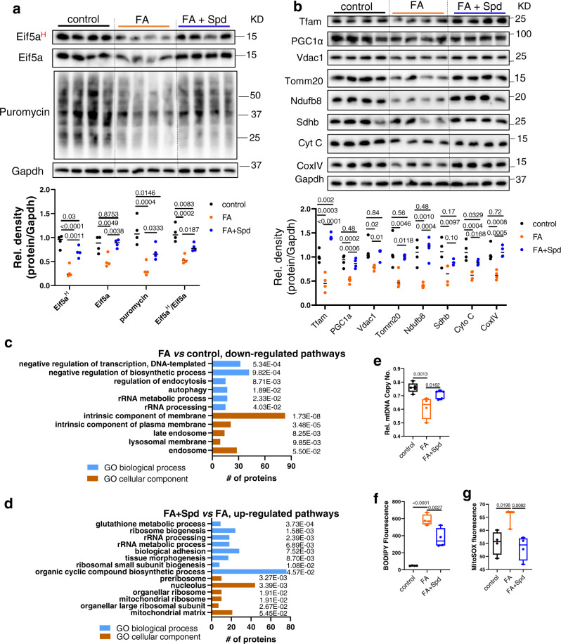
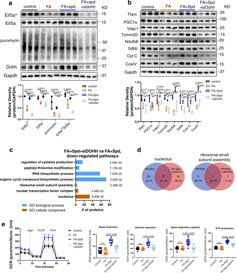
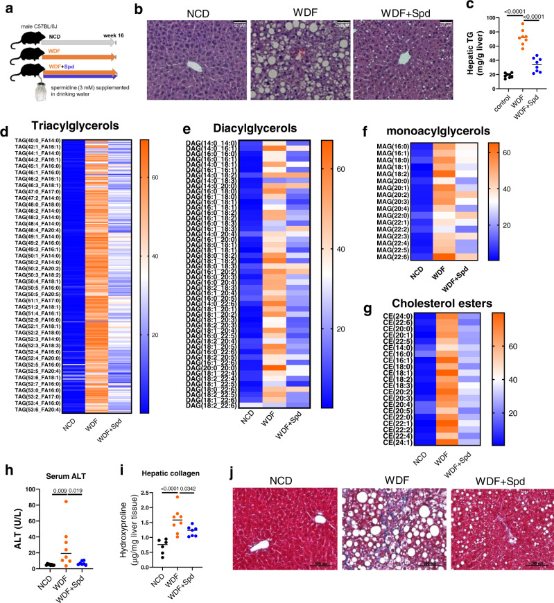
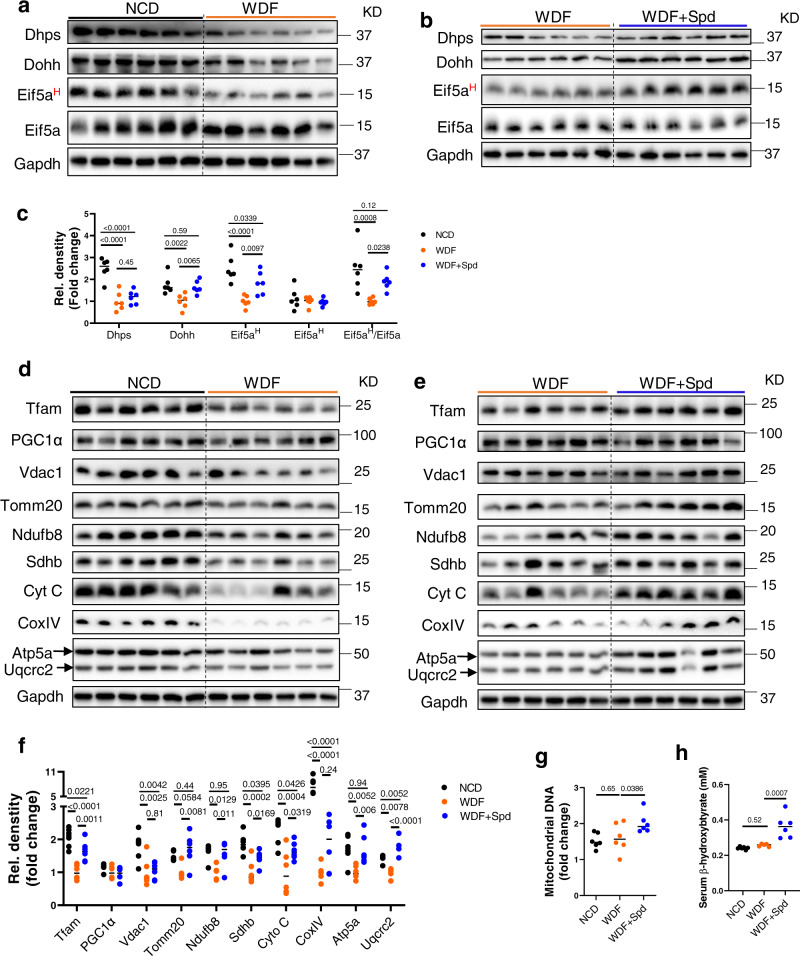
Spermidine is a natural polyamine that has health benefits and extends life span in several species. Deoxyhypusine synthase (DHPS) and deoxyhypusine hydroxylase (DOHH) are key enzymes that utilize spermidine to catalyze the post-translational hypusination of the translation factor EIF5A (EIF5AH). Here, we have found that hepatic DOHH mRNA expression is decreased in patients and mice with non-alcoholic steatohepatitis (NASH), and hepatic cells treated with fatty acids. The mouse and cell culture models of NASH have concomitant decreases in Eif5aH and mitochondrial protein synthesis which leads to lower mitochondrial activity and fatty acid β-oxidation. Spermidine treatment restores EIF5AH, partially restores protein synthesis and mitochondrial function in NASH, and prevents NASH progression in vivo. Thus, the disrupted DHPS-DOHH-EIF5AH pathway during NASH represents a therapeutic target to increase hepatic protein synthesis and mitochondrial fatty acid oxidation (FAO) and prevent NASH progression.


Conflict of interest statement

The authors declare no competing interests.

Figures

Fig. 1
Fig. 1. Decreased DHPS-DOHH-EIF5AH pathway in NAFLD.
a Polyamine synthesis and hypusination of EIF5A pathway in eukaryotic cells. Enzymes ARG1, ODC, SRM are involved in converting arginine to spermidine. DHPS and DOHH use spermidine to hypusinate EIF5A. b Violin plots showing mRNA levels of genes involved in endogenous polyamine biosynthesis and EIF5A hypusination in Control (n = 19), steatosis (n = 10), and NASH (n = 16) from publicly available database (accession number E-MEXP-3291, http://www.webcitation.org/5zyojNu7T). c Violin plots showing mRNA levels of genes involved in endogenous polyamine biosynthesis and EIF5A hypusination in Control (n = 12), steatosis (n = 9), and NASH (n = 17) from publicly available database (GSE48452). b, c Significance was calculated by one-way ANOVA or Kruskal–Wallis test, as appropriate. d Quantitative-PCR analysis of mRNA levels of polyamine metabolism genes in the livers from mice fed with NCD (n = 8) or WDF (n = 8) for 16 weeks. e Western blot and densitometric analysis of protein levels of Dhps, Dohh, eIF5AH, and eIF5A in the liver from mice fed with NCD (n = 7) or WDF (n = 6) for 16 weeks. f, g mRNA expression of genes in polyamine biosynthesis and hypusination pathways (f, n = 5), and protein levels of Dhps, Dohh, eIF5AH, and eIF5A (g, n = 3) in AML12 hepatic cells treated with fatty acids (FA, palmitic acid 0.6 mM, oleic acid 0.17 mM) for 48 h. fg Data were shown as box-and-whisker with median (middle line), 25th–75th percentiles (box), and min-max values (whiskers). dg significance was calculated by two-tailed Student’s t test or Mann–Whitney U test, as appropriate. Source data are provided as a Source Data file.
Fig. 2
Fig. 2. Dohh KD decreased protein synthesis and mitochondrial function in hepatic cells.
AML12 cells were transfected with 20 nM of negative (siNeg), Dohh (siDohh), or eIF5A (siEif5a) siRNA for 48 h. Puromycin (1 µg/ml) was added 1 h before harvesting cell lysate. a Western blot and densitometric analysis of eIF5AH, and proteins after puromycin incorporation. (n = 3). b Western blot and densitometric analysis of Tfam, PGC1α, and mitochondrial proteins. (n = 3). c Proteomics and gene ontology (GO) enrichment analysis (with corrected p value indicated on the bar) of downregulated proteins in Dohh KD (siDohh) vs control AML12 cells. A Bonferroni correction was applied to correct for multiple testing. d Agilent Seahorse XF Mito Stress Test measurement of mitochondrial OXPHOS. Oligomycin (oligo), FCCP, rotenone (R), and antimycin A (A) were used at 1 µM each. (n = 15). Line graph is presented as mean value ± SEM. e Mitochondrial DNA copy number in Dohh or eIF5A knockdown cells. (n = 4). a, b, d, e Data were shown as box-and-whisker with median (middle line), 25th–75th percentiles (box), and min-max values (whiskers). Significance was calculated by one-way ANOVA or Kruskal–Wallis test, as appropriate. Source data are provided as a Source Data file.
Fig. 3
Fig. 3. FA decreased Eif5aH, protein synthesis rate and mitochondrial proteins, which can be rescued by co-treatment with spermidine.
a, b Western blot and densitometric analysis of Eif5aH and total proteins after puromycin incorporation (a), and mitochondrial proteins (b) in control, FA−, and FA+ Spd-treated AML12 cells. (n = 4). c, d Proteomics and gene ontology (GO) enrichment analysis (with corrected p value indicated on the bar) of downregulated proteins in FA-treated vs control AML12 cells (c), and upregulated proteins in FA+ Spd-treated vs FA-treated AML12 cells (d). A Bonferroni correction was applied to correct for multiple testing. eg Relative mitochondrial copy number ((e, control (n = 5), FA (n = 4), FA + Spd (n = 5)), BODIPY fluorescence (f, n = 4), and MitoSOX measurement of mitochondrial ROS ((g, control (n = 4), FA (n = 3), FA + Spd (n = 4)) in control, FA−, FA + Spd-treated AML12 cells. AML12 cells were treated with BSA-conjugated FA (palmitic acid 0.6 mM, oleic acid 0.17 mM) with or without spermidine (Spd, 100 µM) for 48 h. eg Data were shown as box-and-whisker with median (middle line), 25th–75th percentiles (box), and min-max values (whiskers). Significance was calculated by one-way ANOVA or Kruskal–Wallis test as appropriate. Source data are provided as a Source Data file.
Fig. 4
Fig. 4. Spermidine rescued mitochondrial function in Eif5aH-dependent manner.
a, b Western blot and densitometric analysis of Eif5aH, and puromycin corporation (a, n = 3), and mitochondrial proteins (b, n = 3). c Proteomics and gene ontology (GO) enrichment analysis (with corrected p value indicated on the bar) of downregulated proteins in FA + Spd+siDohh vs FA + Spd cells. A Bonferroni correction was applied to correct for multiple testing. d Venn diagram showing the overlap of proteins under identified pathways in 4c and 3d. e Agilent Seahorse XF Mito Stress Test of mitochondrial OXPHOS. AML12 cells were first transfected with 20 nM of negative (siNeg), or Dohh (siDohh) siRNA for 24 h, followed by treatment with BSA-conjugated FA (palmitic acid 0.6 mM, oleic acid 0.17 mM) with or without spermidine (Spd, 100 µM) for 48 h. Line graph is presented as mean value ± SEM. (control (n = 18), FA (n = 22), FA + Spd (n = 18), FA + Spd+siDohh (n = 13)) a, b, e Data were shown as box-and-whisker with median (middle line), 25th–75th percentiles (box), and min-max values (whiskers). Significance was calculated by one-way ANOVA or Kruskal–Wallis test, as appropriate. Source data are provided as a Source Data file.
Fig. 5
Fig. 5. Spermidine supplementation prevented NAFLD progression in a dietary mouse model of NASH.
a Schematic showing administration protocol for spermidine (3 mM in drinking water) in mice fed with WDF for 16 weeks. b Representative images of H&E staining of the liver. (n = 5). c Hepatic triglyceride content was measured by biochemical assay. (n = 8). dg Liquid chromatography coupled with mass spectrometry (LC-MS) lipidomic profiling of hepatic triacylglycerols (TAGs) (d), diacylclycerols (DAGs) (e), monoacylglycerol (MAGs) (f), and cholesterol esters (CEs) (g) levels from mice fed with NCD (n = 6), WDF (n = 8), or WDF supplemented with spermidine (WDF + Spd, n = 6) for 16 weeks. Heat maps represent the average of each condition. h circulating ALT level from mice fed with NCD (n = 8), WDF (n = 8), WDF + Spd (n = 7). i Hepatic collagen content was determined by measurement of hydroxyproline level in the liver from mice fed with NCD (n = 6), WDF (n = 8), WDF + Spd (n = 7). j Representative images of Masson trichrome stain of liver sections. (n = 5). Significance was calculated by One-Way ANOVA. Source data are provided as a Source Data file.
Fig. 6
Fig. 6. Spermidine supplementation restored hepatic Eif5aH and mitochondrial protein levels in a dietary mouse model of NASH.
ac Western blot and densitometric analysis of hepatic Dhps, Dohh, Eif5aH, and Eif5a in NCD vs. WDF (a), and WDF vs. WDF + Spd (b) mice. The blots in a and b were processed in parallel. Densitometric analysis (c) was first normalized with GAPDH, and then calculated the fold change against WDF (NCD vs WDF, and WDF + Spd vs WDF). (n = 6) (df) Western blot and densitometric analysis of Tfam, PGC1α, and mitochondrial proteins in the liver from mice fed with NCD vs. WDF (d), or WDF vs. WDF + Spd (e). The blots in d and e were processed in parallel. Densitometric analysis (f, n = 6) was first normalized with GAPDH, and then calculated the fold change against WDF (NCD vs WDF, and WDF + Spd vs WDF). (g, h) Mitochondrial DNA copy number (g) and circulating β-hydroxybutyrate (h) in NCD (n = 6), WDF (n = 6), and WDF + Spd (n = 6) groups. Significance was calculated by one-way ANOVA or Kruskal–Wallis test, as appropriate. Source data are provided as a Source Data file.

References

    1. Minguet EG, Vera-Sirera F, Marina A, Carbonell J, Blazquez MA. Evolutionary diversification in polyamine biosynthesis. Mol. Biol. Evol. 2008;25:2119–2128. doi: 10.1093/molbev/msn161. - DOI - PubMed
    1. Munoz-Esparza NC, et al. Polyamines in food. Front Nutr. 2019;6:108. doi: 10.3389/fnut.2019.00108. - DOI - PMC - PubMed
    1. Tofalo R, Cocchi S, Suzzi G. Polyamines and gut microbiota. Front Nutr. 2019;6:16. doi: 10.3389/fnut.2019.00016. - DOI - PMC - PubMed
    1. Eisenberg T, et al. Induction of autophagy by spermidine promotes longevity. Nat. Cell Biol. 2009;11:1305–1314. doi: 10.1038/ncb1975. - DOI - PubMed
    1. Eisenberg T, et al. Cardioprotection and lifespan extension by the natural polyamine spermidine. Nat. Med. 2016;22:1428–1438. doi: 10.1038/nm.4222. - DOI - PMC - PubMed

Publication types

MeSH terms