

Colour vision in stomatopod crustaceans
- PMID: 36058241
- PMCID: PMC9441230
- DOI: 10.1098/rstb.2021.0278
Colour vision in stomatopod crustaceans
Abstract
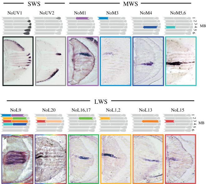
The stomatopod crustaceans, or mantis shrimps, are colourful marine invertebrate predators. Their unusual compound eyes have dorsal and ventral regions resembling typical crustacean apposition designs separated by a unique region called the midband that consists of from two to six parallel rows of ommatidia. In species with six-row midbands, the dorsal four rows are themselves uniquely specialized for colour analysis. Rhabdoms of ommatidia in these rows are longitudinally divided into three distinct regions: an apical ultraviolet (UV) receptor, a shorter-wavelength middle tier receptor and a longer-wavelength proximal tier receptor. Each of the total of 12 photoreceptors has a different spectral sensitivity, potentially contributing to a colour-vision system with 12 channels. Mantis shrimps can discriminate both human-visible and UV colours, but with limited precision compared to other colour-vision systems. Here, we review the structure and function of stomatopod colour vision, examining the types of receptors present in a species, the spectral tuning of photoreceptors both within and across species, the neural analysis of colour and the genetics underlying the multiple visual pigments used for colour vision. Even today, after many decades of research into the colour vision of stomatopods, much of its operation and its use in nature remain a mystery. This article is part of the theme issue 'Understanding colour vision: molecular, physiological, neuronal and behavioural studies in arthropods'.
Keywords: colour vision; filtering; stomatopod; ultraviolet vision; visual ecology; visual genetics.
Figures


Similar articles
-
Behavioural evidence of spectral opponent processing in the visual system of stomatopod crustaceans.J Exp Biol. 2025 Jan 1;228(1):jeb247952. doi: 10.1242/jeb.247952. Epub 2025 Jan 8. J Exp Biol. 2025. PMID: 39670570 Free PMC article.
-
Filtering and polychromatic vision in mantis shrimps: themes in visible and ultraviolet vision.Philos Trans R Soc Lond B Biol Sci. 2014 Jan 6;369(1636):20130032. doi: 10.1098/rstb.2013.0032. Print 2014. Philos Trans R Soc Lond B Biol Sci. 2014. PMID: 24395960 Free PMC article. Review.
-
Colour vision in stomatopod crustaceans: more questions than answers.J Exp Biol. 2022 Mar 15;225(6):jeb243699. doi: 10.1242/jeb.243699. Epub 2022 Mar 28. J Exp Biol. 2022. PMID: 35224643 Free PMC article.
-
The molecular genetics and evolution of colour and polarization vision in stomatopod crustaceans.Ophthalmic Physiol Opt. 2010 Sep;30(5):460-9. doi: 10.1111/j.1475-1313.2010.00762.x. Ophthalmic Physiol Opt. 2010. PMID: 20883329 Review.
-
A unique colour and polarization vision system in mantis shrimps.Nature. 1988 Jun 9;333(6173):557-60. doi: 10.1038/333557a0. Nature. 1988. PMID: 3374602
Cited by
-
High diversity of arthropod colour vision: from genes to ecology.Philos Trans R Soc Lond B Biol Sci. 2022 Oct 24;377(1862):20210273. doi: 10.1098/rstb.2021.0273. Epub 2022 Sep 5. Philos Trans R Soc Lond B Biol Sci. 2022. PMID: 36058249 Free PMC article.
-
Within-host adaptive speciation of commensal yoyo clams leads to ecological exclusion, not co-existence.PeerJ. 2024 Aug 5;12:e17753. doi: 10.7717/peerj.17753. eCollection 2024. PeerJ. 2024. PMID: 39119103 Free PMC article.
-
Ancestral photoreceptor diversity as the basis of visual behaviour.Nat Ecol Evol. 2024 Mar;8(3):374-386. doi: 10.1038/s41559-023-02291-7. Epub 2024 Jan 22. Nat Ecol Evol. 2024. PMID: 38253752 Review.
References
-
- Ahyong ST, Harling C. 2000. The phylogeny of the stomatopod Crustacea. Austr. J. Zool. 48, 607-642. (10.1071/ZO00042) - DOI
-
- Ahyong ST, Jarman SN. 2009. Stomatopod interrelationships: preliminary results based on analysis of three molecular loci. Arthropod. Syst. Phylogeny 67, 91-98.
-
- Harling C. 2000. Reexamination of eye design in the classification of stomatopod crustaceans. J. Crust. Biol. 20, 172-185. (10.1163/20021975-99990026) - DOI
Publication types
MeSH terms
Grants and funding
LinkOut - more resources
Full Text Sources
