. 2022 Oct 24;377(1862):20210274.
doi: 10.1098/rstb.2021.0274. Epub 2022 Sep 5.

Expression of a homologue of a vertebrate non-visual opsin Opn3 in the insect photoreceptors

Affiliations

Expression of a homologue of a vertebrate non-visual opsin Opn3 in the insect photoreceptors

Mitsumasa Koyanagi et al. Philos Trans R Soc Lond B Biol Sci. .

Abstract

Insect vision starts with light absorption by visual pigments based on opsins that drive Gq-type G protein-mediated phototransduction. Since Drosophila, the most studied insect in vision research, has only Gq-coupled opsins, the Gq-mediated phototransduction has been solely focused on insect vision for decades. However, genome projects on mosquitos uncovered non-canonical insect opsin genes, members of the Opn3 or c-opsin group composed of vertebrate and invertebrate non-visual opsins. Here, we report that a homologue of Opn3, MosOpn3 (Asop12) is expressed in eyes of a mosquito Anopheles stephensi. In situ hybridization analysis revealed that MosOpn3 is expressed in dorsal and ventral ommatidia, in which only R7 photoreceptor cells express MosOpn3. We also found that Asop9, a Gq-coupled visual opsin, exhibited co-localization with MosOpn3. Spectroscopic analysis revealed that Asop9 forms a blue-sensitive opsin-based pigment. Thus, the Gi/Go-coupled opsin MosOpn3, which forms a green-sensitive pigment, is co-localized with Asop9, a Gq-coupled opsin that forms a blue-sensitive visual pigment. Since these two opsin-based pigments trigger different phototransduction cascades, the R7 photoreceptors could generate complex photoresponses to blue to green light. This article is part of the theme issue 'Understanding colour vision: molecular, physiological, neuronal and behavioural studies in arthropods'.

Keywords: G protein; mosquito; ommatidium; rhodopsin; signal transduction; vision.


Figures
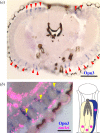
Figure 1.
Figure 1.
The expression pattern of the Opn3 homologue, MosOpn3 in the A. stephensi eye. (a) In situ hybridization analysis of MosOpn3 (Asop12) in the mosquito eye. The signals derived from MosOpn3 mRNA (arrowheads) were detected in a specific kind of photoreceptor cells in dorsal and ventral ommatidia. (b) Visualization of cell nuclei using the Hoechst stain (magenta) in the eye, in which MosOpn3-expressing cells were stained by in situ hybridization. The locations of MosOpn3-expressing cell nuclei (arrowheads) indicated that MosOpn3 is expressed in R7. The schematic drawing of mosquito ommatidium is also shown based on the previous report [32]. The lens is illustrated at the top of the ommatidium. R7 (blue) and other photoreceptor cells (yellow) with nuclei (magenta) are shown. Rhabdomeres were also indicated (grey). The scale bars represent 100 μm in (a) and 30 μm in (b).
Figure 2.
Figure 2.
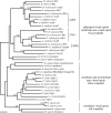
The phylogenetic relationship of insect opsins. The composite tree of the Gq-coupled opsin, the Opn3 and the Gt-coupled opsin groups was inferred with the NJ method. In addition to virtually all opsins of A. stephensi and D. melanogaster, several opsins representing each group are included. For the Opn3 group, representatives of the Opn3 homologues of insects also called c-opsin or pteropsin are included to show their wide distribution in insects. Insect Gq-coupled visual opsins are classified based on the spectral sensitivity, as shown on the right of the tree. LWS, UVS and SWS indicate the subgroups of LWS, UVS and SWS opsins, respectively. The MosOpn3 and Asop9 are highlighted with bold text. Bootstrap probabilities greater than 90% are indicated at each branch node. The scale bar represents 0.1 substitutions per site.
Figure 3.
Figure 3.
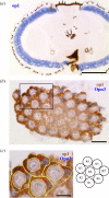
The expression pattern of the LWS visual opsin (Asop1) and MosOpn3 in A. stephensi eye. (a) In situ hybridization analysis of Asop1 in the mosquito eye. The signals derived from Asop1 mRNA (purple) were detected in many ommatidia around the whole eye. (b) Double in situ hybridization analysis of Asop1 and MosOpn3 (Asop12) in the transverse section of the mosquito eye. Expressions of Asop1 (brown) and MosOpn3 (purple) are visualized. (c) The enlarged image from the square in (b). The yellow traces indicate the landmarks of each ommatidium containing R1–R7. Judging from the schematic drawing of the transverse view of mosquito ommatidium modified from the previous report [32], Asop1 and MosOpn3 are exclusively expressed in R1–R6 and R7 (arrows), respectively. The scale bars represent 100 μm in (a), 20 μm in (b) and 10 μm in (c).
Figure 4.
Figure 4.
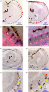
The expression pattern of the UVS (Asop8), the SWS (Asop9) visual opsins and MosOpn3 in the A. stephensi eye. (a,c) In situ hybridization analysis of Asop8 and Asop9 in the mosquito eye. The signals derived from Asop8 ((a), arrowheads) and Asop9 ((c), arrowheads) mRNAs were detected in a specific kind of photoreceptor cells in dorsal and ventral ommatidia and lateral ommatidia, respectively. (b,d) Visualization of cell nuclei using the Hoechst stain (magenta) in the eye, in which Asop8-expressing cells (b) or Asop9-expressing cells (d) were stained by in situ hybridization. The location of nuclei (arrowheads) indicate that Asop8 and Asop9 are expressed in R7 of dorsal and ventral ommatidia and lateral ommatidia, respectively. (e–h) Double in situ hybridization analysis of Asop8 and MosOpn3 (Asop12) (e,f) and Asop9 and MosOpn3 (g,h). (f) An enlarged image of (e), in which expressions of Asop8 (brown) and MosOpn3 (purple) are visualized, indicate the mutually exclusive expression of Asop8 and MosOpn3 in different R7. (h) An enlarged image of (g), in which expressions of Asop9 (brown) and MosOpn3 (purple) were visualized, indicates co-localization of Asop9 and MosOpn3 in the same R7 of dorsal and ventral ommatidia. Note that signals derived from Asop9 (arrows) and MosOpn3 (arrowheads) mRNAs are distributed differently in R7. The scale bars represent 100 μm in (a,c,e,g) and 30 μm in (b,d,f,h).
Figure 5.
Figure 5.
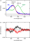
Spectroscopic characteristics of the mosquito blue-sensitive visual opsin (Asop9). (a) The absorption spectrum of Asop9-based pigment (blue), fitted with the rhodopsin-nomogram (red) [39] to estimate that the Asop9 forms a blue-sensitive pigment, having an absorption maximum at approximately 430 nm. The absorption spectrum of MosOpn3 having an absorption maximum at approximately 500 nm was also shown (green) [21]. (b) The difference absorption spectra, showing the spectral changes of the blue-sensitive Asop9-based pigment caused by irradiation with blue light (black) and subsequent yellow light (red). The photoresponses indicate bistability of Asop9, like other insect visual opsins and MosOpn3. Error bars (in light red and grey around the averaged values shown in dark red and black, respectively) indicate s.e. (n = 5). Note that Asop9-based pigment was reconstituted with 11-cis form of A1 retinal.

Similar articles

Cited by

References

    1. Borst A. 2009. Drosophila's view on insect vision. Curr. Biol. 19, R36-R47. (10.1016/j.cub.2008.11.001) - DOI - PubMed
    1. Hardie RC, Juusola M. 2015. Phototransduction in Drosophila. Curr. Opin. Neurobiol. 34, 37-45. (10.1016/j.conb.2015.01.008) - DOI - PubMed
    1. Terakita A. 2005. The opsins. Genome Biol. 6, 213. (10.1186/gb-2005-6-3-213) - DOI - PMC - PubMed
    1. Koyanagi M, Terakita A. 2014. Diversity of animal opsin-based pigments and their optogenetic potential. Biochim. Biophys. Acta 1837, 710-716. (10.1016/j.bbabio.2013.09.003) - DOI - PubMed
    1. Terakita A, Hariyama T, Tsukahara Y, Katsukura Y, Tashiro H. 1993. Interaction of GTP-binding protein Gq with photoactivated rhodopsin in the photoreceptor membranes of crayfish. FEBS Lett. 330, 197-200. (10.1016/0014-5793(93)80272-V) - DOI - PubMed

Publication types

LinkOut - more resources