. 2023 Feb;53(2):249-255.
doi: 10.1007/s00247-022-05498-1. Epub 2022 Sep 5.

Percutaneous treatment of chest wall chondroid hamartomas: the experience of a single center

Affiliations

Percutaneous treatment of chest wall chondroid hamartomas: the experience of a single center

Alessandro Inserra et al. Pediatr Radiol. 2023 Feb.

Abstract

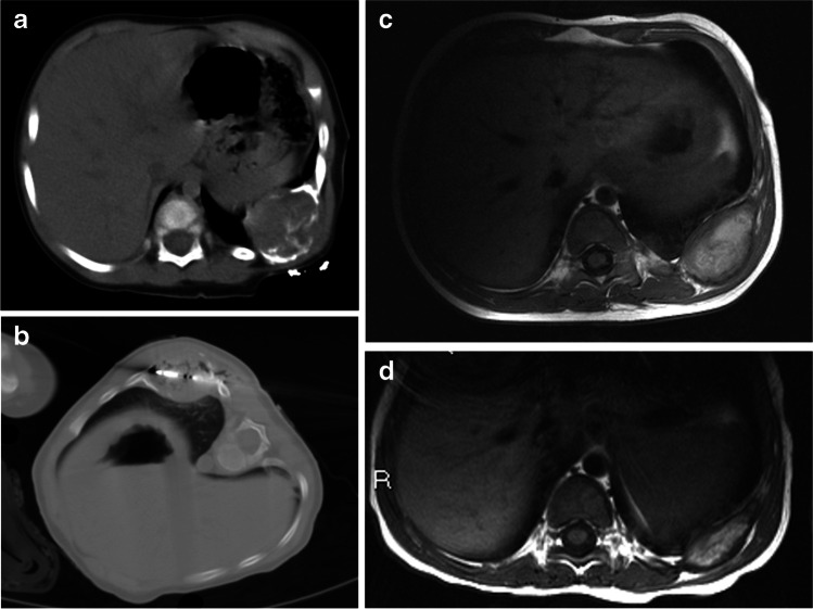
Background: Thoracic mesenchymal hamartomas are rare benign lesions. Rarely symptomatic, they may compress pulmonary parenchyma, leading to respiratory distress. Although spontaneous regression has been documented, the more common outcome is progressive growth. The treatment of choice is en bloc excision of the involved portion of the chest wall, frequently leading to significant deformity.

Objective: The aim of our study was to describe percutaneous techniques to treat these lesions.

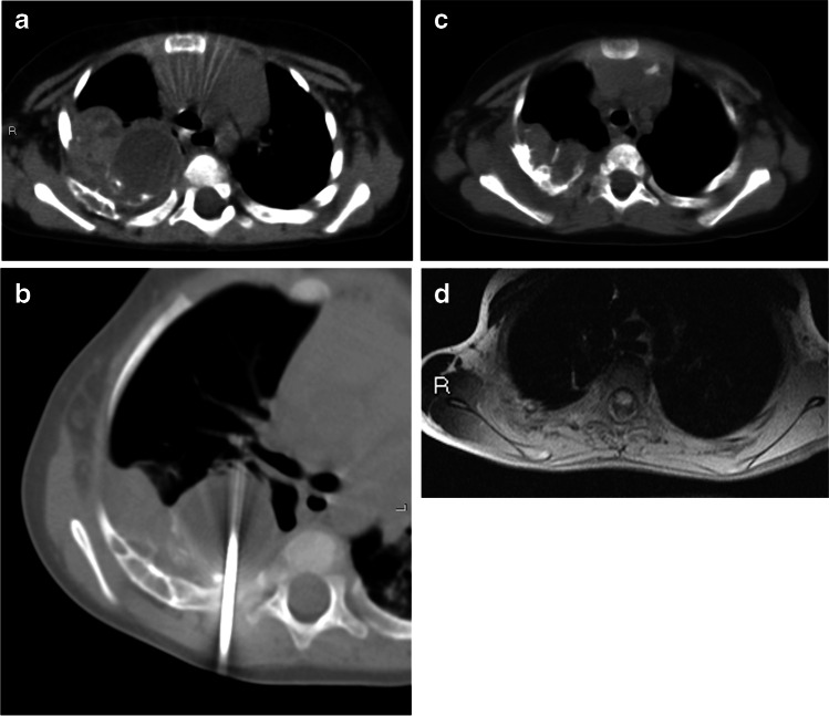
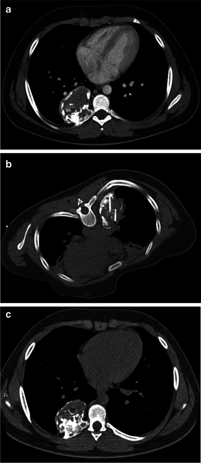
Materials and methods: We collected data of children with thoracic mesenchymal hamartomas who were treated at our institution from 2005 to 2020 using various percutaneous techniques. Techniques included radiofrequency thermoablation, microwave thermoablation (microwave thermoablation) and cryoablation.

Results: Five children were treated for chest wall hamartomas; one child showed bilateral localization of the mass. Two children underwent microwave thermoablation, one radiofrequency thermoablation and two cryoablation; one child treated with cryoablation also had radiofrequency thermoablation because mass volume increased after the cryoablation procedure. The median reduction of tumor volume was 69.6% (24.0-96.5%). One child treated with microwave thermoablation showed volumetric increase of the mass and underwent surgical removal of the tumor. No major complication was reported.

Conclusion: Percutaneous ablation is technically feasible for expert radiologists and might represent a valid and less invasive treatment for chest wall chondroid hamartoma, avoiding skeletal deformities.

Keywords: Chest wall; Children; Computed tomography; Cryosurgery; Hamartoma; Microwave; Radiofrequency ablation; Ribs; Thorax.


Conflict of interest statement

None

Figures

Fig. 1
Fig. 1
Mesenchymal hamartoma in a 2-week-old boy at diagnosis (patient 1) treated with microwave thermoablation. a Axial CT scan with contrast agent shows an inhomogeneously hypodense oval lesion involving the 5th–6th left ribs. b, c The boy underwent two microwave thermoablation sessions (axial CT images) followed by a dimensional increase in the mass, requiring surgical resection. d After 21-month follow-up, axial contrast-enhanced CT scan demonstrates almost complete resection of the lesion
Fig. 2
Fig. 2
Mesenchymal hamartoma in an 11-month-old boy (patient 2) treated with microwave thermoablation. a, b He presented with a clinical mass of the 9th–10th left ribs, as confirmed on axial CT (with contrast agent) at diagnosis (a), and he underwent microwave thermoablation (axial CT, b). c, d Follow-up MRI. At first (1-month) follow-up, axial T1-weighted MRI (c) shows an inhomogeneous hyperintense lesion, which decreased in dimension on the 9-month follow-up axial T1-weighted MRI (d)
Fig. 3
Fig. 3
Mesenchymal hamartoma in a 6-month-old girl (patient 3) treated with radiofrequency thermoablation. She presented with persistent respiratory distress requiring diagnostic studies including chest radiograph and, successively, CT scan. a Axial CT shows a inhomogeneous oval mass arising from the 1st–2nd right ribs. b Two sessions of radiofrequency thermoablation were performed (axial CT). c, d Six-month follow-up images show a significant dimensional decrease of the tumor, as a smaller inhomogeneous mass with calcifications at axial CT (c) and as hypo/isointense residual disease on axial T1-weighted MRI (d)
Fig. 4
Fig. 4
Incidental mesenchymal hamartoma in a 163-month-old boy (patient 4) treated with cryoablation. The mass, involving the 6th–7th–8th right ribs, was found at chest radiography. a Axial chest CT with contrast agent confirms the finding. b, c The boy underwent two sessions of cryoablation (intraoperative axial CT, b) with a slight dimensional increase in the lesion, as shown by the axial CT scan at last 18-month follow-up (c)
Fig. 5
Fig. 5
Mesenchymal hamartomas in a 4-month-old boy (patient 5) who was treated with cryoablation followed by radiofrequency thermoablation. He presented with acute respiratory distress and chest radiograph revealed bilateral masses of the thoracic wall. a, b Contrast-enhanced CT shows inhomogeneously hypodense oval lesions, with calcifications, arising from 4th–5th–6th right ribs and 4th left rib (a, axial; b, coronal). c He had cryoablation (axial CT image). Initial treatment was unsuccessful (tumor development and persistent dyspnea were seen), and he later underwent radiofrequency thermoablation. d, e Results were very good at 2-year follow-up, confirmed by contrast-enhanced axial (d) and coronal (e) CT images

References

    1. Andiran F, Ciftci AO, Senocak ME, Akcoren Z, Gogus S. Chest wall hamartoma: an alarming chest lesion with a benign course. J Pediatr Surg. 1998;33:727–729. doi: 10.1016/S0022-3468(98)90199-3. - DOI - PubMed
    1. Virgone C, Dall'Igna P, Alaggio R, Burnelli R, Zanon GF, Cecchetto G. Management of symptomatic mesenchymal hamartoma of the chest wall: surgical resection only in symptomatic cases. Klin Padiatr. 2013;225:420–422. doi: 10.1055/s-0033-1354355. - DOI - PubMed
    1. Bertocchini A, Falappa P, Accinni A, Devito R, Inserra A. Radiofrequency thermoablation in chest wall mesenchymal hamartoma of an infant. Ann Thorac Surg. 2007;84:2091–2093. doi: 10.1016/j.athoracsur.2007.06.042. - DOI - PubMed
    1. Amstalden EM, Carvalho RB, Pacheco EM, Oliveira-Filho A, Stragea-Neto L, Rosenberg AE. Chondromatous hamartoma of the chest wall: description of 3 new cases and literature review. Int J Surg Pathol. 2006;14:119–126. doi: 10.1177/106689690601400203. - DOI - PubMed
    1. Ayala AG, Ro JY, Bolio-Solis A, Hernandez-Batres F, Eftekhari F, Edeiken J. Mesenchymal hamartoma of the chest wall in infants and children: a clinicopathological study of five patients. Skelet Radiol. 1993;22:569–576. doi: 10.1007/BF00197137. - DOI - PubMed