. 2023 Jun 1;77(6):1968-1982.
doi: 10.1002/hep.32776. Epub 2022 Oct 12.

Interleukin-18 signaling promotes activation of hepatic stellate cells in mouse liver fibrosis

Affiliations

Interleukin-18 signaling promotes activation of hepatic stellate cells in mouse liver fibrosis

Jana Knorr et al. Hepatology. .

Abstract

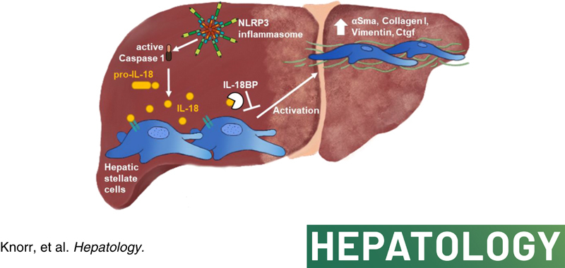
Background and aims: Nucleotide-binding oligomerization domain-like receptor-family pyrin domain-containing 3 (NLRP3) inflammasome activation has been shown to result in liver fibrosis. Mechanisms and downstream signaling remain incompletely understood. Here, we studied the role of IL-18 in hepatic stellate cells (HSCs), and its impact on liver fibrosis.

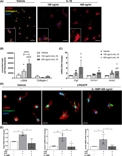
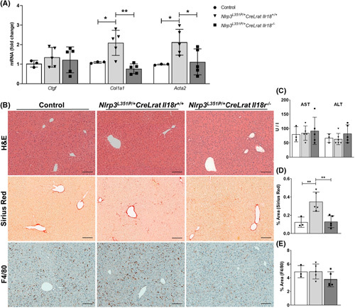
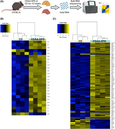
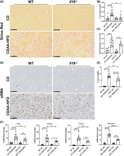
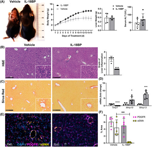
Approach and results: We observed significantly increased serum levels of IL-18 (128.4 pg/ml vs. 74.9 pg/ml) and IL-18 binding protein (BP; 46.50 ng/ml vs. 15.35 ng/ml) in patients with liver cirrhosis compared with healthy controls. Single cell RNA sequencing data showed that an immunoregulatory subset of murine HSCs highly expresses Il18 and Il18r1 . Treatment of cultured primary murine HSC with recombinant mouse IL-18 accelerated their transdifferentiation into myofibroblasts. In vivo , IL-18 receptor-deficient mice had reduced liver fibrosis in a model of fibrosis induced by HSC-specific NLRP3 overactivation. Whole liver RNA sequencing analysis from a murine model of severe NASH-induced fibrosis by feeding a choline-deficient, L-amino acid-defined, high fat diet showed that genes related to IL-18 and its downstream signaling were significantly upregulated, and Il18-/- mice receiving this diet for 10 weeks showed protection from fibrotic changes with decreased number of alpha smooth muscle actin-positive cells and collagen deposition. HSC activation triggered by NLRP3 inflammasome activation was abrogated when IL-18 signaling was blocked by its naturally occurring antagonist IL-18BP. Accordingly, we observed that the severe inflammatory phenotype associated with myeloid cell-specific NLRP3 gain-of-function was rescued by IL-18BP.

Conclusions: Our study highlights the role of IL-18 in the development of liver fibrosis by its direct effect on HSC activation identifying IL-18 as a target to treat liver fibrosis.


Conflict of interest statement

Frank Tacke advises and received grants from Gilead. He consults for Novo Nordisk and Pfizer. He is on the speakers’ bureau for Falk and AbbVie. He received grants from Inventiva, Bristol‐Myers Squibb, and Allergan. Hal M. Hoffman consults for Novartis.

Figures

None
Graphical abstract
FIGURE 1
FIGURE 1
IL‐18 signaling impacts liver fibrosis/cirrhosis in humans and mice. (A) Circulating IL‐18 levels in patients with NASH‐induced liver cirrhosis (128.4 pg/ml; 76.48–209.8 pg/ml) compared with healthy controls (74.9 pg/ml; 47.73–103.1 pg/ml). (B) Serum IL‐18BP of patients with liver cirrhosis (46.50 ng/ml; 34.89–64.07 ng/ml) in comparison with the control group (15.36 ng/ml; 13.50–17.84 ng/ml). Data shown: median; IQR. (Control, n = 50; Liver cirrhosis, n = 38; ****p < 0.0001; Mann–Whitney U test). T‐SNE plots mapping distribution of HSCs expressing (C) Il18 and (D) Il18r1. Activated HSCs from CCl4‐treated mice that induce liver fibrosis were differentiated into four subpopulations (MFB I to MFB IV) according to their different gene expression patterns. T‐SNE plots show that especially HSCs from the immunoregulatory MFB II cluster express Il18 and Il18r1. BP, binding protein; CCl4, carbon tetrachloride; HSC, hepatic stellate cell; IQR, interquartile range; MFB, myofibroblast; t‐SNE, T‐distributed stochastic neighbor embedding.
FIGURE 2
FIGURE 2
Stimulation with recombinant IL‐18 accelerates HSC activation in vitro, whereas blockage of IL‐18 signaling inhibits HSC activation. (A) Representative immunofluorescence images and (B) their quantification by the corrected total cell fluorescence showing αSMA and Collagen I of HSCs after treatment with either 100 ng/ml or 400 ng/ml rmIL‐18 after 24 h. (C) mRNA levels of Ctgf, Col1a1, and Acta2 of primary murine HSCs after rmIL‐18 stimulation. (D) Immunofluorescence images and (E) mRNA levels of Ctgf, Col1a1, and Acta2 of primary HSCs treated with LPS/ATP and additional pretreatment with rmIL‐18BP. αSMA, alpha smooth muscle actin; ATP, adenosine triphosphate; BP, binding protein; Col1a1, collagen type I alpha 1; Ctgf, connective tissue growth factor; HSC, hepatic stellate cell; LPS, lipopolysaccharide; rm, recombinant mouse. 3–4 independent replicates were included for all measured values; upper scale bars represent 50 μm. *p < 0.05; **p < 0.01; ****p < 0.0001.
FIGURE 3
FIGURE 3
IL‐18 receptor deficiency significantly reduced expression of the HSC activation marker in 24‐week‐old mice with HSC‐specific NLRP3 hyperactivation. (A) mRNA levels of Ctgf, Col1a1, and Acta2 in control, Nlrp3 L351P/+ CreLrat Il18r +/+ , and Nlrp3 L351P/+ CreLrat Il18r −/− mice. (B) Representative pictures of H&E‐, Sirius Red‐, and F4/80‐stained liver sections. (C) Liver aminotransferases and quantification of (D) Sirius Red and (E) F4/80‐positive cells in mice with Il‐18R deficiency compared with Nlrp3 L351P/+ CreLrat Il18r +/+ and control mice. Acta2, actin alpha 2; Col1a1, collagen type I alpha 1; Ctgf, connective tissue growth factor; F4/80, murine macrophage marker; H&E, hematoxylin–eosin; HSC, hepatic stellate cell; Lrat, lecithin retinol acyltransferase; NLRP3, nucleotide‐binding oligomerization domain leucine rich repeat containing receptor‐family pyrin domain‐containing 3. n = 3–5 mice per group for all measured values; scale bars represent 100 μm. *p < 0.05; **p < 0.01.
FIGURE 4
FIGURE 4
IL‐18 signaling and downstream regulatory network are activated in CDAA‐HFD–induced fibrotic NASH. Bulk RNA sequencing analysis of liver tissue after 10 weeks of CDAA‐HFD or CD. (A) An overview of experimental design. (B) Clustering of IL‐18 signaling‐related gene transcripts. (C) Clustering of fibrosis‐related gene transcripts. CD, control diet; CDAA‐HFD, choline‐deficient, L‐amino acid‐defined high fat diet. n = 4 mice per group.
FIGURE 5
FIGURE 5
IL‐18 deficiency reduced CDAA‐HFD–induced fibrosis. (A, B) Collagen deposition assessed by Sirius Red staining and (C) hydroxyproline, (D, E) αSMA‐positive cells, and (F) mRNA expression levels of the profibrotic genes Ctgf, Col3a, Timp1, and Vimentin in WT and Il18 −/− mice fed with CDAA‐HFD compared with mice on CD. αSMA, alpha smooth muscle actin; CD, control diet; CDAA‐HFD, choline‐deficient, L‐amino acid‐defined high fat diet; Ctgf, connective tissue growth factor; Col3a, collagen type III alpha 1 chain; Timp1, tissue inhibitor of matrix metalloproteinase 1; WT, wild type. n = 5–8 mice per group for all measured values; scale bars represent 250 μm. *p < 0.05; **p < 0.01; ***p < 0.001; ****p < 0.0001.
FIGURE 6
FIGURE 6
Treatment of Nlrp3 D301N CreL mice with IL‐18BP rescued highly inflammatory phenotype. Nlrp3 D301N CreL mice were treated with 6 μg/g rhIL‐18BP (Tadekinig Alfa) or PBS from day 2 to 14. (A) Growth impairment, rash, and reduced body weight, consistent with the phenotype of Nlrp3 D301N CreL mice, were clearly normalized by IL‐18BP treatment, whereas serum alanine aminotransferase (ALT) did not significantly change. (B) H&E‐stained liver sections and quantification showing less inflammation in IL‐18BP‐treated mice. (C) Sirius Red staining. (D) mRNA expression of ECM regulating enzymes (Timp1, Mmp10, Mmp13) were measured from total liver lysate. (E) Immunofluorescence staining and (F) quantification of PDGFR and αSMA showing more activated HSCs in the nontreated mice. αSMA, alpha smooth muscle actin; BP, binding protein; ECM, extracellular matrix; H&E, hematoxylin–eosin; HSCs, hepatic stellate cells; Mmp, matrix metalloproteinase; Nlrp3, nucleotide‐binding oligomerization domain leucine rich repeat containing receptor‐family pyrin domain‐containing 3; PDGFR, platelet‐derived growth factor receptor; Timp1, tissue inhibitor of matrix metalloproteinase 1; rh, recombinant human. n = 5–6 mice per group for all measured values; black scale bars represent 250 μm; *p < 0.05; **p < 0.01; ***p < 0.001.

References

    1. Okamura H, Tsutsi H, Komatsu T, Yutsudo M, Hakura A, Tanimoto T, et al. . Cloning of a new cytokine that induces IFN‐gamma production by T cells. Nature. 1995;378(6552):88–91. - PubMed
    1. Ghayur T, Banerjee S, Hugunin M, Butler D, Herzog L, Carter A, et al. . Caspase‐1 processes IFN‐gamma‐inducing factor and regulates LPS‐induced IFN‐gamma production. Nature. 1997;386(6625):619–23. - PubMed
    1. Guo H, Callaway JB, Ting JPY. Inflammasomes: mechanism of action, role in disease, and therapeutics. Nat Med. 2015;21(7):677–87. - PMC - PubMed
    1. Puren AJ, Fantuzzi G, Dinarello CA. Gene expression, synthesis, and secretion of interleukin 18 and interleukin 1beta are differentially regulated in human blood mononuclear cells and mouse spleen cells. Proc Natl Acad Sci USA. 1999;96(5):2256–61. - PMC - PubMed
    1. Mehta VB, Hart J, Wewers MD. ATP‐stimulated release of interleukin (IL)‐1beta and IL‐18 requires priming by lipopolysaccharide and is independent of caspase‐1 cleavage. J Biol Chem. 2001;276(6):3820–6. - PubMed

Publication types