. 2022 Jul 28;4(4):e220100.
doi: 10.1148/ryct.220100. eCollection 2022 Aug.

Intrapericardial Paraganglioma

Affiliations

Intrapericardial Paraganglioma

Lucas de Pádua Gomes de Farias et al. Radiol Cardiothorac Imaging. .
No abstract available

Keywords: CT Angiography; Cardiac; Heart; Image Postprocessing; Neoplasms; PET/CT; Pericardium.


Conflict of interest statement

Disclosures of conflicts of interest: L.d.P.G.d.F. No relevant relationships. G.B.d.S.T. No relevant relationships. L.d.P.S.B. No relevant relationships. A.S.d.A. No relevant relationships.

Figures
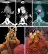
Images in a 56-year-old asymptomatic man referred for an elective coronary
calcium score stratification. (A, B) Axial CT scans, (C) axial two-dimensional
fluorine 18 (18F) fluorodeoxyglucose (FDG) PET/CT scan, and (D, E) tridimensional
reconstructions show a low-attenuation nodule (arrow), measuring 2.6 cm, with
intense and heterogeneous postcontrast enhancement, central nonenhancing areas, and
18F-FDG uptake (maximum standardized uptake value, 9.3) surrounded by the inferior
border of aortic arch, the posterior border of ascending aorta, the upper border of
the right main pulmonary artery, and the anterior border of the trachea and proximal
left main bronchus. Note the paraganglioma relationship with the posterior portion
of the superior aortic recess (arrowheads).
Figure 1:
Images in a 56-year-old asymptomatic man referred for an elective coronary calcium score stratification. (A, B) Axial CT scans, (C) axial two-dimensional fluorine 18 (18F) fluorodeoxyglucose (FDG) PET/CT scan, and (D, E) tridimensional reconstructions show a low-attenuation nodule (arrow), measuring 2.6 cm, with intense and heterogeneous postcontrast enhancement, central nonenhancing areas, and 18F-FDG uptake (maximum standardized uptake value, 9.3) surrounded by the inferior border of aortic arch, the posterior border of ascending aorta, the upper border of the right main pulmonary artery, and the anterior border of the trachea and proximal left main bronchus. Note the paraganglioma relationship with the posterior portion of the superior aortic recess (arrowheads).
Intrapericardial paraganglioma. (A) Photomicrograph shows encapsulated
paraganglioma with tumor cells arranged in solid nests with sustentacular cells,
vascular stroma, and abundant eosinophilic cytoplasm, without mitotic figures,
involvement of intracapsular vessel, or areas of necrosis. (Hematoxylin-eosin stain;
original magnification, ×100; inset original magnification, ×400.) (B)
The tumor is diffusely positive for chromogranin-A. (Chromogranin-A
immunohistochemical staining; original magnification, ×250.) (C) The
sustentacular cells are prominent with S100 staining. (Original magnification,
×400.)
Figure 2:
Intrapericardial paraganglioma. (A) Photomicrograph shows encapsulated paraganglioma with tumor cells arranged in solid nests with sustentacular cells, vascular stroma, and abundant eosinophilic cytoplasm, without mitotic figures, involvement of intracapsular vessel, or areas of necrosis. (Hematoxylin-eosin stain; original magnification, ×100; inset original magnification, ×400.) (B) The tumor is diffusely positive for chromogranin-A. (Chromogranin-A immunohistochemical staining; original magnification, ×250.) (C) The sustentacular cells are prominent with S100 staining. (Original magnification, ×400.)

References

    1. Hamilton BH , Francis IR , Gross BH , et al. . Intrapericardial paragangliomas (pheochromocytomas): imaging features. AJR Am J Roentgenol 1997;168(1):109–113. - PubMed
    1. Araoz PA , Mulvagh SL , Tazelaar HD , Julsrud PR , Breen JF . CT and MR imaging of benign primary cardiac neoplasms with echocardiographic correlation. RadioGraphics 2000;20(5):1303–1319. - PubMed
    1. Yadav PK , Baquero GA , Malysz J , Kelleman J , Gilchrist IC . Cardiac paraganglioma. Circ Cardiovasc Interv 2014;7(6):851–856. - PubMed
    1. Janssen I , Blanchet EM , Adams K , et al. . Superiority of [68Ga]-DOTATATE PET/CT to Other Functional Imaging Modalities in the Localization of SDHB-Associated Metastatic Pheochromocytoma and Paraganglioma. Clin Cancer Res 2015;21(17):3888–3895. - PMC - PubMed
    1. Jha A , Patel M , Saboury B , et al. . Superiority of 68Ga-DOTATATE PET/CT compared to 18F-FDG PET/CT and MRI of the spine in the detection of spinal bone metastases in metastatic pheochromocytoma and/or paraganglioma. J Nucl Med 2020;61(supplement 1):125.https://jnm.snmjournals.org/content/61/supplement_1/125.

LinkOut - more resources