. 2022 Aug 17:13:964263.
doi: 10.3389/fimmu.2022.964263. eCollection 2022.

Immunosuppressant exposure confounds gene expression analysis in systemic lupus erythematosus

Affiliations

Immunosuppressant exposure confounds gene expression analysis in systemic lupus erythematosus

Melissa Northcott et al. Front Immunol. .

Abstract

Objectives: The analysis of gene module expression in SLE is emerging as a tool to identify active biological pathways, with the aim of developing targeted therapies for subsets of patients. Detailed information on the effect of immunosuppressants on gene module expression is lacking. We aimed to examine the impact of medication exposure on gene module expression.

Methods: A set of commercially available disease-relevant gene modules were measured in 730 whole blood samples from a dedicated lupus clinic on whom prospectively collected, contemporaneous clinical data including medication exposure were available.

Results: Compared to heathy controls, SLE patients showed over-expression of IFN and under-expression of B cell, T cell and pDC modules. Neutrophil module over-expression and under-expression of B and T cell modules were observed in patients with active lupus nephritis or highly active disease (SLEDAI-2K > 8), while Lupus Low Disease Activity State (LLDAS) had inverse associations. Disease activity in other organ domains was not associated with specific gene modules. In contrast, medications were associated with multiple effects. Glucocorticoid use was associated with under-expression of T cell, B cell and plasmablast modules, and over-expression of neutrophil modules. Mycophenolate and azathioprine exposure were associated with plasmablast module and B cell module under-expression respectively. Disease activity associations with neutrophil over-expression and lymphocyte module under-expression were attenuated by multivariable adjustment for medication exposure.

Conclusion: Medications have significant effect on gene module expression in SLE patients. These findings emphasize the need to control for medications in studies of gene expression in SLE.

Keywords: gene modules; glucocorticoids; immunosuppressants; systemic lupus erythematosus; transcriptional profiling.


Conflict of interest statement

Outside the scope of this work, EM has received consulting fees from AbbVie, AstraZeneca, Amgen, Biogen, Bristol Myers Squibb, Eli Lilly, EMD Serono, Genetech, Janssen, Servier and UCB, and research grants from AbbVie, AstraZeneca, Bristol Myers Squibb, Eli Lilly, EMD Serono and Janssen. AH has received research grants from AstraZeneca, Merck and GSK, and consulting fees from Abbvie, AstraZeneca, BMS, GSK, Janssen and Pfizer. The remaining authors declare that the research was conducted in the absence of any commercial or financial relationships that could be construed as a potential conflict of interest.

Figures

Figure 1
Figure 1
Gene module expression in SLE patients compared to healthy controls depicted (A) as dot plots and (B) as relative log2 fold change (log2FC). Blue symbols represent modules that were significantly under-expressed, grey symbols modules that were not significantly different and red symbols represent modules that were significantly over-expressed in SLE patients compared with controls. Horizontal bars indicate 95% confidence intervals (95%CI).
Figure 2
Figure 2
Univariate analysis depicting log2FC gene expression in SLE patients associated with (A) demographic factors, (B) disease manifestations at any time point in their documented disease course, (C) medication exposure at the time of sample collection, (D) SLEDAI-2K organ domain disease activity at time of sample collection and (E) composite disease activity measurements at the time of sample collection. * p<0.05 **p<0.01 ***p<0.001. Asian ancestry backgrounds are compared to European ancestry.
Figure 3
Figure 3
Gene module expression associations with disease activity (SLEDAI-2K) (expressed as log2FC per SLEDAI point) in (A) univariate analysis, (B) analysis adjusted for prednisolone dose (pred), (C) analysis adjusted for prednisolone and mycophenolate (MMF) and (D) analysis adjusted for prednisolone and azathioprine (AZA) exposure. Blue symbols represent statistically significant under-expression of gene module, grey symbols represent results that were not significant, and red symbols represent statistically significant over-expression.
Figure 4
Figure 4
(A) Neutrophil module expression in patients with and without active renal disease and their associated prednisolone dose. (B) B cell module expression in patients with and without active renal disease and their associated prednisolone dose. (C) Expression of gene modules in patients with active renal disease compared to those without active renal disease in univariate analysis and (D) analysis adjusted for prednisolone exposure. (E) B cell module expression in patients with and without renal disease and their associated azathioprine use. (F) Expression of gene modules in patients with active renal disease compared to those without active renal disease adjusted for prednisolone and azathioprine (AZA) exposure and (G) prednisolone and mycophenolate (MMF) exposure.
Figure 5
Figure 5
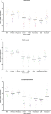
Gene module expression in patients taking (A) rituximab, (B) belimumab and (C) cyclophosphamide. Grey dots represent samples from patients not taking these medications, colored dots represent samples taken from patients taking these medications. On each graph, each color represents samples from a single patient.

References

    1. Apostolopoulos D, Morand EF. It hasn't gone away: the problem of glucocorticoid use in lupus remains. Rheumatol (Oxford England) (2017) 56(suppl_1):i114–i22. doi: 10.1093/rheumatology/kew406 - DOI - PubMed
    1. Nakano M, Iwasaki Y, Fujio K. Transcriptomic studies of systemic lupus erythematosus. Inflammation Regeneration (2021) 41(1):11. doi: 10.1186/s41232-021-00161-y - DOI - PMC - PubMed
    1. Chaussabel D, Quinn C, Shen J, Patel P, Glaser C, Baldwin N, et al. A modular analysis framework for blood genomics studies: application to systemic lupus erythematosus. Immunity (2008) 29(1):150–64. doi: 10.1016/j.immuni.2008.05.012 - DOI - PMC - PubMed
    1. Bennett L, Palucka AK, Arce E, Cantrell V, Borvak J, Banchereau J, et al. Interferon and granulopoiesis signatures in systemic lupus erythematosus blood. J Exp Med (2003) 197(6):711–23. doi: 10.1084/jem.20021553 - DOI - PMC - PubMed
    1. Northcott M, Jones S, Koelmeyer R, Bonin J, Vincent F, Kandane-Rathnayake R, et al. Type 1 interferon status in systemic lupus erythematosus: a longitudinal analysis. Lupus Sci Med (2022) 9(1):1–12. doi: 10.1136/lupus-2021-000625 - DOI - PMC - PubMed

Publication types

MeSH terms

Substances