. 2022 Aug 18:13:978851.
doi: 10.3389/fimmu.2022.978851. eCollection 2022.

MX2: Identification and systematic mechanistic analysis of a novel immune-related biomarker for systemic lupus erythematosus

Affiliations

MX2: Identification and systematic mechanistic analysis of a novel immune-related biomarker for systemic lupus erythematosus

Xiang-Wen Meng et al. Front Immunol. .

Abstract

Background: Systemic lupus erythematosus (SLE) is an autoimmune disease that involves multiple organs. However, the current SLE-related biomarkers still lack sufficient sensitivity, specificity and predictive power for clinical application. Thus, it is significant to explore new immune-related biomarkers for SLE diagnosis and development.

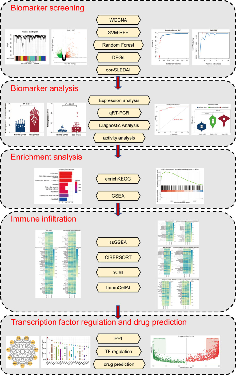
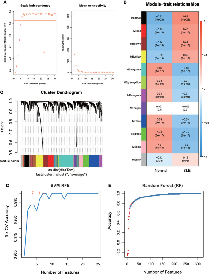
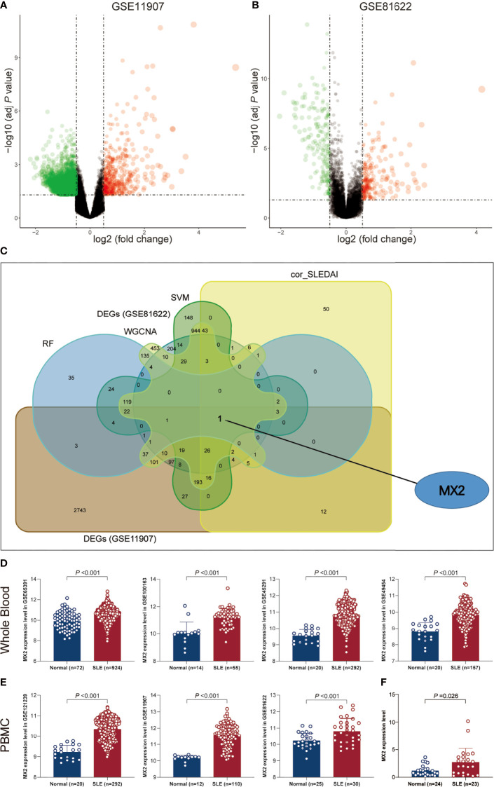
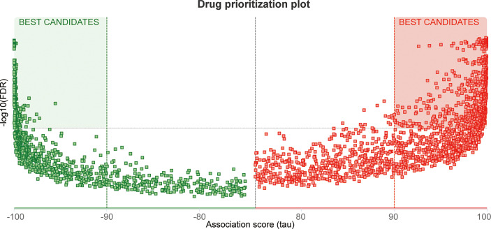
Methods: We obtained seven SLE gene expression profile microarrays (GSE121239/11907/81622/65391/100163/45291/49454) from the GEO database. First, differentially expressed genes (DEGs) were screened using GEO2R, and SLE biomarkers were screened by performing WGCNA, Random Forest, SVM-REF, correlation with SLEDAI and differential gene analysis. Receiver operating characteristic curves (ROCs) and AUC values were used to determine the clinical value. The expression level of the biomarker was verified by RT‒qPCR. Subsequently, functional enrichment analysis was utilized to identify biomarker-associated pathways. ssGSEA, CIBERSORT, xCell and ImmuCellAI algorithms were applied to calculate the sample immune cell infiltration abundance. Single-cell data were analyzed for gene expression specificity in immune cells. Finally, the transcriptional regulatory network of the biomarker was constructed, and the corresponding therapeutic drugs were predicted.

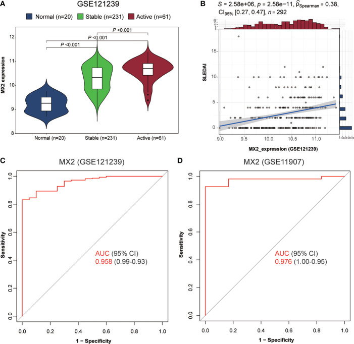
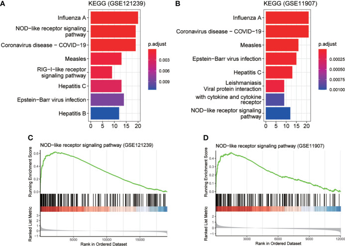
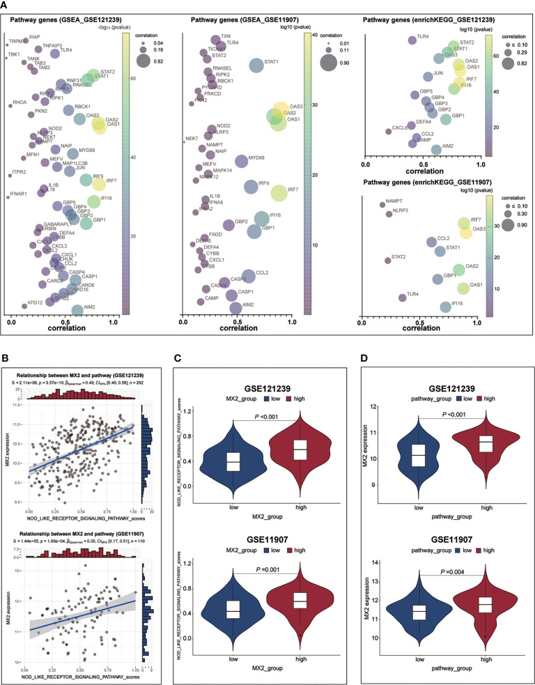
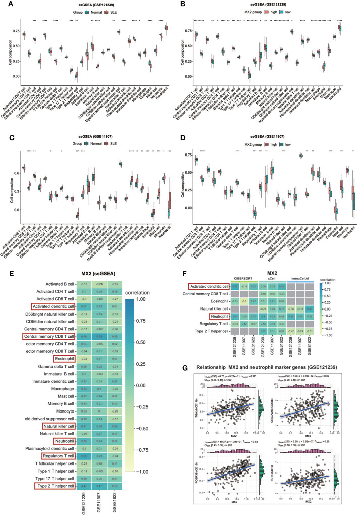
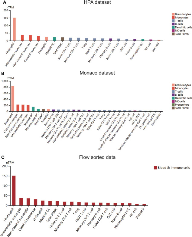
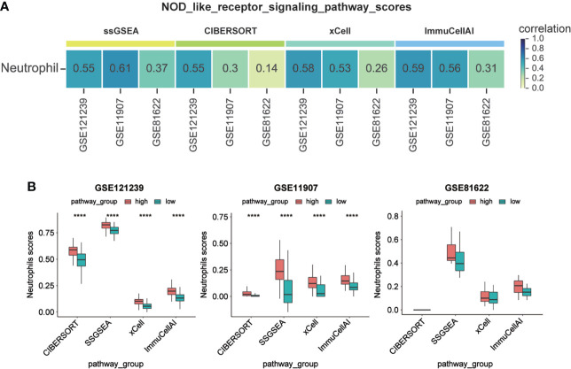
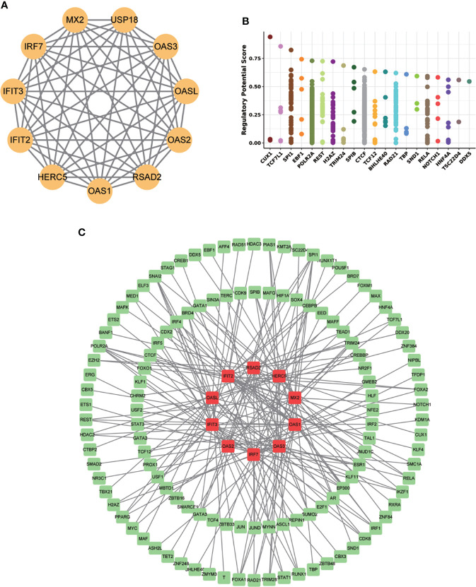
Results: Multiple algorithms were screened together for a unique marker gene, MX2, and expression analysis of multiple datasets revealed that MX2 was highly expressed in SLE compared to the normal group (all P < 0.05), with the same trend validated by RT‒qPCR (P = 0.026). Functional enrichment analysis identified the main pathway of MX2 promotion in SLE as the NOD-like receptor signaling pathway (NES=2.492, P < 0.001, etc.). Immuno-infiltration analysis showed that MX2 was closely associated with neutrophils, and single-cell and transcriptomic data revealed that MX2 was specifically expressed in neutrophils. The NOD-like receptor signaling pathway was also remarkably correlated with neutrophils (r >0.3, P < 0.001, etc.). Most of the MX2-related interacting proteins were associated with SLE, and potential transcription factors of MX2 and its related genes were also significantly associated with the immune response.

Conclusion: Our study found that MX2 can serve as an immune-related biomarker for predicting the diagnosis and disease activity of SLE. It activates the NOD-like receptor signaling pathway and promotes neutrophil infiltration to aggravate SLE.

Keywords: MX2; biomarker; immune infiltration; machine learning; systemic lupus erythematosus.


Conflict of interest statement

The authors declare that the research was conducted in the absence of any commercial or financial relationships that could be construed as a potential conflict of interest.

Figures

Figure 1
Figure 1
The flowchart of the analysis process.
Figure 2
Figure 2
SLE biomarker screening. (A) Analysis of the scale-free fit index and the mean connectivity for various soft-thresholding powers. (B) Relationships of consensus modules with samples. It contains a set of highly linked genes. Each specified color represents a specific gene module. (C) Clustering dendrogram of differentially expressed genes related to SLE. (D), (E) Based on support vector machine-recursive feature elimination (SVM -RFE) and random forest (RF) algorithm to screen biomarkers.
Figure 3
Figure 3
SLE biomarker screening. (A) Volcano plots of DEGs distribution in GSE11907. (B) Volcano plots of DEGs distribution in GSE81622. (C) Venn diagram showed the intersection of candidate biomarkers obtained by the five algorithms. (D) Expression levels of MX2 in SLE and normal donors in different whole blood datasets. (E) Expression levels of MX2 in SLE and normal donors in different PBMC datasets. (F) The MX2 expression was validated by qRT-PCR.
Figure 4
Figure 4
MX2 was highly expressed in SLE and served as a biomarker for diagnosis and disease activity. (A) Comparison of MX2 expression in normal and different stages of SLE. (B) Correlation of MX2 with SLEDAI. (C) The ROC curve for diagnostic efficacy validation of MX2 in GSE121239. (D) The ROC curve for diagnostic efficacy validation of MX2 in GSE11907.
Figure 5
Figure 5
MX2 activated NOD-like receptor signaling pathway in SLE. (A), (B) enrichKEGG pathway analysis results of MX2. (C), (D) GSEA analysis results of MX2.
Figure 6
Figure 6
MX2 activated NOD-like receptor signaling pathway in SLE. (A) Correlation between MX2 and NOD-like receptor signaling pathway genes. (B) Correlation between MX2 and NOD-like receptor signaling pathway scores. (C) Comparison of the distribution of NOD-like receptor signaling pathway scores between high and low MX2 expression subgroups. (D) Comparison of MX2 expression between high and low subgroups of the NOD-like receptor signaling pathway scores.
Figure 7
Figure 7
MX2 was mainly positively associated with neutrophil infiltration. (A), (C) Comparison of the ssGSEA scores for immune cells estimated between all samples with Normal and SLE in the GSE121239. (B), (D) Comparison of the ssGSEA scores for immune cells estimated between patients with high and low MX2 expression subgroups in the GSE121239. (E) Correlation between MX2 and infiltrating immune cells in three datasets (ssGSEA). (F) Correlation between MX2 and seven types of infiltrating immune cells in three datasets (CIBERSORT, xCell and ImmuCellAI). The gray area indicates that the algorithm does not contain this type of cell. (G) Correlation between MX2 and neutrophil marker genes. (*P <0.05, **P <0.01, ***P <0.001, ****P <0.0001).
Figure 8
Figure 8
MX2 was mainly positively associated with neutrophil infiltration. (A) Expression of MX2 in immune cells in the HPA dataset. (B) Expression of MX2 in immune cells in the Monaco dataset. (C) Expression of MX2 in immune cells in the flow sorted data from HPA database.
Figure 9
Figure 9
Activation of NOD-like receptor signaling pathway promoted neutrophil infiltration. (A) Correlation between NOD-like receptor signaling pathway scores and Neutrophil infiltration scores. (B) Comparison of Neutrophil infiltration scores between high and low subgroups of the NOD-like receptor signaling pathway scores. (****P <0.0001).
Figure 10
Figure 10
MX2 regulatory mechanism network. (A) The MX2 PPI network constructed by the STRING database. (B) The top 20 transcription factors regulating MX2 in the Cistrome DB database. (C) Protein-interacting gene-transcription factor (TF) network. Red represents genes and green represents TFs.
Figure 11
Figure 11
Drug screening. Heatmap of the distribution of potential therapeutic drugs.

Similar articles

Cited by

References

    1. Ruiz-Irastorza G, Khamashta MA, Castellino G, Hughes GR. Systemic lupus erythematosus. Lancet (2001) 357(9261):1027–32. doi: 10.1016/S0140-6736(00)04239-2 - DOI - PubMed
    1. Barber MRW, Drenkard C, Falasinnu T, Hoi A, Mak A, Kow NY, et al. . Global epidemiology of systemic lupus erythematosus. Nat Rev Rheumatol (2021) 17(9):515–32. doi: 10.1038/s41584-021-00668-1 - DOI - PMC - PubMed
    1. Guo G, Chen A, Ye L, Wang H, Chen Z, Yan K, et al. . TCONS_00483150 as a novel diagnostic biomarker of systemic lupus erythematosus. Epigenomics (2020) 12(11):973–88. doi: 10.2217/epi-2019-0265 - DOI - PubMed
    1. Guo G, Wang H, Ye L, Shi X, Yan K, Lin K, et al. . Hsa_circ_0000479 as a novel diagnostic biomarker of systemic lupus erythematosus. Front Immunol (2019) 10:2281. doi: 10.3389/fimmu.2019.02281 - DOI - PMC - PubMed
    1. Leu CM, Hsu TS, Kuo YP, Lai MZ, Liu PC, Chen MH, et al. . Deltex1 suppresses T cell function and is a biomarker for diagnosis and disease activity of systemic lupus erythematosus. Rheumatol (Oxford) (2019) 58(4):719–28. doi: 10.1093/rheumatology/key418 - DOI - PubMed

MeSH terms