Review
. 2022 Aug 17:12:968351.
doi: 10.3389/fonc.2022.968351. eCollection 2022.

Metabolic management of microenvironment acidity in glioblastoma

Affiliations
Review

Metabolic management of microenvironment acidity in glioblastoma

Thomas N Seyfried et al. Front Oncol. .

Abstract

Glioblastoma (GBM), similar to most cancers, is dependent on fermentation metabolism for the synthesis of biomass and energy (ATP) regardless of the cellular or genetic heterogeneity seen within the tumor. The transition from respiration to fermentation arises from the documented defects in the number, the structure, and the function of mitochondria and mitochondrial-associated membranes in GBM tissue. Glucose and glutamine are the major fermentable fuels that drive GBM growth. The major waste products of GBM cell fermentation (lactic acid, glutamic acid, and succinic acid) will acidify the microenvironment and are largely responsible for drug resistance, enhanced invasion, immunosuppression, and metastasis. Besides surgical debulking, therapies used for GBM management (radiation, chemotherapy, and steroids) enhance microenvironment acidification and, although often providing a time-limited disease control, will thus favor tumor recurrence and complications. The simultaneous restriction of glucose and glutamine, while elevating non-fermentable, anti-inflammatory ketone bodies, can help restore the pH balance of the microenvironment while, at the same time, providing a non-toxic therapeutic strategy for killing most of the neoplastic cells.

Keywords: fermentation; glutamate; glutaminolysis; glycolysis; ketogenic diet; ketogenic metabolic therapy; lactate; succinate.


Conflict of interest statement

Author KM was employed by the company BERG LLC. Author SL was employed by the company Matterworks. The remaining authors declare that the research was conducted in the absence of any commercial or financial relationships that could be construed as the potential conflict of interest.

Figures

Figure 1
Figure 1
Kaplan-Meier plots for overall survival of GBM patients across five (–5), Canadian surgical institutions. Each line represents patient survival for a particular institution as described (8). The GBM survival statistics recorded for these Canadian institutions are similar to those recorded in surgical institutions of other countries (2, 9, 10). These findings support the view of no major improvement of GBM patient survival in almost 100 years (8, 11). Image reproduced under a Creative Commons license from (8).
Figure 2
Figure 2
Morphological abnormalities seen in GBM mitochondria from the work of Deighton et al. (15). The morphology of 150 mitochondria was assessed in six GBM samples and in seven peri-tumoral control samples using Electron Microscopy (EM). (A) Percentage of normal mitochondria where cristae were visible throughout the mitochondria in peri-tumoral control and GBM samples (each bar represents one sample; *** p-value = 0.0001); (B) Percentage of abnormal mitochondria where cristae were sparse and abnormal in peri-tumoral control and GBM samples; *** p-value = 0.0001). (C, D) Representative EM images of normal and abnormal mitochondria, respectively. Cristolysis was significantly greater in mitochondria from GBM tissue than in mitochondria from normal surrounding brain tissue. The scale bars represent 0.5 um. The authors reported 117 mitochondrial proteins altered in GBM in association with ultrastructural mitochondrial abnormalities, similar to those described previously by Arismendi-Morillo et al. (16). ATP synthesis through OxPhos cannot be normal in tumor cells with these abnormalities. Image reproduced under a Creative Commons license from Deighton et al. (15).
Figure 3
Figure 3
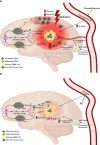
Origin and management of microenvironment acidity in GBM. (A) Glucose and glutamine are the primary energy metabolites necessary for driving rapid GBM growth. Glucose is the metabolic fuel necessary for nearly all brain functions under normal physiological conditions and is the major source of carbons for biomass synthesis through the glycolytic and pentose phosphate pathways in tumor cells. Tumor cells metabolize glutamine to glutamate, which is then metabolized to alpha-ketoglutarate. Significant energy is generated from the succinyl CoA ligase reaction (substrate level phosphorylation) in the glutaminolysis pathway using alpha-ketoglutarate-derived succinyl CoA as substrate (see Figure 7 ). In contrast to extracranial tissues, where glutamine is the most available amino acid, glutamine is tightly regulated in the brain through its involvement in the glutamate-glutamine cycle of neurotransmission (1, 78, 79). Glutamate is a major excitatory neurotransmitter that must be cleared rapidly following synaptic release in order to prevent excitotoxic damage to neurons (, –81). Glial cells possess transporters for the clearance of extracellular glutamate, which is then metabolized to glutamine for delivery back to neurons. Neurons metabolize the glutamine to glutamate, which is then repackaged into synaptic vesicles for synaptic release (1). This cycle maintains low extracellular levels of both glutamate and glutamine in the normal neural parenchyma. Disruption of the cycle can provide neoplastic GBM cells access to glutamine. Besides serving as a metabolic fuel for the neoplastic tumor cells, glutamine is also an important fuel for cells of myeloid linage, which include macrophages, monocytes, microglia, and especially the invasive mesenchymal cells in GBM (, , –84). In contrast to the proliferative GBM stem cells, the neoplastic GBM mesenchymal cells are thought to be derived from microglia or from microglia-stem cell fusion hybrids, which would have immuno-suppressive properties (82, 85). As long as GBM cells have access to glucose and glutamine, the tumor will grow and acidify the microenvironment making long-term management difficult. The current treatments for GBM (radiation and TMZ chemotherapy) will further increase glucose and glutamine availability, creating an unnecessary metabolic storm that will enhance microenvironment acidification and rapid tumor recurrence. The red hue is indicative of the inflammation and acidification of the tumor microenvironment (see text for further details). (B) The simultaneous restriction of glucose and glutamine, while elevating non-fermentable, anti-inflammatory ketone bodies, will reduce acidification, restore the pH balance of the microenvironment, and growth arrest or kill most of the neoplastic cells (–13). RAC, reactive astrocytes; TAM, tumor-associated macrophages; Gln, glutamine; Glu, glutamate. These images were modified from that in (86).
Figure 4
Figure 4
Influence of diet on the intracerebral growth of the CT-2A malignant astrocytoma. Dietary treatment was initiated 1 day after tumor implantation and was continued for 13 days. The visual representation (A) and quantitative assessment (B) of tumor growth in C57BL/6J mice receiving either the standard diet (SD) or ketogenic diet (KD) under either unrestricted (UR) or restricted (R) feeding. The asterisk indicates that the dry weights of the tumours in R groups were significantly lower than those in the UR groups at P < 0.01. (C) Linear regression analysis of plasma glucose and CT-2A-tumor growth in mice from both the SD and KD dietary groups combined (n = 34). These analyses included the values for plasma glucose and tumor growth of individual mice from both the UR and R-fed groups. The linear regression was highly significant at P < 0.001, indicating that blood glucose levels predict CT-2A tumor growth rate (100). The failure of the KD-UR to reduce blood glucose levels and tumor growth could be due to insulin insensitivity in this mouse strain (102). Images reproduced under a Creative Commons license.
Figure 5
Figure 5
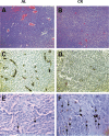
Influence of calorie restriction (CR) on microvessel density and apoptosis in the CT-2A malignant astrocytoma. CR was initiated 7 days before intracerebral tumor implantation and was continued for 11 days. H & E stained tumor sections in an ad libitum (AL) mouse (A) and in a CR mouse (B) (100X). Factor VIII immunostaining from the tumor grown in an AL mouse (C) and in a CR mouse (D) (200X). TUNEL positive apoptotic cells (arrows) from the tumor grown in an AL mouse (E) and in a CR mouse (F) (400X). Each stained section was representative of the entire tumor. All images were produced from digital photography. Image reproduced under a Creative Commons license from (119).
Figure 6
Figure 6
Influence of diet/drug therapy on overall survival of VM/Dk mice with the VM-M3 invasive GBM. A calorie restricted ketogenic diet (KD-R) was administered together with the glutaminase inhibitor, 6-diazo-5-oxo-L-norleucine (DON) as we described (13). Overall survival was significantly longer in the tumor bearing mice receiving the diet/drug combination (KD-R + DON) than in the mice receiving the standard high-carbohydrate diet (SD-UR), the KD-R alone, or DON alone. It is important to mention that 2-3x more DON was delivered to the tumor of the mice fed the KD-R than to the mice fed the SD-UR indicating that the KD facilitates a non-toxic delivery of small drug molecules through the blood brain barrier (13, 188). Image reproduced under a Creative Commons license from (13).
Figure 7
Figure 7
The metabolic pathways responsible for the acidification of the GBM microenvironment. GBM growth is dependent on glucose carbons for biomass synthesis through glycolysis and glutamine carbons for ATP synthesis through glutaminolysis. The glutamine nitrogen is necessary for protein and nucleic acid synthesis. The waste products of the glycolytic and the glutaminolysis pathways (lactic acid, glutamic acid, and succinic acid) will acidify the GBM microenvironment. The oxygen consumption is linked to ROS production, not to ATP synthesis. Excessive ROS produce somatic mutations and further increase inflammation and acidification of the microenvironment (7, 12, 14, 194). A calorie restricted KD will reduce glucose availability for glycolysis while also interfering with the glutaminolysis pathway (11). Glutamine-driven mSLP in the glutaminolysis pathway is a major source of ATP synthesis for GBM cells (7, 14). The glutaminolysis pathway (red) becomes dominant in tumor cells with inefficient OxPhos and that express the dimeric PKM2 isoform. PKM2 is expressed in GBM and produces less ATP through glycolysis than does the PKM1 isoform (73, 75, 195, 196). The elevation of ketone bodies (D-β-hydroxybutyrate and acetoacetate) through KD will indirectly reduce ATP synthesis through the succinate CoA ligase (SUCL) reaction by diverting CoA from succinate to acetoacetate. The IDH1 mutation could reduce ATP synthesis through mSLP by increasing synthesis of 2-hydroxyglutarate that is derived from α-ketoglutarate and thus reduce the succinyl CoA substrate for the SUCL reaction (11, 14, 197). Besides its potential effect in reducing glutaminolysis, 2-hydroxyglutarate can also target multiple HIF1α-responsive genes and enzymes in the glycolysis pathway thus limiting synthesis of metabolites and one-carbon metabolism needed for rapid tumor growth (7, 14, 68, 198). The down regulation of Hif1-α-regulated lactate dehydrogenase A (LDHA), through the action of both the KD and the IDH1 mutation, will reduce extracellular lactate levels thus further reducing microenvironment inflammation, acidification, and tumor cell invasion. Hence, the simultaneous inhibition of glycolysis and glutaminolysis through the synergistic effects KMT and the IDH1 mutation will reduce the majority of signaling pathways necessary for rapid GBM growth and acidification of the microenvironment. BDH, β-hydroxybutyrate dehydrogenase; FAD, flavin adenine dinucleotide; GLSc, glutaminase cytosolic; GLSm, glutaminase mitochondrial; GLUD, glutamate dehydrogenase; GOT2, aspartate aminotransferase; KGDHC, α-ketoglutarate dehydrogenase complex; LDHA, lactate dehydrogenase A; NME, nucleoside diphosphate kinase; OXCT1, succinyl-CoA:3-ketoacid coenzyme A transferase 1; PC, pyruvate carboxylase; PDH, pyruvate dehydrogenase; PEP, phosphoenolpyruvate; PKM2, pyruvate kinase M2; SDH, succinate dehydrogenase; SUCL, succinate-CoA ligase. KMT, Ketogenic metabolic therapy. Reprinted with modifications from (14).

References

    1. Seyfried TN, Shelton L, Arismendi-Morillo G, Kalamian M, Elsakka A, Maroon J, et al. . Provocative question: Should ketogenic metabolic therapy become the standard of care for glioblastoma? Neurochem Res (2019) 44(10):2392–404. doi: 10.1007/s11064-019-02795-4 - DOI - PubMed
    1. Efremov L, Abera SF, Bedir A, Vordermark D, Medenwald D. Patterns of glioblastoma treatment and survival over a 16-years period: pooled data from the german cancer registries. J Cancer Res Clin Oncol (2021) 147(11):3381–90. doi: 10.1007/s00432-021-03596-5 - DOI - PMC - PubMed
    1. Polivka J, Jr., Polivka J, Holubec L, Kubikova T, Priban V, Hes O, et al. . Advances in experimental targeted therapy and immunotherapy for patients with glioblastoma multiforme. Anticancer Res (2017) 37(1):21–33. doi: 10.21873/anticanres.11285 - DOI - PubMed
    1. Fabbro-Peray P, Zouaoui S, Darlix A, Fabbro M, Pallud J, Rigau V, et al. . Association of patterns of care, prognostic factors, and use of radiotherapy-temozolomide therapy with survival in patients with newly diagnosed glioblastoma: a French national population-based study. J Neurooncol (2018) 142(1):91–101. doi: 10.1007/s11060-018-03065-z - DOI - PMC - PubMed
    1. Geraldo LHM, Garcia C, da Fonseca ACC, Dubois LGF, de Sampaio ESTCL, Matias D, et al. . Glioblastoma therapy in the age of molecular medicine. Trends Cancer (2019) 5(1):46–65. doi: 10.1016/j.trecan.2018.11.002 - DOI - PubMed

LinkOut - more resources