. 2022 Aug 18:9:897188.
doi: 10.3389/fmed.2022.897188. eCollection 2022.

Podocyte-specific deletion of miR-146a increases podocyte injury and diabetic kidney disease

Affiliations

Podocyte-specific deletion of miR-146a increases podocyte injury and diabetic kidney disease

Xiaobo Li et al. Front Med (Lausanne). .

Abstract

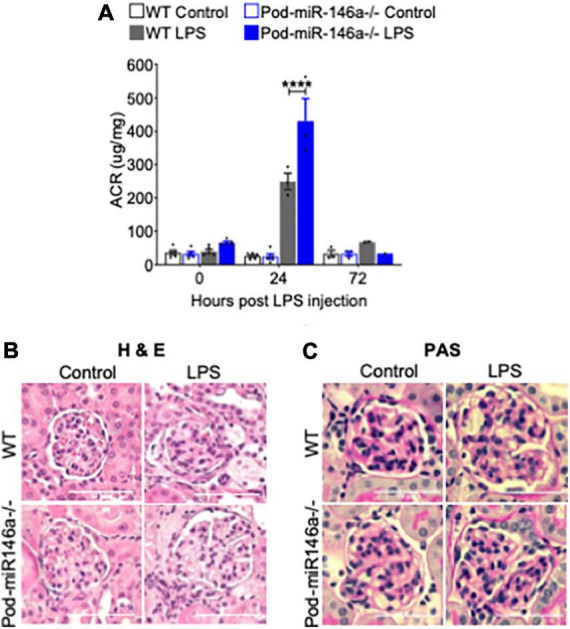
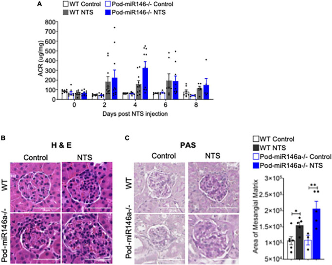
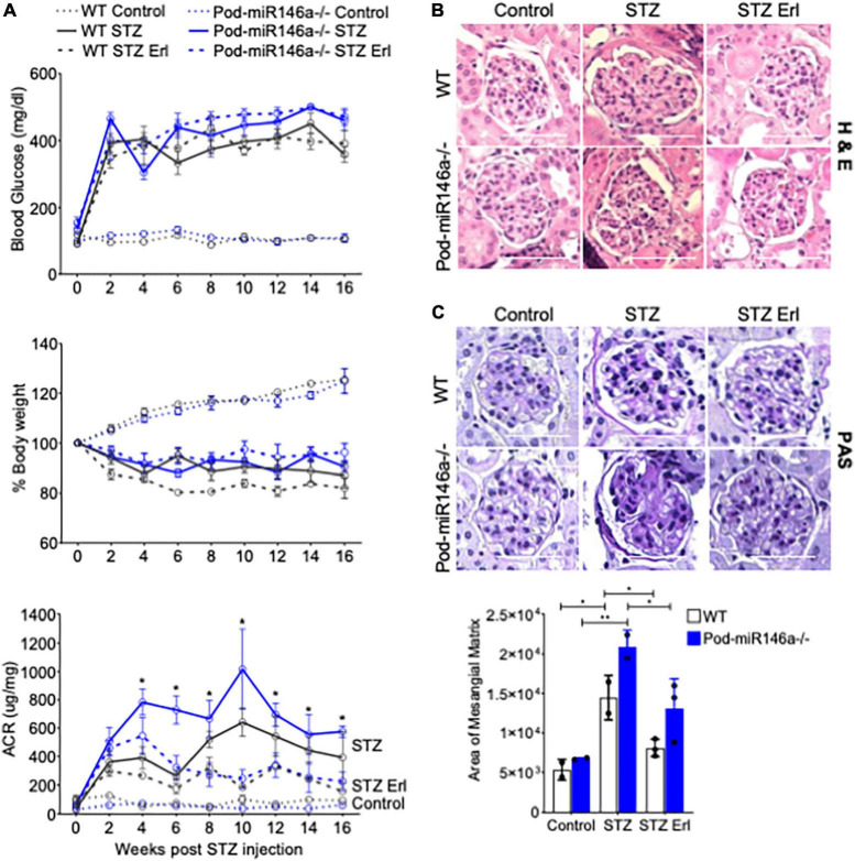
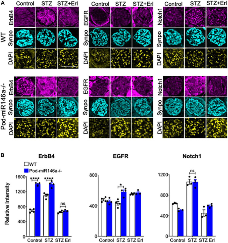
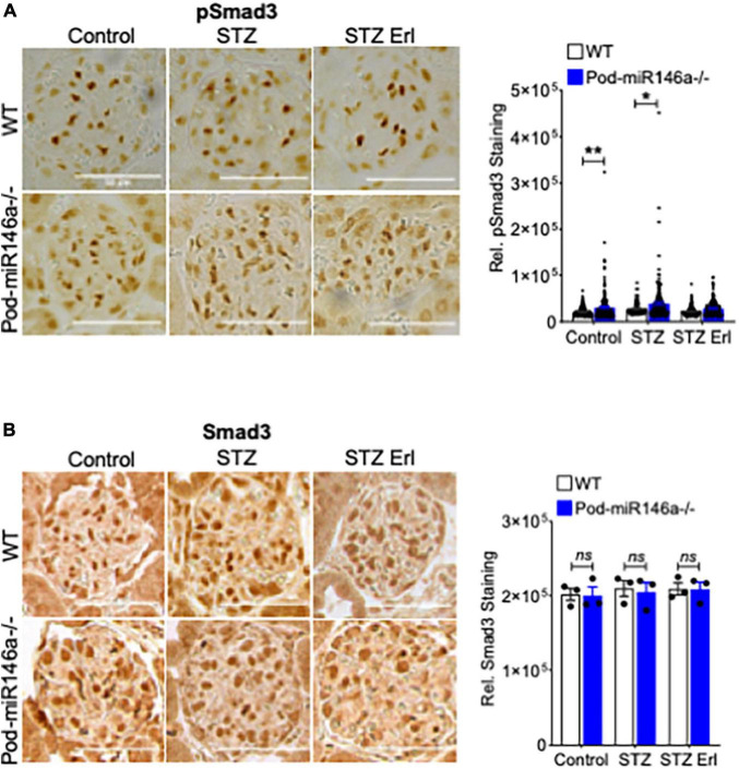
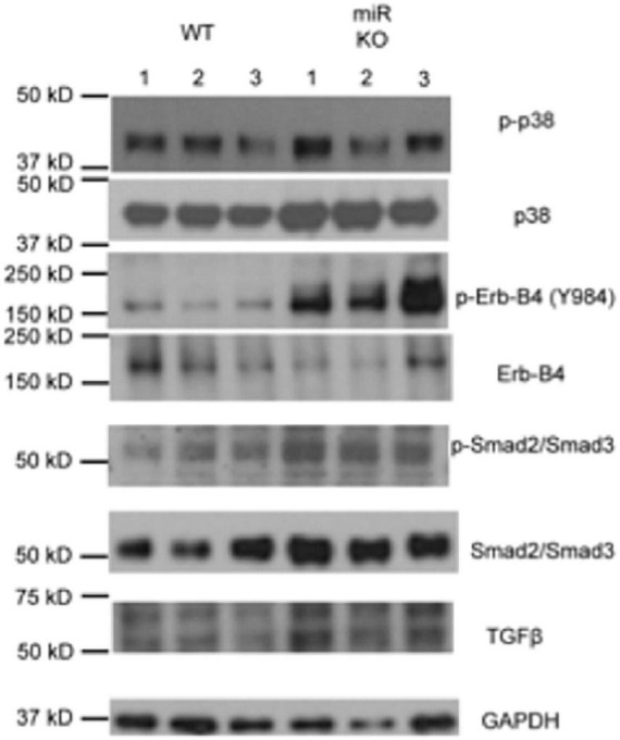
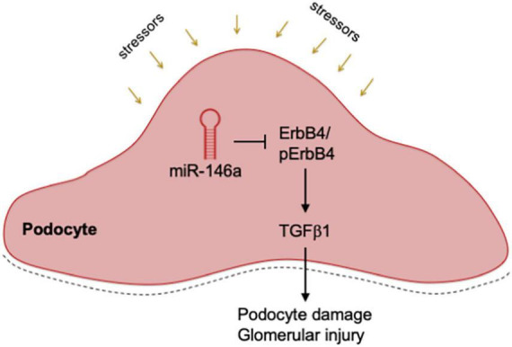
Diabetic glomerular injury is a major complication of diabetes mellitus and is the leading cause of end stage renal disease (ESRD). Healthy podocytes are essential for glomerular function and health. Injury or loss of these cells results in increased proteinuria and kidney dysfunction and is a common finding in various glomerulopathies. Thus, mechanistic understanding of pathways that protect podocytes from damage are essential for development of future therapeutics. MicroRNA-146a (miR-146a) is a negative regulator of inflammation and is highly expressed in myeloid cells and podocytes. We previously reported that miR-146a levels are significantly reduced in the glomeruli of patients with diabetic nephropathy (DN). Here we report generation of mice with selective deletion of miR-146a in podocytes and use of these mice in models of glomerular injury. Induction of glomerular injury in C57BL/6 wildtype mice (WT) and podocyte-specific miR-146a knockout (Pod-miR146a-/-) animals via administration of low-dose lipopolysaccharide (LPS) or nephrotoxic serum (NTS) resulted in increased proteinuria in the knockout mice, suggesting that podocyte-expressed miR-146a protects these cells, and thus glomeruli, from damage. Furthermore, induction of hyperglycemia using streptozotocin (STZ) also resulted in an accelerated development of glomerulopathy and a rapid increase in proteinuria in the knockout animals, as compared to the WT animals, further confirming the protective role of podocyte-expressed miR-146a. We also confirmed that the direct miR-146a target, ErbB4, was significantly upregulated in the diseased glomeruli and erlotinib, an ErbB4 and EGFR inhibitor, reducedits upregulation and the proteinuria in treated animals. Primary miR146-/- podocytes from these animals also showed a basally upregulated TGFβ-Smad3 signaling in vitro. Taken together, this study shows that podocyte-specific miR-146a is imperative for protecting podocytes from glomerular damage, via modulation of ErbB4/EGFR, TGFβ, and linked downstream signaling.

Keywords: MicroRNA; diabetic nephropathy; glomerular disease; miR-146a; podocytes.


Conflict of interest statement

VG was an inventor on patent applications related to these studies. AR recently completed her post-doctoral fellowship and was employed by Genentech. The remaining authors declare that the research was conducted in the absence of any commercial or financial relationships that could be construed as a potential conflict of interest.

Figures

FIGURE 1
FIGURE 1
Generation of podocyte-specific miR-146a deleted mice. (A) Schematic showing steps used in the generation of podocyte-specific miR146a knockout mice by crossing miR146aflox/flox mice with Flp-mice and subsequently with podocin-cre mice. (B) miR-146a expression levels measured by quantitative RT-qPCR from primary podocytes, spleen and whole kidney isolated from WT or Pod-miR146a mice. Statistics were performed using the Student’s t-test. Data shown are mean ± SEM (n = 3). ***p < 0.0001; ns, no significant difference.
FIGURE 2
FIGURE 2
Pod-miR-146a mice show increased impairment of kidney function in response to LPS. (A) Graph showing the albumin to creatinine ratio (μg/mg) in the urine of WT (gray bars) and Pod-miR-146a (blue bars) mice after 0, 24, and 72 h post administration of either vehicle (open bars) or LPS (shaded bars). Statistics were performed using two-way ANOVA. Data shown are mean ± SEM (n = 5). ****p < 0.0001. (B,C) Representative images showing histochemical analyses with hematoxylin-eosin (H&E), periodic acid-Schiff (PAS) staining of kidney tissues from 24 h post LPS treatment. Scale bar, 50 μm.
FIGURE 3
FIGURE 3
Increased albuminuria in Pod-miR-146a–/– mice after NTS treatment. (A) Graph showing the albumin to creatinine ratio (μg/mg) in the urine of WT (gray bars) and Pod-miR-146a (blue bars) mice post administration of either vehicle (open bars) or NTS (shaded bars) at various time-points, as indicated. Statistics were performed using two-way ANOVA. Data shown are mean ± SEM (n = 5). Representative images showing histochemical analyses with (B) H&E and (C) PAS staining of kidney tissues from each of the four groups of animals from 8th day post NTS treatment. Scale bar, 50 μm. Graph showing quantified mesangial matrix from the PAS-stained sections. Statistics were performed using the Mann-Whitney test. Data shown are normalized to the level of staining in control tissue and are mean ± SEM (n = 3). *p < 0.05; **p < 0.01.
FIGURE 4
FIGURE 4
STZ accelerates glomerular injury in Pod-miR-146a–/– mice that is attenuated by erlotinib. (A) (Top) Graph showing levels of hyperglycemia in various animals, as measured by serum glucose levels (mg/dL) in each of the six groups, as indicated, post STZ administration and at various time-points, as indicated. Data shown are mean ± SEM (n = 5). Treatment with erlotinib starting at 4 weeks after STZ-induction did not result in any change in level of hyperglycemia in either strain. Blood glucose levels remained unchanged in the non-STZ treated mice; (Middle) Graph showing levels of weight loss upon in various animals in each of the six groups, as indicated, post STZ administration and at various time-points, as indicated. Data shown are mean ± SEM (n = 5). WT and Pod-miR146a animals displayed equal levels of weight loss upon STZ-induced hyperglycemia that was unaffected by treatment with erlotinib; (Bottom) Graph showing the albumin to creatinine ratio (μg/mg) in the urine of mice from each of the six groups, as indicated, post STZ administration and at various time-points, as indicated. Statistics were performed using one-way ANOVA. Data shown are mean ± SEM (n = 5). *p < 0.05. (B) Representative images showing histochemical analyses with H&E staining of kidney tissues from each of the six groups of animals at end point. Scale bar, 50 μm. (C) Representative images showing histochemical analyses with PAS staining of kidney tissues from each of the six groups of animals at end point. Scale bar, 50 μm. Graph showing quantified mesangial matrix from the PAS-stained sections. Data shown are normalized to the level of staining in control tissue and are mean ± SEM (n = 3). *p < 0.05; **p < 0.01.
FIGURE 5
FIGURE 5
Immunofluorescence imaging-based analyses of glomerular sections shows erlotinib administration protects WT and Pod-miR-146a–/– mice from STZ injury via reduction in ErbB4 and EGFR. (A) Representative confocal microscopy images of immunofluorescently labeled glomeruli from WT (top three panels) and Pod-miR146a (bottom three panels) mice treated with vehicle alone (Control), with STZ and vehicle (STZ) or with STZ and erlotinib (STZ Erl). Tissue sections were imaged after staining with DAPI (nuclear marker) and antibodies against ErbB4, EGFR, Notch-1 and Synaptopodin (Synpo, podocyte marker) (as indicated). Scale bar, 50 μm. (B) Bar graphs showing quantification of relative glomerular signal intensity of ErbB4, EGFR and Notch-1 in tissue samples from A. Statistics were performed using two-way ANOVA. Data shown are mean ± SEM (n = 5/group). *p < 0.05; ***p < 0.001; ns, no significant difference.
FIGURE 6
FIGURE 6
Increase in glomerular pSmad3 levels by STZ treatment is suppressed by erlotinib. (A) Representative immunohistochemical images of glomeruli stained with an antibody against pSmad3 from WT (top row) and Pod-miR146a mice (bottom row) treated with vehicle alone (Control), with STZ and vehicle (STZ) or with STZ and erlotinib (STZ Erl). Scale bar, 50 μm. Graph on the right shows quantification of pSmad3 positive cells per glomeruli in these samples. Statistics were performed using two-way ANOVA. Data shown are mean ± SEM (n = 3). *p < 0.05, **p < 0.01. (B) Representative immunohistochemical images of glomeruli stained with an antibody against total Smad3 from WT (top row) and Pod-miR146a mice (bottom row) treated with vehicle alone (Control), with STZ and vehicle (STZ) or with STZ and erlotinib (STZ Erl). Scale bar, 50 μm. Graph on the right shows quantification of total Smad3 positive cells per glomeruli in these samples. Statistics were performed using two-way ANOVA. Data shown are mean ± SEM (n = 3). ns, no significant difference.
FIGURE 7
FIGURE 7
Deletion of miR146a in podocytes upregulates ErbB4/TGFβ /Smad3 signaling. Immunoblot analysis of various phosphorylated (p-) and total proteins in the lysates from primary podocytes isolated from the WT and Pod-miR146a mice. Data presented is from three independent samples from each group. GAPDH was used as the loading control. Relative position of the molecular weight markers is shown on the left.
FIGURE 8
FIGURE 8
Mechanistic model. A diagram showing a mechanistic working model. Podocyte expressed miR-146a represses expression of ErbB4 and Notch-1 during homeostatic conditions, thereby controlling the ErbB4/EGFR and TGFβ 1 signaling pathways. Various external stressors or deletion of miR-146a result in de-repression of ErbB4 and Notch-1, thereby driving the harmful ErbB4/EGFR signaling and inducing TGFβ 1. An autocrine feed-forward loop via TGFβ 1 induces the downstream TGFR/Smad3 signaling, that result in podocyte damage, glomerular injury and proteinuria.

References

    1. Centers for Disease Control and Prevention. National diabetes statistics report website. Atlanta, GA: Centers for Disease Control and Prevention; (2022).
    1. USRDS: The United States renal data system. Am J Kidney Dis. (2003) 42:1–230. 10.1053/j.ajkd.2003.09.004 - DOI - PubMed
    1. Wiggins RC. The spectrum of podocytopathies: A unifying view of glomerular diseases. Kidney Int. (2007) 71:1205–14. 10.1038/sj.ki.5002222 - DOI - PubMed
    1. Reiser J, Altintas MM. Podocytes. F1000Res. (2016) 5:F1000FacultyRev–114. 10.12688/f1000research.7255.1 - DOI - PMC - PubMed
    1. Bartel DP. Metazoan micrornas. Cell. (2018) 173:20–51. 10.1016/j.cell.2018.03.006 - DOI - PMC - PubMed