. 2022 Jul:101:57-120.
doi: 10.3114/sim.2022.101.02. Epub 2022 Mar 30.

Generic boundaries in the Ophiostomatales reconsidered and revised

Affiliations

Generic boundaries in the Ophiostomatales reconsidered and revised

Z W de Beer et al. Stud Mycol. 2022 Jul.

Abstract

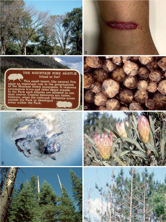
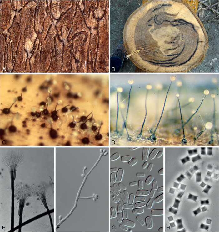
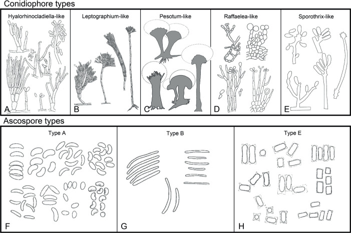
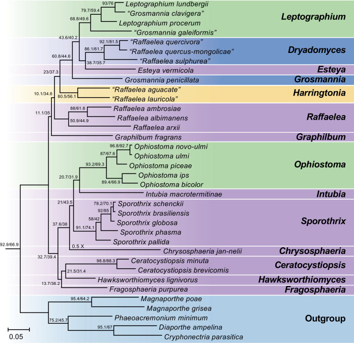
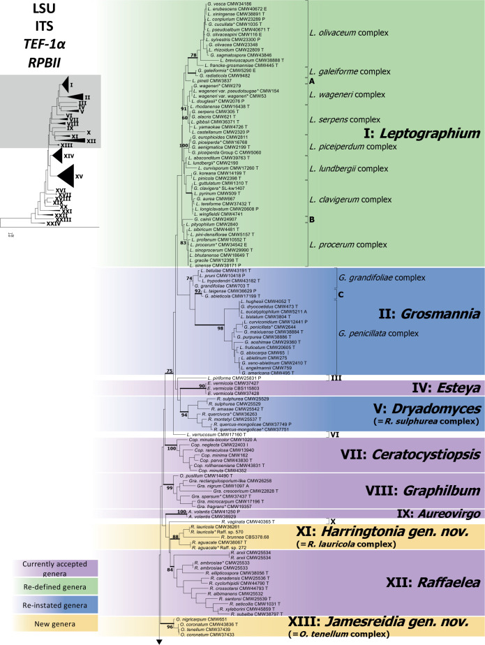
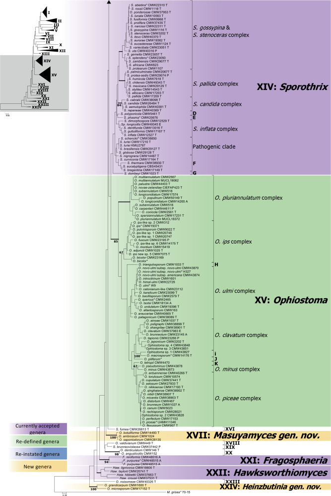
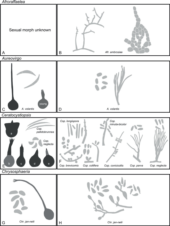
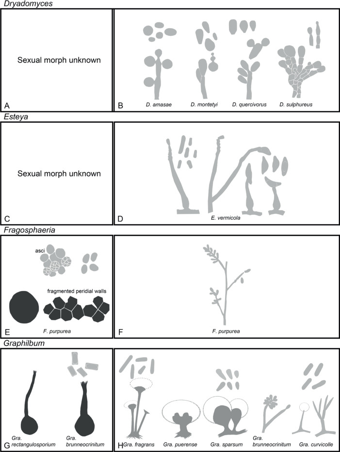
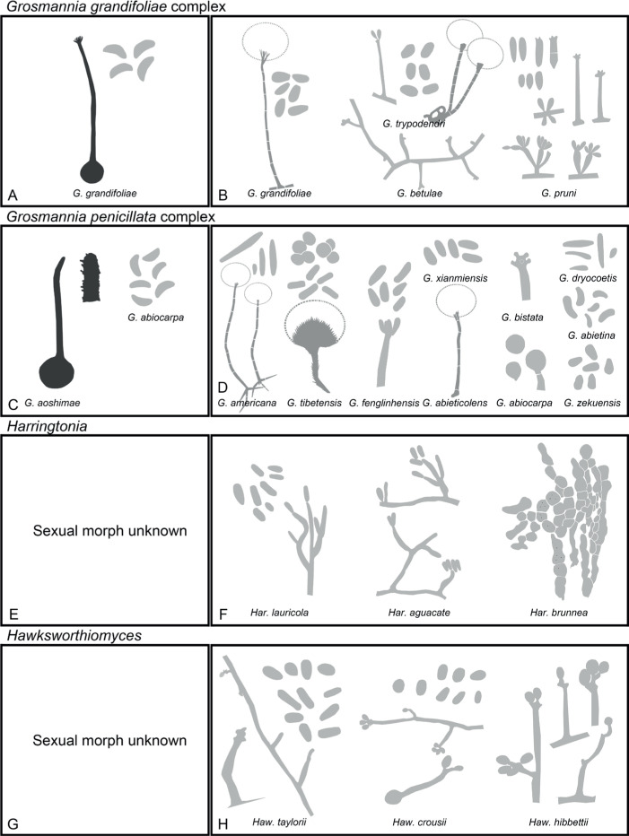
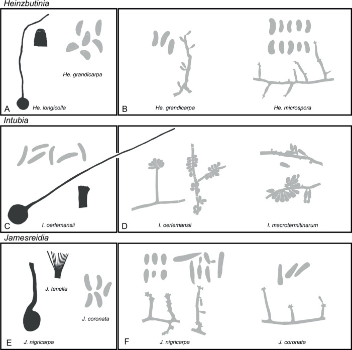
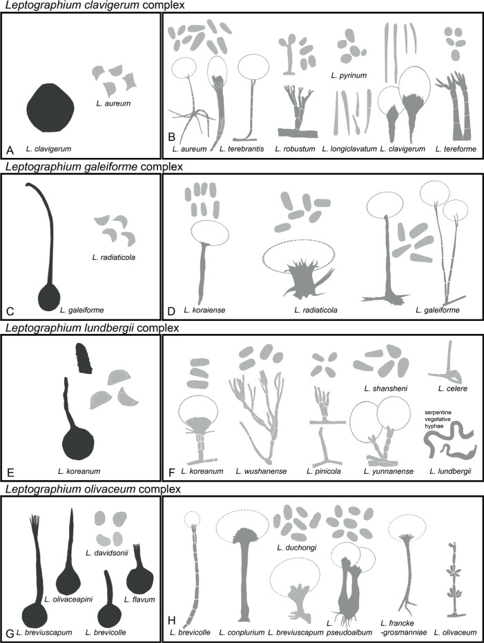
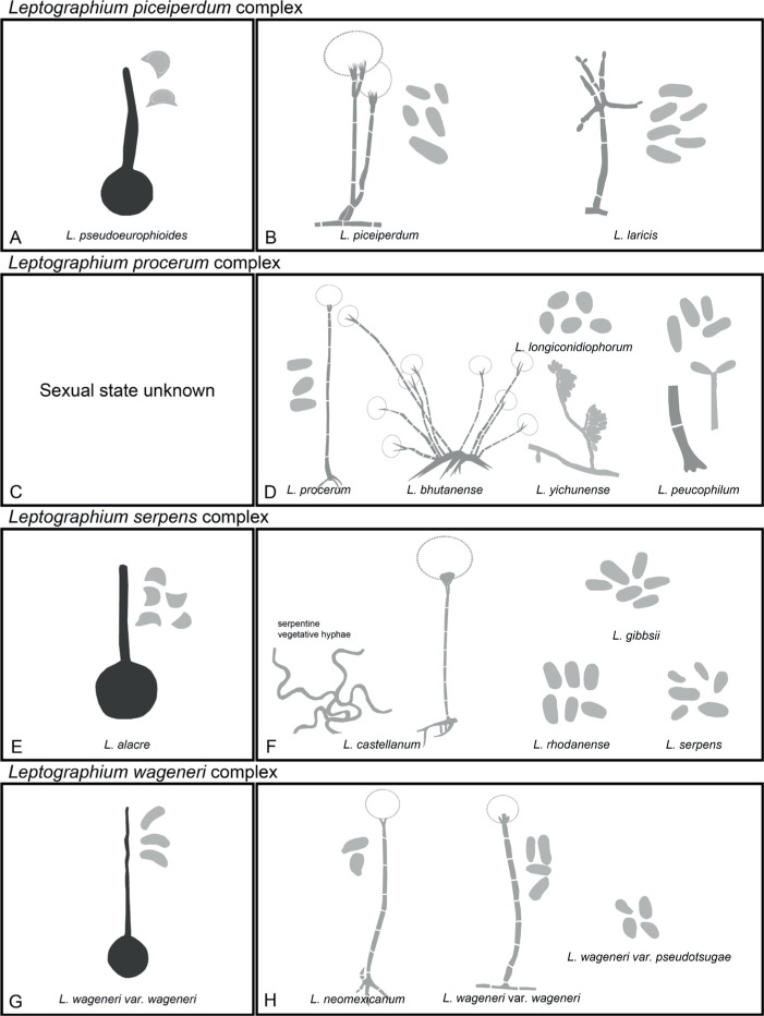
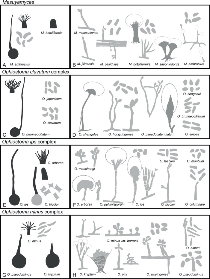
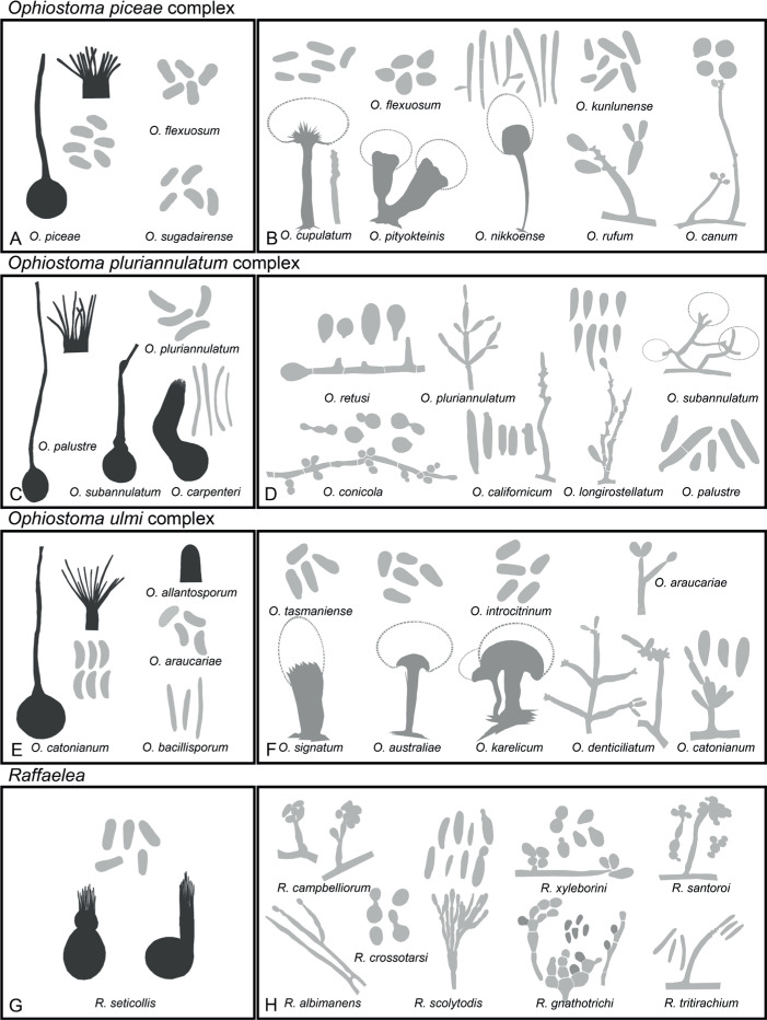
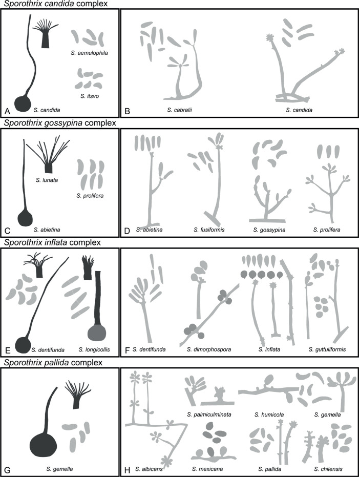
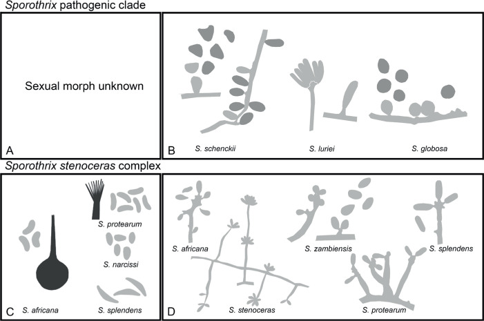
The Ophiostomatales was erected in 1980. Since that time, several of the genera have been redefined and others have been described. There are currently 14 accepted genera in the Order. They include species that are the causal agents of plant and human diseases and common associates of insects such as bark beetles. Well known examples include the Dutch elm disease fungi and the causal agents of sporotrichosis in humans and animals. The taxonomy of the Ophiostomatales was confused for many years, mainly due to the convergent evolution of morphological characters used to delimit unrelated fungal taxa. The emergence of DNA-based methods has resolved much of this confusion. However, the delineation of some genera and the placement of various species and smaller lineages remains inconclusive. In this study we reconsidered the generic boundaries within the Ophiostomatales. A phylogenomic framework constructed from genome-wide sequence data for 31 species representing the major genera in the Order was used as a guide to delineate genera. This framework also informed our choice of the best markers from the currently most commonly used gene regions for taxonomic studies of these fungi. DNA was amplified and sequenced for more than 200 species, representing all lineages in the Order. We constructed phylogenetic trees based on the different gene regions and assembled a concatenated data set utilising a suite of phylogenetic analyses. The results supported and confirmed the delineation of nine of the 14 currently accepted genera, i.e. Aureovirgo, Ceratocystiopsis, Esteya, Fragosphaeria, Graphilbum, Hawksworthiomyces, Ophiostoma, Raffaelea and Sporothrix. The two most recently described genera, Chrysosphaeria and Intubia, were not included in the multi-locus analyses. This was due to their high sequence divergence, which was shown to result in ambiguous taxonomic placement, even though the results of phylogenomic analysis supported their inclusion in the Ophiostomatales. In addition to the currently accepted genera in the Ophiostomatales, well-supported lineages emerged that were distinct from those genera. These are described as novel genera. Two lineages included the type species of Grosmannia and Dryadomyces and these genera are thus reinstated and their circumscriptions redefined. The descriptions of all genera in the Ophiostomatales were standardised and refined where this was required and 39 new combinations have been provided for species in the newly emerging genera and one new combination has been provided for Sporothrix. The placement of Afroraffaelea could not be confirmed using the available data and the genus has been treated as incertae sedis in the Ophiostomatales. Paleoambrosia was not included in this study, due to the absence of living material available for this monotypic fossil genus. Overall, this study has provided the most comprehensive and robust phylogenies currently possible for the Ophiostomatales. It has also clarified several unresolved One Fungus-One Name nomenclatural issues relevant to the Order. Taxonomic novelties: New genera: Harringtonia Z.W. de Beer & M. Procter, Heinzbutinia Z.W. de Beer & M. Procter, Jamesreidia Z.W. de Beer & M. Procter, Masuyamyces Z.W. de Beer & M. Procter. New species: Masuyamyces massonianae M. Procter & Z.W. de Beer. New combinations: Dryadomyces montetyi (M. Morelet) M. Procter & Z.W. de Beer, Dryadomyces quercivorus (Kubono & Shin. Ito) M. Procter & Z.W. de Beer, Dryadomyces quercus-mongolicae (K.H. Kim et al.) M. Procter & Z.W. de Beer, Dryadomyces sulphureus (L.R. Batra) M. Procter & Z.W. de Beer, Graphilbum pusillum (Masuya) M. Procter & Z.W. de Beer, Grosmannia abieticolens (K. Jacobs & M.J. Wingf.) M. Procter & Z.W. de Beer, Grosmannia altior (Paciura et al.) M. Procter & Z.W. de Beer, Grosmannia betulae (Jankowiak et al.) M. Procter & Z.W. de Beer, Grosmannia curviconidia (Paciura et al.) M. Procter & Z.W. de Beer, Grosmannia euphyes (K. Jacobs & M.J. Wingf.) M. Procter & Z.W. de Beer, Grosmannia fenglinhensis (R. Chang et al.) M. Procter & Z.W. de Beer, Grosmannia gestamen (de Errasti & Z.W. de Beer) M. Procter & Z.W. de Beer, Grosmannia innermongolica (X.W. Liu et al.) M. Procter & Z.W. de Beer, Grosmannia pistaciae (Paciura et al.) M. Procter & Z.W. de Beer, Grosmannia pruni (Masuya & M.J. Wingf.) M. Procter & Z.W. de Beer, Grosmannia taigensis (Linnak. et al.) M. Procter & Z.W. de Beer, Grosmannia trypodendri (Jankowiak et al.) M. Procter & Z.W. de Beer, Harringtonia aguacate (D.R. Simmons et al.) M. Procter & Z.W. de Beer, Harringtonia brunnea (L.R. Batra) M. Procter & Z.W. de Beer, Harringtonia lauricola (T.C. Harr. et al.) Z.W. de Beer & M. Procter, Heinzbutinia grandicarpa (Kowalski & Butin) Z.W. de Beer & M. Procter, Heinzbutinia microspora (Arx) M. Procter & Z.W. de Beer, Heinzbutinia solheimii (B. Strzałka & Jankowiak) Z.W. de Beer & M. Procter, Jamesreidia coronata (Olchow. & J. Reid) M. Procter & Z.W. de Beer, Jamesreidia nigricarpa (R.W. Davidson) M. Procter & Z.W. de Beer, Jamesreidia rostrocoronata (R.W. Davidson & Eslyn) M. Procter & Z.W. de Beer, Jamesreidia tenella (R.W. Davidson) Z.W. de Beer & M. Procter, Leptographium cainii (Olchow. & J. Reid) M. Procter & Z.W. de Beer, Leptographium europioides (E.F. Wright & Cain) M. Procter & Z.W. de Beer, Leptographium galeiforme (B.K. Bakshi) M. Procter & Z.W. de Beer, Leptographium pseudoeurophioides (Olchow. & J. Reid) M. Procter & Z.W. de Beer, Leptographium radiaticola (J.J. Kim et al.) M. Procter & Z.W. de Beer, Masuyamyces acarorum (R. Chang & Z.W. de Beer) M. Procter & Z.W. de Beer, Masuyamyces ambrosius (B.K. Bakshi) M. Procter & Z.W. de Beer, Masuyamyces botuliformis (Masuya) Z.W. de Beer & M. Procter, Masuyamyces jilinensis (R. Chang et al.) M. Procter & Z.W. de Beer, Masuyamyces lotiformis (Z. Wang & Q. Lu) M. Procter & Z.W. de Beer, Masuyamyces pallidulus (Linnak. et al.) M. Procter & Z.W. de Beer, Masuyamyces saponiodorus (Linnak. et al.) M. Procter & Z.W. de Beer, Sporothrix longicollis (Massee & E.S. Salmon) M. Procter & Z.W. de Beer. Citation: de Beer W, Procter M, Wingfield MJ, Marincowitz S, Duong TA (2022). Generic boundaries in the Ophiostomatales reconsidered and revised. Studies in Mycology 101: 57-120. doi: 10.3114/sim.2022.101.02.

Keywords: Generic boundaries; Ophiostomataceae; Ophiostomatales; Sordariomycetidae; new taxa; nomenclature; taxonomy.


Conflict of interest statement

The authors declare that there is no conflict of interest.

Figures

Fig. 1.
Fig. 1.
Ecological niches in which genera and species residing in the Ophiostomatales are found. A. Ulmus americana street trees dying as a result of Dutch Elm Disease (photo: D.W. French). B. Symptoms of infection by the human pathogen Sporothix schenckii (photo: Prof. Dr Flávio de Queiroz Telles Filho, Federal University of Paraná, Brazil). C. Signage in Yellowstone National Park emphasising the important role that bark beetles (and by extension their fungal symbionts) play in the ecology of conifer ecosystems. D. Blue stain in conifer timber caused by numerous species of Ophiostomatoid fungi. E. Hylobius rhizophagus (root collar weevil) squashed onto the surface of agar medium containing cycloheximide selective for many genera and species of Ophiostomatales and in this case Leptographium procerum. F. Infructescences of a Protea species in which numerous species of Ophiostomatoid fungi can be found. G. Douglas fir (Pseudotsuga menzesii) trees dying as a result of black stain root disease caused by Leptographium wageneri var. pseudotsugae (Photo: F.W. Cobb). H. Pinus resinosa trees dying as a result of mass infestation by Ips pini and associated Ophiostoma minus.
Fig. 2.
Fig. 2.
Ecological niches of Ophiostomatoid fungi and micrographs providing examples of structures typical of these fungi. A. Transverse stellate gallery systems of Ips schmutzenhoferi in the bark of a Pinus spinulosa tree, showing blue-stain around nuptial chambers and female galleries. B. Section through a Eucalyptus stem infested by the ambrosia beetle Megaplatypus mutatus showing tunnels in which species of Ophiostomatales occur. C, D. Ascomata of Ophiostoma ulmi (C) (photo: D.W. French) and O. pilliferum (D) (photo: Z.W. de Beer) with sticky ascospores masses at their apices, illustrating the manner by which these fungi easily attach to the insects that carry them. E. Conidiophores of Leptographium procerum, illustrating asexual structures well suited to being vectored by insects. F. Conidiogenous cells of a Sporothrix sp. G. Typical single-celled conidia found in most species of Ophiostomatales. H. Many species of Ophiostomatales have ascospores with sheaths such as these pillow-shaped spores in Ophiostoma ips.
Fig. 3.
Fig. 3.
Conidiophore and ascospore types mentioned in the paper. A–E. Conidiophore types. F–H. Ascospore types sensu De Beer & Wingfield (2013). A, B, D, F–H. Adapted from illustrations in De Beer & Wingfield (2013). Shades of grey depict colours of various structures ranging from hyaline to dematiaceous.
Fig. 4.
Fig. 4.
Phylogenomic tree obtained from supertree analysis with ASTRAL using gene trees constructed from 3 548 BUSCO genes (identified using the sordariomycetes_odb10 dataset; BUSCO v. 4.0.5). All 31 species in the Ophiostomatales for which genome sequence currently available were included in the analysis. Cryphonectria parasitica, Diaporthe ampelina, Magnaporthe grisea, Magnaporthe poae and Phaeoacremonium minimum were included as outgroup taxa. Gene concordance factors (gCF) and site concordance factors (sCF), which indicate the percentage of genes and sites that support a parcular nodes respectively, were determined using IQ-TREE2 are presented at nodes as gCF/sCF. Species names presented in double quotes denote old names which have been changed to their new respective genera subsequent to this study.
Fig. 5.
Fig. 5.
Phylogenetic tree depicting the boundaries of currently accepted genera in the Ophiostomatales. This tree was generated using maximum likelihood analysis of the concatenated dataset of LSU, ITS, TEF1-α and RPBII gene regions. The dataset consisted of 264 isolates and 2 360 characters (including gaps). Bootstrap values above 60 % are shown. Bold lines indicate Bayesian posterior probabilities values above 0.8. Bootstrap values and Bayesian posterior probabilities values below the species complex level were removed for simplification. Purple blocks indicate existing genera, yellow blocks new genera described in this study, green blocks, genera that we redefine here, and blue blocks indicate genera that that have been reinstated and re-defined. (T = ex-type, E = ex-epitype, P = ex-paratype; L = ex-lectotype; A = authentic isolate, used in the original study; * Genome sequenced).
Fig. 5.
Fig. 5.
Phylogenetic tree depicting the boundaries of currently accepted genera in the Ophiostomatales. This tree was generated using maximum likelihood analysis of the concatenated dataset of LSU, ITS, TEF1-α and RPBII gene regions. The dataset consisted of 264 isolates and 2 360 characters (including gaps). Bootstrap values above 60 % are shown. Bold lines indicate Bayesian posterior probabilities values above 0.8. Bootstrap values and Bayesian posterior probabilities values below the species complex level were removed for simplification. Purple blocks indicate existing genera, yellow blocks new genera described in this study, green blocks, genera that we redefine here, and blue blocks indicate genera that that have been reinstated and re-defined. (T = ex-type, E = ex-epitype, P = ex-paratype; L = ex-lectotype; A = authentic isolate, used in the original study; * Genome sequenced).
Fig. 6.
Fig. 6.
Genera of the Ophiostomatales redrawn from published images with sexual morphs (if known) on the left and asexual morphs on the right. A, B. Afroraffaelea. C, D. Aureovirgo. E, F. Ceratocystiopsis (a. Cop. collifera; b. Cop. ochracea; c. Cop. manitobensis; d. Cop. concentrica; e. Cop. rollhanseniana). G, H. Chrysosphaeria. (Pale grey shading reflects hyaline to subhyaline colouration, medium-tone grey brown to dark brown and dark grey reflects fuscous black to dark black colouration).
Fig. 7.
Fig. 7.
Genera of the Ophiostomatales redrawn from published images with sexual morphs (if known) on the left and asexual morphs on the right. A, B. Dryadomyces. C, D. Esteya. E, F. Fragosphaeria. G, H. Graphilbum. (Pale grey shading reflects hyaline to subhyaline colouration, medium-tone grey brown to dark brown and dark grey reflects fuscous black to dark black colouration).
Fig. 8.
Fig. 8.
Genera of the Ophiostomatales redrawn from published images with sexual morphs (if known) on the left and asexual morphs on the right. A, B. Grosmannia grandifoliae complex. C, D. Grosmannia penicillata complex. E, F. Harringtonia. G, H. Hawksworthiomyces. (Pale grey shading reflects hyaline to subhyaline colouration, medium-tone grey brown to dark brown and dark grey reflects fuscous black to dark black colouration).
Fig. 9.
Fig. 9.
Genera of the Ophiostomatales redrawn from published images with sexual morphs (if known) on the left and asexual morphs on the right. A, B. Heinzbutinia. C, D. Intubia. E, F. Jamesreidia. (Pale grey shading reflects hyaline to subhyaline colouration, medium-tone grey brown to dark brown and dark grey reflects fuscous black to dark black colouration).
Fig. 10.
Fig. 10.
Genera of the Ophiostomatales redrawn from published images with sexual morphs (if known) on the left and asexual morphs on the right. A, B. Leptographium clavigerum complex. C, D. Leptographium galeiforme complex. E, F. Leptographium lundbergii complex. G, H. Leptographium olivaceum complex. (Pale grey shading reflects hyaline to subhyaline colouration, medium-tone grey brown to dark brown and dark grey reflects fuscous black to dark black colouration).
Fig. 11.
Fig. 11.
Genera of the Ophiostomatales redrawn from published images with sexual morphs (if known) on the left and asexual morphs on the right. A, B. Leptographium piceiperdum complex. C, D. Leptographium procerum complex. E, F. Leptographium serpens complex. G, H. Leptographium wageneri complex. (Pale grey shading reflects hyaline to subhyaline colouration, medium-tone grey brown to dark brown and dark grey reflects fuscous black to dark black colouration).
Fig. 12.
Fig. 12.
Genera of the Ophiostomatales redrawn from published images with sexual morphs (if known) on the left and asexual morphs on the right. A, B. Masuyamyces. C, D. Ophiostoma clavatum complex. E, F. Ophiostoma ips complex. G, H. Ophiostoma minus complex. (Pale grey shading reflects hyaline to subhyaline colouration, medium-tone grey brown to dark brown and dark grey reflects fuscous black to dark black colouration).
Fig. 13.
Fig. 13.
Genera of the Ophiostomatales redrawn from published images with sexual morphs (if known) on the left and asexual morphs on the right. A, B. Ophiostoma piceae complex. C, D. Ophiostoma pluriannulatum complex. E, F. Ophiostoma ulmi complex. G, H. Raffaelea. (Pale grey shading reflects hyaline to subhyaline colouration, medium-tone grey brown to dark brown and dark grey reflects fuscous black to dark black colouration).
Fig. 14.
Fig. 14.
Genera of the Ophiostomatales redrawn from published images with sexual morphs (if known) on the left and asexual morphs on the right. A, B. Sporothrix candida complex. C, D. Sporothrix gossypina complex. E, F. Sporothrix inflata complex. G, H. Sporothrix pallida complex. (Pale grey shading reflects hyaline to subhyaline colouration, medium-tone grey brown to dark brown and dark grey reflects fuscous black to dark black colouration).
Fig. 15.
Fig. 15.
Genera of the Ophiostomatales redrawn published images with sexual morphs (if known) on the left and asexual morphs on the right. A, B. Sporothrix pathogenic clade. C, D. Sporothrix stenoceras complex. (Pale grey shading reflects hyaline to subhyaline colouration, medium-tone grey brown to dark brown and dark grey reflects fuscous black to dark black colouration).

Similar articles

Cited by

References

    1. Aghayeva DN, Wingfield MJ, De Beer ZW, et al. (2004). Two new Ophiostoma species with Sporothrix anamorphs from Austria and Azerbaijan. Mycologia 96: 866–878. - PubMed
    1. Ando Y, Masuya H, Motohashi K, et al. (2016). Phylogenetic relationship of Japanese isolates belonging to the Grosmannia piceiperda complex (Ophiostomatales). Mycoscience 57: 123–135.
    1. Bakshi BK. (1950). Fungi associated with ambrosia beetles in Great Britain. Transactions of the British Mycological Society 33: 111–120.
    1. Bakshi BK. (1951). Studies on four species of Ceratocystis, with a discussion on fungi causing sap-stain in Britain. Mycological Paper 35: 1–16.
    1. Bateman C, Huang Y-T, Simmons DR, et al. (2017). Ambrosia beetle Premnobius cavipennis (Scolytinae: Ipini) carries highly divergent ascomycotan ambrosia fungus, Afroraffaelea ambrosiae gen. nov. et sp. nov. (Ophiostomatales). Fungal Ecology 25: 41–49.

LinkOut - more resources