. 2022 Aug 5;25(9):104882.
doi: 10.1016/j.isci.2022.104882. eCollection 2022 Sep 16.

Optical control of the β2-adrenergic receptor with opto-prop-2: A cis-active azobenzene analog of propranolol

Affiliations

Optical control of the β2-adrenergic receptor with opto-prop-2: A cis-active azobenzene analog of propranolol

Reggie Bosma et al. iScience. .

Abstract

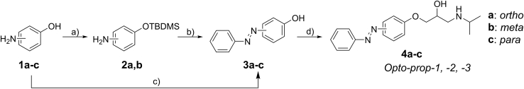
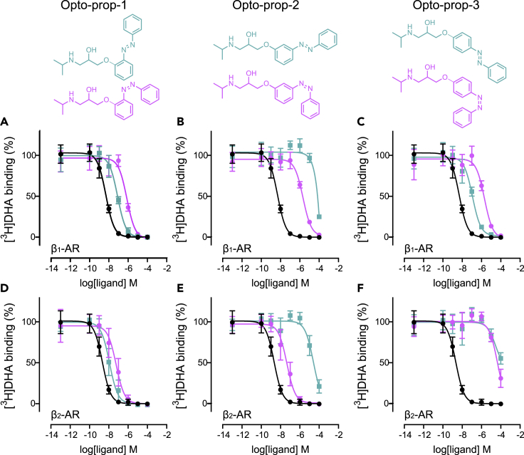
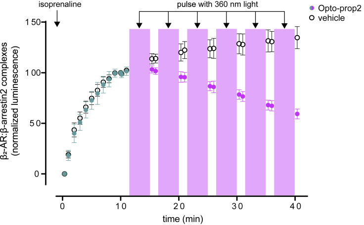
In this study, we synthesized and evaluated new photoswitchable ligands for the beta-adrenergic receptors β1-AR and β2-AR, applying an azologization strategy to the first-generation beta-blocker propranolol. The resulting compounds (Opto-prop-1, -2, -3) have good photochemical properties with high levels of light-induced trans-cis isomerization (>94%) and good thermal stability (t 1/2 > 10 days) of the resulting cis-isomer in an aqueous buffer. Upon illumination with 360-nm light to PSS cis , large differences in binding affinities were observed for photoswitchable compounds at β1-AR as well as β2-AR. Notably, Opto-prop-2 (VUF17062) showed one of the largest optical shifts in binding affinities at the β2-AR (587-fold, cis-active), as recorded so far for photoswitches of G protein-coupled receptors. We finally show the broad utility of Opto-prop-2 as a light-dependent competitive antagonist of the β2-AR as shown with a conformational β2-AR sensor, by the recruitment of downstream effector proteins and functional modulation of isolated adult rat cardiomyocytes.

Keywords: Biochemical engineering; Biochemical research method; Photomedicine.


Conflict of interest statement

The authors declare no competing interests.

Figures

None
Graphical abstract
Figure 1
Figure 1
Design of propranolol-based photoswitchable inhibitors of the β2-AR Azobenzene was selected as a photoswitchable moiety and propranolol was taken as the standard antagonist of the β2-AR. Photoswitchable antagonists were designed by azologization, replacing the naphthalene ring of propranolol with an azobenzene. The propranolol side chain was positioned at either the ortho-, meta-, or para-position of the azobenzene.
Scheme 1
Scheme 1
Synthesis of photoswitchable compounds 4a-c (Opto-prop-1, -2, -3) Key: (a) TBDMS-Cl, imidazole, DMF, RT, 3 h – overnight, 99%. (b) [1] PhNO, AcOH, RT, overnight; [2] TBAF, THF, RT, 30 min, 51–70% two steps. (c) PhNO, AcOH, RT, 24 h, 26%. (d) [1] NaOH, EtOH, rac-epichlorohydrin, 50 °C, overnight; [2] iPrNH2, RT, 1 h, 22–41% two steps. Detailed synthesis procedures can be found in Data S1.
Figure 2
Figure 2
Light-mediated isomer switching of Opto-prop-2 (A) UV-Vis spectroscopy analysis of a sample of trans-Opto-prop-2 (25 μM in 1% DMSO/Trs-HCl buffer) after illumination at the indicated wavelengths for 5 min. Panels (B–E) depict the kinetic analysis of Opto-prop-2 isomerization (10 mM in DMSO-d6) upon illumination with 360- and 434-nm light. Trans- Opto-prop-2 was illuminated for 1,000 s with 360-nm light and the resulting solution of Opto-prop-2 at PSScis was illuminated for another 1,000 s with 434-nm light. At various time points, a sample was extracted and the amount of cis- Opto-prop-2 was determined by (B) the respective NMR peak area or (C) LCMS absorbance peak area at at the isosbestic wavelength (263 nm). (D) NMR-spectrum of Opto-prop-2 before illumination (t = 0, upper panel) and after 1,000 s of illuminating with 360-nm light (lower panel). The x axis shows only a part of the aromatic region for clarity. The key resonances used for area integration in panel B (analyzing the chemical shift for the hydrogen atom depicted in bold in the structure) are highlighted. (E) LCMS analysis of Opto-prop-2 before illumination (t = 0, upper panel) and after 1,000 s of illuminating with 360-nm light (lower panel). Samples were withdrawn, diluted with MeCN and subjected to LCMS analysis at the isosbestic wavelength (263 nm).
Figure 3
Figure 3
Light-induced modulation of ligand affinity at the β1-AR and β2-AR Binding of the radioligand [3H]DHA to membranes of HEK293T-cells expressing the β1-AR (A–C) or the β2-AR (D–F) was determined in the presence of increasing concentrations of competing ligands. The competing ligands Opto-prop-1 (A and D), Opto-prop-2 (B and E), and Opto-prop-3 (C and F) were used without prior illumination (trans-isomer; green squares) or at PSScis (>94% cis-isomer; magenta circles). The reference ligand propranolol (black circles) is depicted in all graphs for comparison. All data points represent the mean ± SD of >3 experiments with individual triplicates.
Figure 4
Figure 4
Light-dependent effects of Opto-prop-2 on β2-AR activity in cell-based experiments Conformational changes of the β2-AR can be monitored using an intramolecular BRET sensor (A). Upon binding of an agonist, the β2-AR adopts an active conformation in which the distance between the BRET donor Nluc and BRET acceptor (NanoBRET-618-labeled HaloTag) is enlarged, resulting in a reduced BRET signal. Conformational changes of the β2-AR were determined when incubating cells with epinephrine alone (B) or in the presence of the ligands (C) propranolol (black circles), trans-Opto-prop-2 (teal squares) or Opto-prop-2 at PSScis (>94% cis; magenta circles). The agonist-induced recruitment of intracellular signaling molecules to the β2-AR was determined by nanoluc luciferase-complementation proximity sensors (D–I). The LgBit-fused β2-AR recruits the SmBit-fused signaling molecules mini-Gαs (D–F) or β-arrestin2 (G–I) upon stimulation with an agonist. Recruitment for both the mini-Gαs (D and E) and β-arrestin2 (G and H) were determined for increasing concentrations of the agonist isoprenaline in the absence (black circles) or presence of increasing concentrations trans-Opto-prop-2 (D and G) or Opto-prop-2 at PSScis (>94% cis; E and H). Schild-analysis of the cis-Opto-prop-2 induced dextral shifts of the isoprenaline concentration–response curve are depicted for mini-Gαs recruitment (F) and β-arrestin2 recruitment (I) All data points represent the mean ± SD of three experiments.
Figure 5
Figure 5
Temporal regulation of β2-AR activity using Opto-prop-2 HEK293T-cells transiently expressing β2-AR-SmBit and βarrestin2-LgBit were stimulated at time point 0 using 32 nM isoprenaline with or without Opto-prop-2 (1 μM) as depicted by closed and open circles, respectively. β-arrestin2 binding to β2-AR was monitored by measuring the luminsecence for 10 min after which cells were pulsed with 360 nm for six cycles of 5 min with an intermittent readout of luminescence. Cells incubated with Opto-prop-2 are depicted in green and magenta before and after illumination, respectively. Data points represent the mean ± SEM of the three experiments, normalized to the luminescence at 10 min as 100%.
Figure 6
Figure 6
Light-dependent betablocker activity of Opto-prop-2 on freshly isolated adult rat cardiomyocytes Electrical pacing of rat cardiomyocytes resulted in transient shortening of the sarcomeres, which was monitored over time as depicted in panel (A) for cells treated with vehicle (DMSO, gray line), 15-nM isoprenaline (black line), or isoprenaline in combination with trans-Opto-prop-2 (teal line) or cis-Opto-prop-2 (PSScis; magenta line) at 1 μM. A representative graph is depicted, with the average sarcomere length (line) and SEM (shading) of cardiomyocytes measured on the same day. In panel (B), the normalized maximal shortening of cardiomyocytes is depicted for each perturbation with the mean ± SEM The number of replicate measurements is depicted per condition in the respective bar graph. Bar graphs were compared by one-way ANOVA with Dunnett’s multiple comparison. ns = no significant difference; ∗∗∗∗significant difference with p < 0.001.

References

    1. Ahmed Z., Siiskonen A., Virkki M., Priimagi A. Controlling azobenzene photoswitching through combined ortho -fluorination and -amination. Chem. Commun. 2017;53:12520–12523. doi: 10.1039/C7CC07308A. - DOI - PubMed
    1. Al-Haddad C., El Salloukh N.A., El Moussawi Z. β-blockers in the treatment of periocular infantile hemangioma. Curr. Opin. Ophthalmol. 2019;30:319–325. doi: 10.1097/ICU.0000000000000591. - DOI - PubMed
    1. Arunlakshana O., Schild H.O. Some quantitative uses of drug antagonists. Br. J. Pharmacol. Chemother. 1959;14:48–58. doi: 10.1111/j.1476-5381.1959.tb00928.x. - DOI - PMC - PubMed
    1. Baker J.G. The selectivity of β -adrenoceptor antagonists at the human β 1, β 2 and β 3 adrenoceptors. Br. J. Pharmacol. 2005;144:317–322. doi: 10.1038/sj.bjp.0706048. - DOI - PMC - PubMed
    1. Bandara H.M.D., Burdette S.C. Photoisomerization in different classes of azobenzene. Chem. Soc. Rev. 2012;41:1809–1825. doi: 10.1039/C1CS15179G. - DOI - PubMed