

Rapid resolution of a traumatic venous epidural hematoma in a 3-year-old child: illustrative case
- PMID: 36060428
- PMCID: PMC9435549
- DOI: 10.3171/CASE21413
Rapid resolution of a traumatic venous epidural hematoma in a 3-year-old child: illustrative case
Abstract
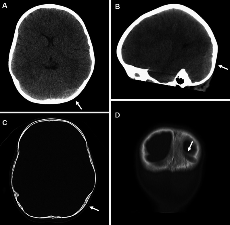
Background: Posterior fossa epidural hematoma rarely occurs in children after traumatic head injury. There is ongoing discussion about appropriate treatment, yet the radiological features regarding the time to resorption of the hematoma or required follow-up imaging are rarely discussed.
Observations: The authors presented the case of a 3-year-old child who was under clinical observation and receiving analgetic and antiemetic treatment in whom near-complete hematoma resorption was shown by magnetic resonance imaging as soon as 60 hours after diagnosis. The child was neurologically stable at all times and showed no deficit after observational treatment. Hematoma resorption was much faster than expected. The authors discussed hematoma drainage via the sigmoid sinus.
Lessons: Epidural hematomas in children can be treated conservatively and are resorbed in a timely manner.
Keywords: CT = computed tomography; EDH = epidural hematoma; MRI = magnetic resonance imaging; PFEDH = posterior fossa epidural hematoma; epidural hematoma; observation; pediatric; resorption; trauma.
© 2021 The authors.
Conflict of interest statement
Disclosures The authors report no conflict of interest concerning the materials or methods used in this study or the findings specified in this paper.
Figures

References
-
- Sencer A, Aras Y, Akcakaya MO, Goker B, Kiris T, Canbolat AT. Posterior fossa epidural hematomas in children: clinical experience with 40 cases. J Neurosurg Pediatr. 2012;9(2):139–143. - PubMed
-
- Jamous MA, Samara QA, Jbarah OF, Ahmed YB. Management of traumatic posterior fossa epidural hematomas in pediatrics: our experience and review of the literature. Childs Nerv Syst. 2021;37(9):2839–2846. - PubMed
-
- Baş NS, Karacan M, Doruk E, Karagoz Guzey F. Management of traumatic epidural hematoma in infants younger than one year: 50 cases—single center experience. Pediatr Neurosurg. 2021;56(3):213–220. - PubMed
Publication types
LinkOut - more resources
Full Text Sources
