

Role of spike compensatory mutations in the interspecies transmission of SARS-CoV-2
- PMID: 36060458
- PMCID: PMC9420691
- DOI: 10.1016/j.onehlt.2022.100429
Role of spike compensatory mutations in the interspecies transmission of SARS-CoV-2
Abstract
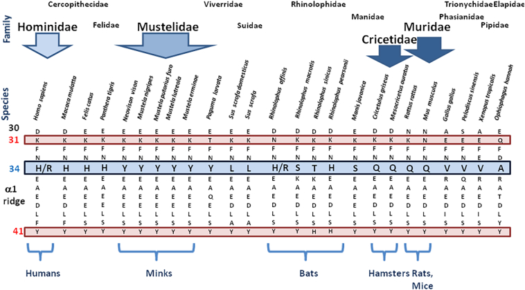
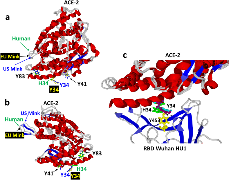
SARS-CoV-2, the virus responsible for COVID-19 in humans, can efficiently infect a large number of animal species. Like any virus, and particularly RNA viruses, SARS-CoV-2 undergoes mutations during its life cycle some of which bring a selective advantage, leading to the selection of a given lineage. Minks are very susceptible to SARS-CoV-2 and owing to their presence in mass rearing, they make a good model for studying the relative importance of mutations in viral adaptation to host species. Variants, such as the mink-selected SARS-CoV-2 Y453F and D614G or H69del/V70del, Y453F, I692V and M1229I were identified in humans after spreading through densely caged minks. However, not all mink-specific mutations are conserved when the virus infects human populations back. Many questions remain regarding the interspecies evolution of SARS-CoV-2 and the dynamics of transmission leading to the emergence of new variant strains. We compared the human and mink ACE2 receptor structures and their interactions with SARS-CVoV-2 variants. In minks, ACE2 presents a Y34 amino acid instead of the H34 amino acid found in the human ACE2. H34 is essential for the interaction with the Y453 residue of the SARS-CoV-2 Spike protein. The Y453F mink mutation abolishes this conflict. A series of 18 mutations not involved in the direct ACE2 interaction was observed in addition to the Y453F and D614G in 16 different SARS-CoV-2 strains following bidirectional infections between humans and minks. These mutations were not random and were distributed into five different functional groups having an effect on the kinetics of ACE2-RD interaction. The interspecies transmission of SARS-CoV-2 from humans to minks and back to humans, generated specific mutations in each species which improved the affinity for the ACE2 receptor either by direct mutation of the core 453 residue or by associated compensatory mutations.
Keywords: ACE2; COVID-19 vaccine; Coronavirus; Mink; SARS-CoV-2 variants; hamster.
© 2022 The Authors.
Conflict of interest statement
The authors declare that the research was conducted in the absence of any commercial of financial relationships that could be construed as a potential conflict of interest.
Figures
Similar articles
-
Mutation Y453F in the spike protein of SARS-CoV-2 enhances interaction with the mink ACE2 receptor for host adaption.PLoS Pathog. 2021 Nov 8;17(11):e1010053. doi: 10.1371/journal.ppat.1010053. eCollection 2021 Nov. PLoS Pathog. 2021. PMID: 34748603 Free PMC article.
-
Molecular Basis of Mink ACE2 Binding to SARS-CoV-2 and Its Mink-Derived Variants.J Virol. 2022 Sep 14;96(17):e0081422. doi: 10.1128/jvi.00814-22. Epub 2022 Aug 24. J Virol. 2022. PMID: 36000849 Free PMC article.
-
Spread of Mink SARS-CoV-2 Variants in Humans: A Model of Sarbecovirus Interspecies Evolution.Front Microbiol. 2021 Sep 20;12:675528. doi: 10.3389/fmicb.2021.675528. eCollection 2021. Front Microbiol. 2021. PMID: 34616371 Free PMC article. Review.
-
Highly conserved binding region of ACE2 as a receptor for SARS-CoV-2 between humans and mammals.Vet Q. 2020 Dec;40(1):243-249. doi: 10.1080/01652176.2020.1823522. Vet Q. 2020. PMID: 32921279 Free PMC article.
-
The basis of mink susceptibility to SARS-CoV-2 infection.J Appl Genet. 2022 Sep;63(3):543-555. doi: 10.1007/s13353-022-00689-w. Epub 2022 Apr 9. J Appl Genet. 2022. PMID: 35396646 Free PMC article. Review.
Cited by
-
Unravelling Antigenic Cross-Reactions toward the World of Coronaviruses: Extent of the Stability of Shared Epitopes and SARS-CoV-2 Anti-Spike Cross-Neutralizing Antibodies.Pathogens. 2023 May 13;12(5):713. doi: 10.3390/pathogens12050713. Pathogens. 2023. PMID: 37242383 Free PMC article. Review.
-
An update on angiotensin-converting enzyme 2 structure/functions, polymorphism, and duplicitous nature in the pathophysiology of coronavirus disease 2019: Implications for vascular and coagulation disease associated with severe acute respiratory syndrome coronavirus infection.Front Microbiol. 2022 Nov 28;13:1042200. doi: 10.3389/fmicb.2022.1042200. eCollection 2022. Front Microbiol. 2022. PMID: 36519165 Free PMC article. Review.
-
Critical amino acid residues in human ACE2 for SARS-CoV-2 spike protein binding and virus entry.Microbiol Spectr. 2025 Aug 5;13(8):e0324424. doi: 10.1128/spectrum.03244-24. Epub 2025 Jun 20. Microbiol Spectr. 2025. PMID: 40539804 Free PMC article.
-
Possible contribution of rare alleles of human ACE2 in the emergence of SARS-CoV-2 variants escaping the immune response.Front Immunol. 2023 Oct 10;14:1252367. doi: 10.3389/fimmu.2023.1252367. eCollection 2023. Front Immunol. 2023. PMID: 37885880 Free PMC article.
-
Convergent Evolution Dynamics of SARS-CoV-2 and HIV Surface Envelope Glycoproteins Driven by Host Cell Surface Receptors and Lipid Rafts: Lessons for the Future.Int J Mol Sci. 2023 Jan 18;24(3):1923. doi: 10.3390/ijms24031923. Int J Mol Sci. 2023. PMID: 36768244 Free PMC article. Review.
References
LinkOut - more resources
Full Text Sources
Miscellaneous
