. 2022 Aug 19:13:980147.
doi: 10.3389/fmicb.2022.980147. eCollection 2022.

AC5 protein encoded by squash leaf curl China virus is an RNA silencing suppressor and a virulence determinant

Affiliations

AC5 protein encoded by squash leaf curl China virus is an RNA silencing suppressor and a virulence determinant

Huijie Wu et al. Front Microbiol. .

Abstract

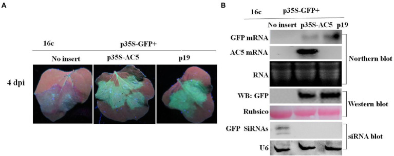
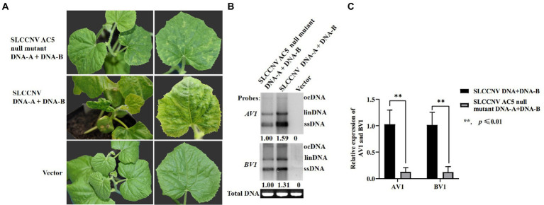
Squash leaf curl China virus (SLCCNV) is a bipartite Begomovirus. The function of the protein AC5, which is encoded by SLCCNV, is unknown. Here, we confirmed that the 172-amino acids (aa) long AC5 protein of SLCCNV could suppress single-stranded RNA but not double-stranded RNA-induced post-transcriptional gene silencing (PTGS). Furthermore, we determined that the C-terminal domain (96-172 aa) of the AC5 protein was responsible for RNA silencing suppressor (RSS) activity via deletion mutant analysis. The AC5 protein can reverse GFP silencing and inhibit systemic silencing of GFP by interfering with the systemic spread of the GFP silencing signal. The SLCCNV AC5 protein was localized to both the nucleus and cytoplasm of Nicotiana benthamiana cells. Furthermore, deletion analysis showed that the putative nuclear localization signal (NLS, 102-155 aa) was crucial for the RNA silencing suppression activity of AC5. In addition, the AC5 protein elicited a hypersensitive response and enhanced potoao virus X (PVX) RNA accumulation in infected N. benthamiana plants. Using the infectious clones of the SLCCNV and SLCCNV-AC5 null mutants, mutational analysis confirmed that knockout of the AC5 gene abolished SLCCNV-induced leaf curl symptoms, showing SLCCNV AC5 is also a virulence determinant.

Keywords: AC5 protein; RNA silencing suppressor; SLCCNV; subcellular localization; virulence determinant.


Conflict of interest statement

The authors declare that the research was conducted in the absence of any commercial or financial relationships that could be construed as a potential conflict of interest.

Figures

Figure 1
Figure 1
Phylogenetic analysis and domain structure of the squash leaf curl China virus (SLCCNV) AC5 protein. (A) The genomic organization of SLCCNV DNA-A. The common region (CR) and all the putative open reading frames (ORFs) located in the viral and complementary strands are indicated. (B) The conserved domain structures Gemini AC5-1 and Gemini AC5-2 were found in SLCCNV AC5 proteins, as determined using the conserved domain database (CDD). Note that the schematic domains of the AC5 protein may not be proportional to the aa scale bar shown at the top because of the varying lengths of the AC5 proteins. (C) Phylogenetic relationships of the AC5 amino acid (aa) sequences of representative begomoviruses. The AC5 aa sequences from begomoviruses were aligned using the Neighbor-Joining method in the MEGA6 program with 1,000 replications. Hides values were lower than 50%.
Figure 2
Figure 2
GFP silencing in GFP-transgenic N. benthamiana 16c plants. (A) Representative leaf patches or plants were co-infiltrated with A. tumefaciens cultures harboring the p35S-GFP, p35S-AC5, and expression vectors pGreenII 62-SK or the p19 vector. Images were photographed under UV light at 4 dpi. (B) Northern blot of GFP and AC5 mRNA accumulation, western blot of GFP protein accumulation and siRNA blot of GFP siRNA accumulation in Agrobacterium-infiltrated leaf patches as indicated in (A). SYBR Safe DNA Gel staining of rRNA and Ponceau S staining of the subunit of Rubisco served as loading controls. U6 was the loading control in the siRNA blot.
Figure 3
Figure 3
Identification of the AC5 region with RNA silencing suppressor activity. (A) Diagrams of the AC5 mutants. (B) Leaf patches were co-infiltrated with A. tumefaciens cultures harboring the p35S-GFP and empty pGreenII 62-SK vector, p35S-GFP + p35S-AC5, p35S-GFP + p35S-AC5(1–132), p35S-GFP + p35S-AC5(135–187), p35S-GFP + p35S-AC5(188–519), p35S-GFP + P19, p35S-AC5 and its mutants were photographed under UV light at 4 dpi. (C) Northern blot of GFP accumulation, GFP protein accumulation and siRNA blot of GFP siRNA accumulation in the Agrobacterium-infiltrated leaf patches as indicated in (B). SYBR Safe DNA Gel staining of rRNA and Ponceau S staining of the subunit of Rubisco served as loading controls. U6 was the loading control in the siRNA blot.
Figure 4
Figure 4
The SLCCNV AC5 could not inhibit GFP silencing of double-stranded dsRNA. (A) Representative 16c leaves were co-infiltrated with A. tumefaciens cultures harboring the p35S-GFP, double-stranded GFP (p35S-dsFP) and vector pGreenII 62-SK, p35S-AC5-expressing vectors or p19. Images were photographed under UV light at 4 dpi. (B) Northern blot of GFP and AC5 mRNA accumulation, western blot of GFP protein accumulation, and siRNA blot of GFP siRNA accumulation in Agrobacterium-infiltrated leaf patches. Ethidium bromide staining of rRNA and Ponceau S staining of the subunit of Rubisco served as loading controls. U6 was the loading control in the siRNA blot.
Figure 5
Figure 5
The effect of SLCCNV AC5 on suppressing systemic RNA silencing. (A) GFP-transgenic 16c plants were used to determine systemic silencing at 25 dpi. The GFP-transgenic 16c plants co-infiltrated at the seven-leaf stage with the mixture of carrying p35S-GFP, vector, p35S-AC5 expressing vectors or p19 were observed. (B) Northern blot analysis of GFP mRNA and western blot analyses of GFP, RNA, and proteins isolated from the systemic leaves at 25 dpi. SYBR Safe DNA Gel staining of rRNA and Ponceau S staining of the subunit of Rubisco served as loading controls.
Figure 6
Figure 6
Analysis of the subcellular localization of SLCCNV AC5, the AC5 nuclear localization signal (NLS), and PTGS. (A) Schematic representation of SLCCNV AC5 and a deletion mutant of its NLS. (B) Cellular and subcellular localization of SLCCNV AC5 and its derivatives in cells. H2B-RFP represents RFP fused at the C-terminus of the nuclear marker histone 2B. The white arrows indicate the nuclei. Scale bar = 34 μm. (C) Suppression of GFP silencing in GFP-transgenic 16c plants. Leaf patches were co-infiltrated with A. tumefaciens cultures harboring p35S-GFP + p19, p35S-GFP + p35S-AC5, p35S-GFP+ p35S-AC5-Δ102-115 aa, and p35S-GFP+ empty vector. The leaves were photographed under UV light at 4 dpi. (D) Western blot analyses of dsRNA-induced GFP gene silencing in infiltrated leaf samples.
Figure 7
Figure 7
Symptoms exhibited by plants following inoculation with pGR106 or pGR106-AC5. (A) N. benthamiana plants inoculated with pGR106 or pGR106-AC5 were photographed at 10 day-post Agrobacterium-infiltration (dpai). Upper infected leaves were photographed directly at 10 dpi and 20 dpi or photographed after 3,3′- diaminobenzidine (DAB) staining. (B) Leaf tissues were harvested from pGR106- or pGR106-AC5-systematically infected N. benthamiana plants at 10 dpai. The mRNA accumulation of potato virus X (PVX) was analyzed using real-time qPCR. *p ≤ 0.05, Student’s t-test. (C) Northern blot was used to detect mRNA accumulation, with the PVX cp gene as probe. The positions of genomic RNA (gRNA), TGB subgenomic RNA (TGB sgRNA), CP subgenomic RNA (CP sgRNA) were indicated.
Figure 8
Figure 8
Symptoms of melon plants infected with SLCCNV and SLCCNV-AC5 null mutants. (A) Chlorotic mosaic symptoms of melon plants infected with SLCCNV-AC5 null mutant and DNA-B infectious clones (SLCCNV AC5 null mutant DNA-A + DNA-B). Curling and chlorotic mosaic symptoms were observed in melon plants infected with SLCCNV DNA-A and DNA-B wild-type infectious clones (SLCCNV DNA + DNA-B). No symptoms were observed in plants inoculated with an empty vector (negative control). (B) Southern blot analysis of viral accumulation. Leaves inoculated with SLCCNV AC5 null mutant DNA-A + DNA-B, and SLCCNV wild-type were detected at 14 dpi using DNA-A AV1 and DNA-B BV1 genes as probes. The empty vector served as the negative control. Viral open circular (ocDNA), linear (linDNA), and single-stranded DNAs (ssDNA) are indicated. ImageJ software was used to analyze the relative ssDNA expression of DNA-A AV1 and DNA-B BV1 in infected melon plants. (C) Viral DNA accumulation in the SLCCNV-AC5 null mutant DNA-A + DNA-B infected plants and the SLCCNV wild-type DNA-A + DNA-B infected melon plants was detected through quantitative PCR (qPCR). **p ≤ 0.01, Student’s t-test.

References

    1. Asigul I., Yakupjan H., Wang Y., Li H., Qian L., Han T., et al. . (2018). Cotton leaf curl Multan virus C4 protein suppresses both transcriptional and post-transcriptional gene silencing by interacting with sam synthetase. PLoS Pathog. 14:e1007282. doi: 10.1371/journal.ppat.1007282 - DOI - PMC - PubMed
    1. Chandran S. A., Jeyabharathy C., Usha R. (2014). The C2 protein of bhendi yellow vein mosaic virus plays an important role in symptom determination and virus replication. Virus Genes 48, 203–207. doi: 10.1007/s11262-013-0992-1, PMID: - DOI - PubMed
    1. Ding S. W., Voinnet O. (2007). Antiviral immunity directed by small RNAs. Cell 130, 413–426. doi: 10.1016/j.cell.2007.07.039, PMID: - DOI - PMC - PubMed
    1. Feng M., Zuo D., Jiang X., Shuai L., Jing C., Jiang L., et al. . (2018). Identification of strawberry vein banding virus encoded p6 as an RNA silencing suppressor. Virology 520, 103–110. doi: 10.1016/j.virol.2018.05.003, PMID: - DOI - PubMed
    1. Fondong V. N. (2013). Geminivirus protein structure and function. Mol. Plant Pathol. 14, 635–649. doi: 10.1111/mpp.12032, PMID: - DOI - PMC - PubMed

LinkOut - more resources