. 2022 Aug 17:10:967765.
doi: 10.3389/fcell.2022.967765. eCollection 2022.

In depth functional characterization of human induced pluripotent stem cell-derived beta cells in vitro and in vivo

Affiliations

In depth functional characterization of human induced pluripotent stem cell-derived beta cells in vitro and in vivo

Federica Fantuzzi et al. Front Cell Dev Biol. .

Abstract

In vitro differentiation of human induced pluripotent stem cells (iPSCs) into beta cells represents an important cell source for diabetes research. Here, we fully characterized iPSC-derived beta cell function in vitro and in vivo in humanized mice. Using a 7-stage protocol, human iPSCs were differentiated into islet-like aggregates with a yield of insulin-positive beta cells comparable to that of human islets. The last three stages of differentiation were conducted with two different 3D culture systems, rotating suspension or static microwells. In the latter, homogeneously small-sized islet-like aggregates were obtained, while in rotating suspension size was heterogeneous and aggregates often clumped. In vitro function was assessed by glucose-stimulated insulin secretion, NAD(P)H and calcium fluctuations. Stage 7 aggregates slightly increased insulin release in response to glucose in vitro. Aggregates were transplanted under the kidney capsule of NOD-SCID mice to allow for further in vivo beta cell maturation. In transplanted mice, grafts showed glucose-responsiveness and maintained normoglycemia after streptozotocin injection. In situ kidney perfusion assays showed modulation of human insulin secretion in response to different secretagogues. In conclusion, iPSCs differentiated with equal efficiency into beta cells in microwells compared to rotating suspension, but the former had a higher experimental success rate. In vitro differentiation generated aggregates lacking fully mature beta cell function. In vivo, beta cells acquired the functional characteristics typical of human islets. With this technology an unlimited supply of islet-like organoids can be generated from human iPSCs that will be instrumental to study beta cell biology and dysfunction in diabetes.

Keywords: aggregate, beta cell; human induced pluripotent stem cell; insulin secretion; islet; microwell.


Conflict of interest statement

The authors declare that the research was conducted in the absence of any commercial or financial relationships that could be construed as a potential conflict of interest.

Figures

FIGURE 1
FIGURE 1
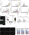
Human iPSC differentiation into beta cells. (A), Human iPSCs were differentiated into beta cells following a 7-stage stepwise protocol. Until stage 4 (St4) cells were cultured in 2D and then transferred to 3D culture, either in suspension (SP) or microwells (MW). Media specification is shown in boxes. (B), Morphology and size of aggregates differentiating into beta cells in rotating suspension (upper panels) or static microwells (bottom panels) at 1, 5 and 12 days post-detachment from 2D culture. Scale bar is 400 µm. (C), Diameter of the aggregates generated with 500, 750, 1000 or 2000 cells per microwell. Diameters were measured at day 1, 4 and 12 post-detachment from 2D culture. (D), Quantification of immunochemical analysis of total or insulin-positive cells (co-)expressing the proliferation marker Ki67 (n = 5). (E), Microwell and suspension aggregates stained for insulin (green), glucagon (red) and PDX1 (purple). Scale bar is 100 µm. Symbols represent different cell models (Hel115.6, circles; 1023A, squares). N, number of independent experiments, defined as one iPSC-beta cell differentiation, shown as individual data points.
FIGURE 2
FIGURE 2
Characterization of human iPSC-derived islet-like organoids. (A), Differentiation markers were measured by quantitative RT-PCR. White bars represent cells until stage 4 (St4, 2D culture), grey bars represent microwell aggregates (MW), red bars suspension aggregates (SP), green bars EndoC-βH1 cells and yellow bars human islets (HI). (B), Representative pictures of dispersed aggregates stained for insulin (green) and glucagon (red). Nuclei are visualized with DAPI (blue). (C), Quantification of insulin (INS), glucagon (GCG) and insulin/glucagon (INS/GCG) expressing cells from immunochemical analysis (MW n = 29; SP n = 22, HI n = 205). (D), Representative flow cytometry analysis of dispersed MW and SP aggregate cells. (E), Representative pictures of dispersed aggregate cells stained for insulin (green) and chromogranin A (CHGA), PDX1 or NKX6.1 (all in red). (F), Quantification of immunochemical analyses of cells expressing insulin only (yellow), chromogranin A, PDX1 or NKX6.1 only (purple), and both (blue) (MW n = 8–11; SP n = 6–7). Median (bold dotted line) and quartiles (light dotted line) are shown and dots represent independent iPSC-beta differentiations or human islet preparations. Symbols represent different cell models (Hel115.6, circles; 1023A, squares, EndoC-βH1, black circles; human islets, triangles). N, number of independent experiments, defined as one iPSC-beta cell differentiation or one human islet preparation, shown as individual data points. Mixed-effects analysis followed by Bonferroni correction for multiple comparisons, *p < 0.05, **p < 0.01, ***p < 0.005.
FIGURE 3
FIGURE 3
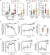
In vitro function of human iPSC-derived beta cells. (A), Glucose-stimulated insulin release, (B), insulin content (pmol/L) normalized by total protein (μg) and (C), proinsulin content (pmol/L) normalized by the total protein (μg) from microwell (MW, grey bars) or suspension aggregates (SP, red bars) or human islets (HI, yellow bars). To stimulate insulin secretion, aggregates were exposed to low (1.6 mmol/L) or high (16.7 mmol/L) glucose or high glucose plus forskolin (1 μmol/L) (MW n = 13; SP n = 12; HI n = 6–8). (D), NAD(P)H autofluorescence during perifusion of MW aggregates at glucose concentrations (Gn, n mmol/L), as indicated on top of the figure (n = 6). (E), Fura-2 LR fluorescence ratio during perifusion of MW aggregates with KRB containing Gn and added compounds (Dz, 250 μmol/L diazoxide; GCZ, 25 μmol/L gliclazide; K30, 30 mmol/L extracellular K+). Data are means ± SEM for 7 preparations, each with 1-2 aggregates of each kind. (F), Insulin secretion in response to G2 or G20 with or without 25 μmol/L GCZ (n = 3). (G,H), Glucose-induced changes in NAD(P)H autofluorescence and fura-2 LR fluorescence ratio after 2-h glucose starvation (n = 5–6). (I), Insulin secretion by glucose-starved MW aggregates during perifusion with Krebs containing G0, G20, Dz or K30 as indicated (n = 4). (A–C), Median (bold dotted line) and quartiles (light dotted line) are shown; dots represent independent iPSC-beta differentiations, and symbols different cell models (Hel115.6, circles; 1023A, squares; human islets, triangles). N, number of independent experiments, defined as one iPSC-beta cell differentiation or one human islet preparation, as indicated. Mixed-effects analysis followed by Bonferroni correction for multiple comparisons, *p < 0.05, **p < 0.01, ***p < 0.005 or exact p value shown.
FIGURE 4
FIGURE 4
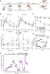
In vivo function of human iPSC-derived beta cells. (A), Stage 7 microwell or suspension aggregates were transplanted under the kidney capsule of NOD-SCID mice. Intraperitoneal glucose tolerance test (IPGTT) was done at week 7, 14 and 20. At 21–22 weeks, mice were injected with streptozotocin (STZ) to selectively ablate mouse beta cells. Glycemia was recorded for 1 week before nephrectomy or in situ kidney perfusion. (B), Mouse glycemia (top panels) and human plasma C-peptide (bottom panels) during IPGTT in suspension (left panels) and microwell (right panels). (C), Dose-response of streptozotocin toxicity in human islets (n = 3), human iPSC-derived beta cells (n = 5) and mouse islets (n = 5) after 24-h exposure to the drug. Mixed-effects analysis followed by Bonferroni’s correction for multiple comparisons, **p < 0.01, ***p < 0.005. (D), Twenty-one weeks after transplantation, a single dose of streptozotocin (200 mg/kg) was administered intraperitoneally. Non-implanted mice rapidly develop diabetes (blue, n = 5). Mice transplanted with stage 7 aggregates remain normoglycemic until graft removal by nephrectomy (black, n = 5). (E), Kidney perifusion was performed 22 weeks after transplantation in two mice STZ-injected and one mouse non-STZ-injected. The grafted kidney was perifused with medium containing 0 (G0) or 20 mmol/L glucose (G20). Forskolin (1 μmol/L, FK), gliclazide (25 μmol/L, GCZ), diazoxide (250 μmol/L, DZ) and KCl (30 mmol/L, K30) were added. Secretion from each graft was normalized to the maximum secretion during the first 12-min in K30-Dz. N, number of independent experiments, defined as one iPSC-beta cell differentiation or one human or mouse islet preparation or one humanized mouse, as indicated. **p < 0.01, ***p < 0.001 vs basal condition (time 0-min) using mixed-effects analysis followed by Dunnett’s (b) or Sidak’s (e) correction for multiple comparison.

Similar articles

Cited by

References

    1. An D., Chiu A., Flanders J. A., Song W., Shou D., Lu Y. C., et al. (2018). Designing a retrievable and scalable cell encapsulation device for potential treatment of type 1 diabetes. Proc. Natl. Acad. Sci. U. S. A. 115 (2), E263-E272. 10.1073/pnas.1708806115 - DOI - PMC - PubMed
    1. Augsornworawat P., Maxwell K. G., Velazco-Cruz L., Millman J. R. (2020). Single-cell transcriptome profiling reveals beta cell maturation in stem cell-derived islets after transplantation. Cell. Rep. 32 (8), 108067. 10.1016/j.celrep.2020.108067 - DOI - PMC - PubMed
    1. Balboa D., Barsby T., Lithovius V., Saarimaki-Vire J., Omar-Hmeadi M., Dyachok O., et al. (2022). Functional, metabolic and transcriptional maturation of human pancreatic islets derived from stem cells. Nat. Biotechnol. 10.1038/s41587-022-01219-z - DOI - PMC - PubMed
    1. Balboa D., Saarimaki-Vire J., Borshagovski D., Survila M., Lindholm P., Galli E., et al. (2018). Insulin mutations impair beta-cell development in a patient-derived iPSC model of neonatal diabetes. Elife 7, e38519. 10.7554/eLife.38519 - DOI - PMC - PubMed
    1. Balboa D., Saarimaki-Vire J., Otonkoski T. (2019). Concise review: human pluripotent stem cells for the modeling of pancreatic beta-cell pathology. Stem Cells 37 (1), 33–41. 10.1002/stem.2913 - DOI - PMC - PubMed

LinkOut - more resources