. 2022 Aug 17:10:901320.
doi: 10.3389/fcell.2022.901320. eCollection 2022.

Proteasomal subunit depletions differentially affect germline integrity in C. elegans

Affiliations

Proteasomal subunit depletions differentially affect germline integrity in C. elegans

Lourds Michelle Fernando et al. Front Cell Dev Biol. .

Abstract

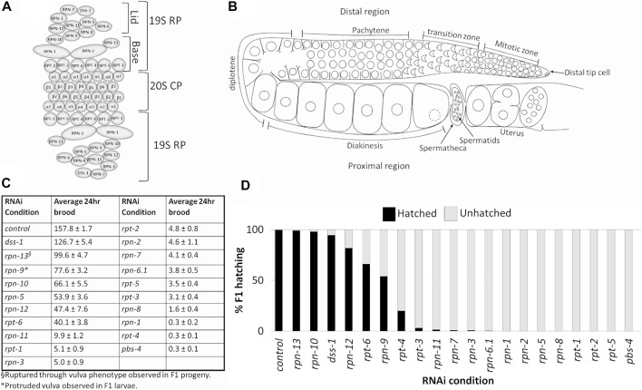
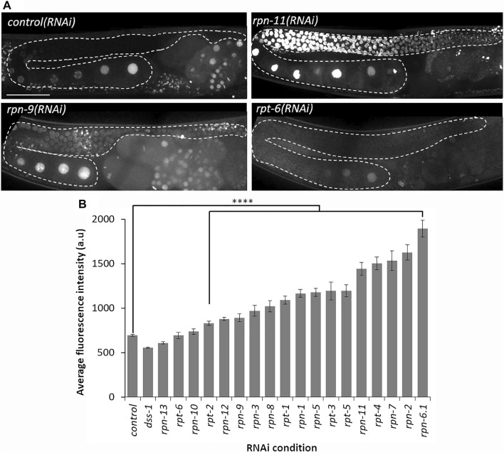
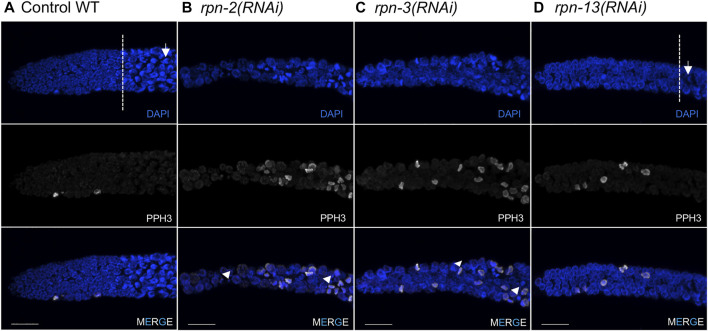
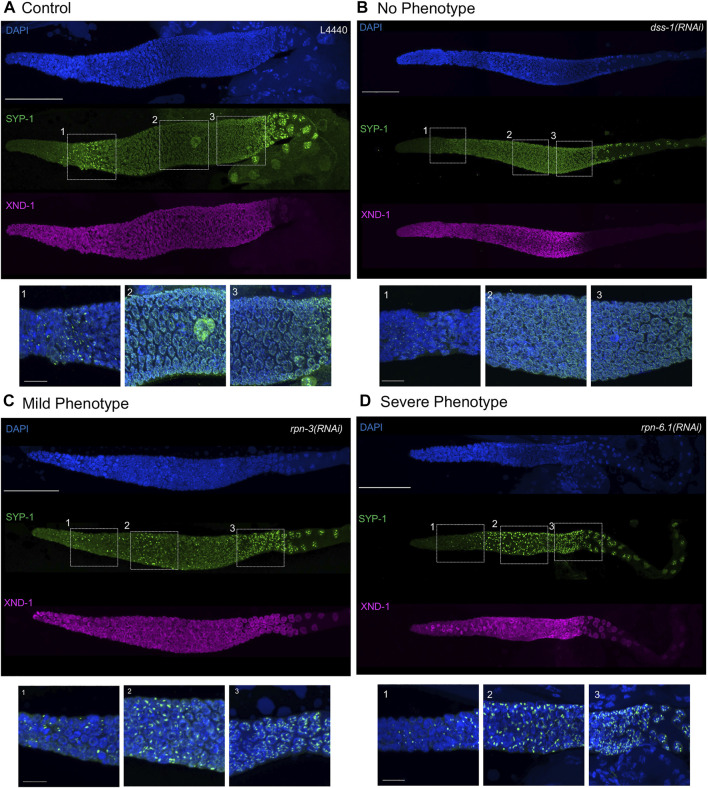
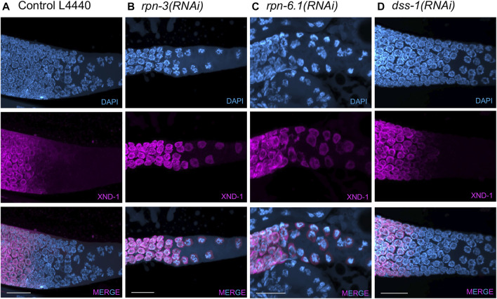
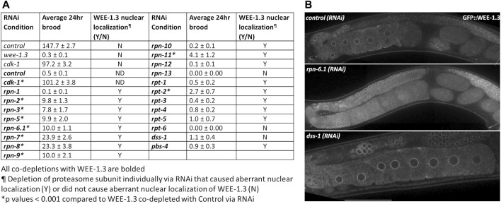
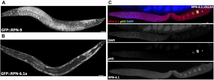
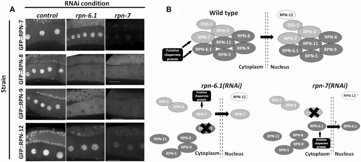
The 26S proteasome is a multi-subunit protein complex that is canonically known for its ability to degrade proteins in cells and maintain protein homeostasis. Non-canonical or non-proteolytic roles of proteasomal subunits exist but remain less well studied. We provide characterization of germline-specific functions of different 19S proteasome regulatory particle (RP) subunits in C. elegans using RNAi specifically from the L4 stage and through generation of endogenously tagged 19S RP lid subunit strains. We show functions for the 19S RP in regulation of proliferation and maintenance of integrity of mitotic zone nuclei, in polymerization of the synaptonemal complex (SC) onto meiotic chromosomes and in the timing of SC subunit redistribution to the short arm of the bivalent, and in turnover of XND-1 proteins at late pachytene. Furthermore, we report that certain 19S RP subunits are required for proper germ line localization of WEE-1.3, a major meiotic kinase. Additionally, endogenous fluorescent labeling revealed that the two isoforms of the essential 19S RP proteasome subunit RPN-6.1 are expressed in a tissue-specific manner in the hermaphrodite. Also, we demonstrate that the 19S RP subunits RPN-6.1 and RPN-7 are crucial for the nuclear localization of the lid subunits RPN-8 and RPN-9 in oocytes, further supporting the ability to utilize the C. elegans germ line as a model to study proteasome assembly real-time. Collectively, our data support the premise that certain 19S RP proteasome subunits are playing tissue-specific roles, especially in the germ line. We propose C. elegans as a versatile multicellular model to study the diverse proteolytic and non-proteolytic roles that proteasome subunits play in vivo.

Keywords: 19S regulatory particle; C. elegans; germ line; meiosis; proteasome.


Conflict of interest statement

The authors declare that the research was conducted in the absence of any commercial or financial relationships that could be construed as a potential conflict of interest.

Figures

FIGURE 1
FIGURE 1
Depletion of 19S RP subunits of the 26S proteasome in C. elegans hermaphrodites causes reduced 24 h brood and/or embryonic lethality. (A) Schematic of eukaryotic 26S proteasome and its subunits. (B) Schematic of an adult C. elegans hermaphrodite germ line (one gonad arm). (C) Average 24 h brood of C. elegans hermaphrodites RNAi-depleted of either a control gene (n = 152), any of the 19 subunits of the 19S RP (n = 10–83), or a 20S CP subunit, PBS-4 (n = 36). Brood is shown ±SEM and calculated from at least three independent trials. All RNAi conditions compared to control exhibit a p-value < 0.0001. (D) Percent of hatched (black bars) and unhatched (grey bars) progeny of hermaphrodites treated with either control(RNAi) or the indicated proteasome subunit(RNAi).
FIGURE 2
FIGURE 2
Depletion of most 19S RP subunits severely decreases proteolytic activity. (A) Representative images of germ line from Ub(G76V)::GFP::H2B animals treated with the indicated RNAi. Representative images of normal germline proteolytic activity [control(RNAi) and rpt-6(RNAi)], severe dysfunction of proteolytic activity [rpn-11(RNAi)], and moderate dysfunction of proteolytic activity [rpn-9(RNAi)]. A gonad arm is outlined with white dashed lines. (B) Average fluorescence intensity of Ub(G76V)::GFP::H2B germ lines treated with either RNAi against a control (n = 122) or any of the various 19 subunits of the 19S RP (n = 10–52). Fluorescence intensity (a. u) was measured in the region outlined with the white dashed lines as indicated in (A). All images taken at the same laser intensity and PMT gain, and then the same post-image modifications made to each image. **** represents p-values < 0.0001 compared to control(RNAi) condition. Error bars represent SEM. Scale bar, 50 µm.
FIGURE 3
FIGURE 3
Defects in the mitotic germ line result from 19S RP subunit knockdown. Representative images of the distal tip of the C. elegans germ line visualized with DAPI (blue) and phospho-H3 Ser10 (white). (A) Wild type N2 controls [white dash line indicates start of transition zone with characteristic crescent shape nuclei (white arrow)]. (B,C) Worms treated with rpn-2(RNAi) or rpn-3(RNAi) presented an increased number of cells in M phase and the presence of small or fragmented nuclei (white arrowheads). Both also had shorter mitotic tips with no clear transition zone. (D) rpn-13(RNAi) resulted in no cell cycle defects, presenting mitotic tips comparable to WT worms and obvious transition zone (white dashed line) and TZ nuclei (white arrow). Images show max projections of Z stacks halfway through each gonad. Distal is to the left in all images. Scale bar, 10 μm.
FIGURE 4
FIGURE 4
Synaptonemal complex defects are observed upon knockdown of 19S proteasome subunits. Representative images of germ lines visualized with anti-SYP-1 to mark the synaptonemal complex (green), anti-XND-1 (purple), and DAPI to mark DNA (blue). (A) Control, empty vector, shows the expected formation of a few SC polycomplexes (PCs) in the TZ. (B) No phenotype: full polymerization of SYP-1 throughout pachytene stage and correct timing of polarization to the short arm of the chromosome at diplotene comparable to control. (C) Mild-phenotype: extended region of PCs reaching early pachytene, with an abundant number of nuclei with fully polymerized SC in mid-pachytene. Premature polarization is also observed. (D) Severe phenotype: extended region of PCs into mid-pachytene, with almost all nuclei having at least one PC and no polymerization of SYP-1. Premature polarization of SYP-1 was present at late pachytene. Whole gonad scale bar, 50 m. Zoom in boxes correspond to: (1) Transition Zone, (2) Early-Mid Pachytene, (3) Late Pachytene. Scale bar, 10 m.
FIGURE 5
FIGURE 5
XND-1 turnover is affected by knockdown of a subset of 19S RP non-ATPase subunits. Representative images showing defects in XND-1 turnover after depletion of a specific group of non-ATPase proteasome subunits. Anti-XND-1 (magenta); DAPI stained DNA (cyan). (A) Vector control. (B) rpn-3(RNAi), and (C) rpn-6.1(RNAi) are examples of two subunits whose knockdown causes persistence of high levels of nucleoplasmic XND-1 in late pachytene nuclei. (D) dss-1(RNAi) is representative of the class of subunits who depletion does not affect XND-1. Scale bar, 50 µm.
FIGURE 6
FIGURE 6
WEE-1.3 function and localization are altered by depletion of specific proteasome subunits. (A) Average 24 h brood and WEE-1.3 nuclear localization status of hermaphrodites treated with either control(RNAi), wee-1.3(RNAi), cdk-1(RNAi) individually or co-depleted with WEE-1.3, or 19S RP subunits co-depleted with WEE-1.3 via RNAi (all co-depletions with WEE-1.3 are bolded). All co-depletion conditions were compared to WEE-1.3 co-depleted with the control RNAi condition. * represents p values < 0.001, Y (yes) or N (No) represents whether or not aberrant nuclear localization of WEE-1.3 occur when control or proteasome subunits depleted individually. ND indicated not determined. (B) Live imaging of gonads from strain WDC2 wee-1.3[ana2(gfp::wee-1.3)] treated with either control(RNAi), rpn-6.1(RNAi) or dss-1(RNAi). All images were taken at the same laser intensity and PMT gain. Scale bar, 100 µm.
FIGURE 7
FIGURE 7
The two RPN-6.1 isoforms exhibit different spatial localization. Live imaging of hermaphrodites expressing endogenously GFP-tagged (A) RPN-9 and (B) RPN-6. Strains are WDC5 rpn-9[ana5 (gfp::rpn-9)] and WDC3 rpn-6.1a [ana3(gfp::rpn-6.1a)]. (C) Immunofluorescence image of rpn-6.1[ana12(rpn-6.1::ollas)] strain dissected gonad co-stained with anti-OLLAS (red), anti-pH3 (green, condensed chromatin) and DAPI for DNA (blue). Anti-pH3 was used solely to confirm presence of maturing oocytes. Bright nuclear and relatively dim cytoplasmic RPN-6.1b expression shown throughout germ line. Scale bar, 50 µm.
FIGURE 8
FIGURE 8
RPN-6.1 and RPN-7 are required for the nuclear localization of RPN-8 and RPN-9. (A) Live imaging of hermaphrodite oocytes from endogenously GFP tagged strains rpn-7[ana1(gfp::rpn7)], rpn-8[ana4(gfp::rpn-8)], rpn-9[ana5(gfp::rpn-9)] and rpn-12[ana6(gfp::rpn-12)] treated with either control(RNAi), rpn-6.1(RNAi) or rpn-7(RNAi) (n = 15–42). Scale bar represents 25 µm. (B) Model for role of RPN-6.1 and RPN-7 in nuclear localization of 19S RP lid combining existing information on eukaryotic proteasome assembly model (Budenholzer et al., 2017; Bai et al., 2019).

References

    1. Adams J., Palombella V. J., Sausville E. A., Johnson J., Destree A., Lazarus D. D., et al. (1999). Proteasome inhibitors: a novel class of potent and effective antitumor agents. Cancer Res. 59 (11), 2615–2622. - PubMed
    1. Ahuja J. S., Sandhu R., Mainpal R., Lawson C., Henley H., Hunt P. A., et al. (2017). Control of meiotic pairing and recombination by chromosomally tethered 26S proteasome. Science 355 (6323), 408–411. 10.1126/science.aaf4778 - DOI - PMC - PubMed
    1. Albert S., Schaffer M., Beck F., Mosalaganti S., Asano S., Thomas H. F., et al. (2017). Proteasomes tether to two distinct sites at the nuclear pore complex. Proc. Natl. Acad. Sci. U. S. A. 114 (52), 13726–13731. 10.1073/pnas.1716305114 - DOI - PMC - PubMed
    1. Allen A. K., Nesmith J. E., Golden A. (2014). An RNAi-based suppressor screen identifies interactors of the Myt1 ortholog of Caenorhabditis elegans . G3 (Bethesda) 4 (12), 2329–2343. 10.1534/g3.114.013649 - DOI - PMC - PubMed
    1. Arribere J. A., Bell R. T., Fu B. X. H., Artiles K. L., Hartman P. S., Fire A. Z. (2014). Efficient marker-free recovery of custom genetic modifications with CRISPR/Cas9 in Caenorhabditis elegans . Genetics 198 (3), 837–846. 10.1534/genetics.114.169730 - DOI - PMC - PubMed

LinkOut - more resources