. 2022 Aug 19:13:909317.
doi: 10.3389/fendo.2022.909317. eCollection 2022.

Vibration therapy as an effective approach to improve bone healing in diabetic rats

Affiliations

Vibration therapy as an effective approach to improve bone healing in diabetic rats

Maysa S Campos et al. Front Endocrinol (Lausanne). .

Abstract

Objective: To investigate the effects of vibration therapy on fracture healing in diabetic and non-diabetic rats.

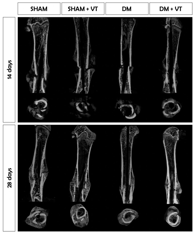
Methods: 148 rats underwent fracture surgery and were assigned to four groups: (1) SHAM: weight-matched non-diabetic rats, (2) SHAM+VT: non-diabetic rats treated with vibration therapy (VT), (3) DM: diabetic rats, and (4) DM+VT: diabetic rats treated with VT. Thirty days after diabetes induction with streptozotocin, animals underwent bone fracture, followed by surgical stabilization. Three days after bone fracture, rats began VT. Bone healing was assessed on days 14 and 28 post-fracture by serum bone marker analysis, and femurs collected for dual-energy X-ray absorptiometry, micro-computed tomography, histology, and gene expression.

Results: Our results are based on 88 animals. Diabetes led to a dramatic impairment of bone healing as demonstrated by a 17% reduction in bone mineral density and decreases in formation-related microstructural parameters compared to non-diabetic control rats (81% reduction in bone callus volume, 69% reduction in woven bone fraction, 39% reduction in trabecular thickness, and 45% in trabecular number). These changes were accompanied by a significant decrease in the expression of osteoblast-related genes (Runx2, Col1a1, Osx), as well as a 92% reduction in serum insulin-like growth factor I (IGF-1) levels. On the other hand, resorption-related parameters were increased in diabetic rats, including a 20% increase in the callus porosity, a 33% increase in trabecular separation, and a 318% increase in serum C terminal telopeptide of type 1 collagen levels. VT augmented osteogenic and chondrogenic cell proliferation at the fracture callus in diabetic rats; increased circulating IGF-1 by 668%, callus volume by 52%, callus bone mineral content by 90%, and callus area by 72%; and was associated with a 19% reduction in circulating receptor activator of nuclear factor kappa beta ligand (RANK-L).

Conclusions: Diabetes had detrimental effects on bone healing. Vibration therapy was effective at counteracting the significant disruption in bone repair induced by diabetes, but did not improve fracture healing in non-diabetic control rats. The mechanical stimulus not only improved bone callus quality and quantity, but also partially restored the serum levels of IGF-1 and RANK-L, inducing bone formation and mineralization, thus creating conditions for adequate fracture repair in diabetic rats.

Keywords: bone turnover; bony callus; diabetes mellitus; fracture healing; vibration.


Conflict of interest statement

The authors declare that the research was conducted in the absence of any commercial or financial relationships that could be construed as a potential conflict of interest.

Figures

Figure 1
Figure 1
Schematic figure representing the experimental design of this study. Animals were subjected to diabetes induction by injection of streptozotocin (STZ) on study entry (day 0). On day 30, diabetic and non-diabetic rats were subjected to a mid-femur fracture. Vibration therapy (VT) was administered to SHAM+VT and DM+VT rats from day 3 post-fracture until the endpoint assessment: day 44 or day 58, in which bones and serum blood were analyzed.
Figure 2
Figure 2
Comparison of body weight (in grams) among groups. Upon study entry (day 0, diabetes/sham induction), body weights were similar among all groups (p>0.05). On day 30, a closed femoral fracture procedure was performed. On day 14 post-fracture (day 44), the body weight was lower in diabetic rats than in sham rats (p<0.05). Similarly, on day 28 post-fracture (day 58), diabetic rats exhibited lower body weights than sham rats (p<0.05). Sample sizes are n=14/group. Lines represent the averages and shaded areas the standard deviation.
Figure 3
Figure 3
Comparison of BMD (A, in g/cm2), BMC (B, in g), and the area (C, in cm2) of the bone callus between each group at both 14 and 28 days post-fracture (DXA assessment). Sample sizes are n=7/group. Asterisks indicate significant differences (p < 0.05).
Figure 4
Figure 4
Comparison of callus volume (in mm3), mineralization (in %), thickness (in mm), number, separation (in mm), and porosity (in %) among the groups on days 14 and 28 post-fracture. Sample sizes are n = 7/group. Diabetes disrupted bone callus formation and quality by decreasing the formation-related microstructural parameters (volume, mineralization, thickness, and number) and increasing the resorption-related parameters (porosity and separation). Asterisks indicate significant differences (p < 0.05).
Figure 5
Figure 5
Coronal and axial tridimensional μCT images of fractured femurs. Diabetes impaired bone healing, and bone calluses were smaller and less mineralized than in sham rats 14 and 28 days post-surgery. Vibration therapy significantly increased bone callus volume and mineralization at 14 and 28 days post-surgery, but only in diabetic rats. Sample sizes are n=7/group, in which representative images from each group were selected for this figure.
Figure 6
Figure 6
Histological slides of fractured femurs stained with HE at magnifications of 12.5, 50, and 200x. Sample sizes are n = 7/group. Diabetes induced a delay in cell proliferation and differentiation during fracture healing. At 14 days after fracture, sham rats exhibited a bone callus formed by trabeculae (represented by *), while diabetic rats only showed connective tissue with fibroblasts (represented by f). The delay persisted in the later stage of healing, where sham rats had dense trabeculae forming a bone callus (*), and diabetic rats only showed thin and well-spaced trabeculae. Vibration therapy accelerated cell differentiation during fracture healing in diabetic rats in both stages but did not cause changes in normal bone healing. Calluses were formed by cartilaginous tissue (c) and newly formed trabeculae (*) on day 14 and thick and dense trabeculae (*) on day 28.
Figure 7
Figure 7
Histological slides of fractured femurs stained with Sirius red solution (A) and TRAP (B), with a magnification of 12.5x. (A): Collagen deposition (shown by asterisk marks) in the bone calluses of diabetic rats was significantly lower than in sham rats. Vibration therapy increased collagen deposition in bone calluses of diabetic rats but not in sham rats. (B): Osteoclastic activity (shown by hashtag marks) (#) in diabetic rats was higher than in sham animals on day 14 after fracture but lower on day 28. Vibration therapy increased osteoclastic activity in bone calluses of diabetic rats on day 28 post-fracture but did not exert a desirable effect in control rats. Sample sizes are n=7/group, in which representative images from each group were selected for this figure. Asterisks (*) indicate collagen deposition.
Figure 8
Figure 8
Diabetes leads to down expression of osteoblast-related genes (Col1a1, Runx2, and Osx) on day 28 post-fracture. Sample sizes are n = 4/group. Asterisks (*) indicate significant difference (p<0.05).

References

    1. Kayal RA, Alblowi J, McKenzie E, Krothapalli N, Silkman L, Gerstenfeld L, et al. . Diabetes causes the accelerated loss of cartilage during fracture repair which is reversed by insulin treatment. Bone (2009) 44:357–63. doi: 10.1016/j.bone.2008.10.042 - DOI - PMC - PubMed
    1. Korres N, Tsiridis E, Pavlou G, Mitsoudis A, Perrea DN, Zoumbos AB. Biomechanical characteristics of bone in streptozotocin-induced diabetic rats: An in-vivo randomized controlled experimental study. World J Orthop (2013) 4:124–9. doi: 10.5312/wjo.v4.i3.124 - DOI - PMC - PubMed
    1. Motyl K, McCabe LR. Streptozotocin, type I diabetes severity and bone. Biol Proced Online (2009) 11:296–315. doi: 10.1007/s12575-009-9000-5 - DOI - PMC - PubMed
    1. Fazzalari NL. Bone fracture and bone fracture repair. Osteoporos Int (2011) 22:2003–6. doi: 10.1007/s00198-011-1611-4 - DOI - PubMed
    1. Krakauer JC, McKenna MJ, Buderer NF, Rao DS, Whitehouse FW, Parfitt AM. Bone loss and bone turnover in diabetes. Diabetes (1995) 44:775–82. doi: 10.2337/diab.44.7.775 - DOI - PubMed

Publication types

Substances