

Sleep deprivation and recovery sleep affect healthy male resident's pain sensitivity and oxidative stress markers: The medial prefrontal cortex may play a role in sleep deprivation model
- PMID: 36061364
- PMCID: PMC9434020
- DOI: 10.3389/fnmol.2022.937468
Sleep deprivation and recovery sleep affect healthy male resident's pain sensitivity and oxidative stress markers: The medial prefrontal cortex may play a role in sleep deprivation model
Abstract
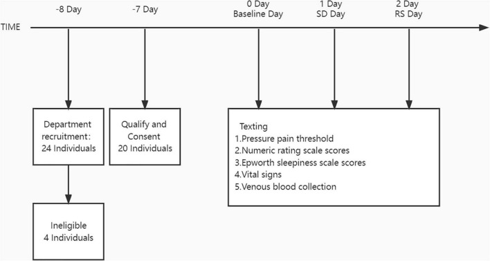
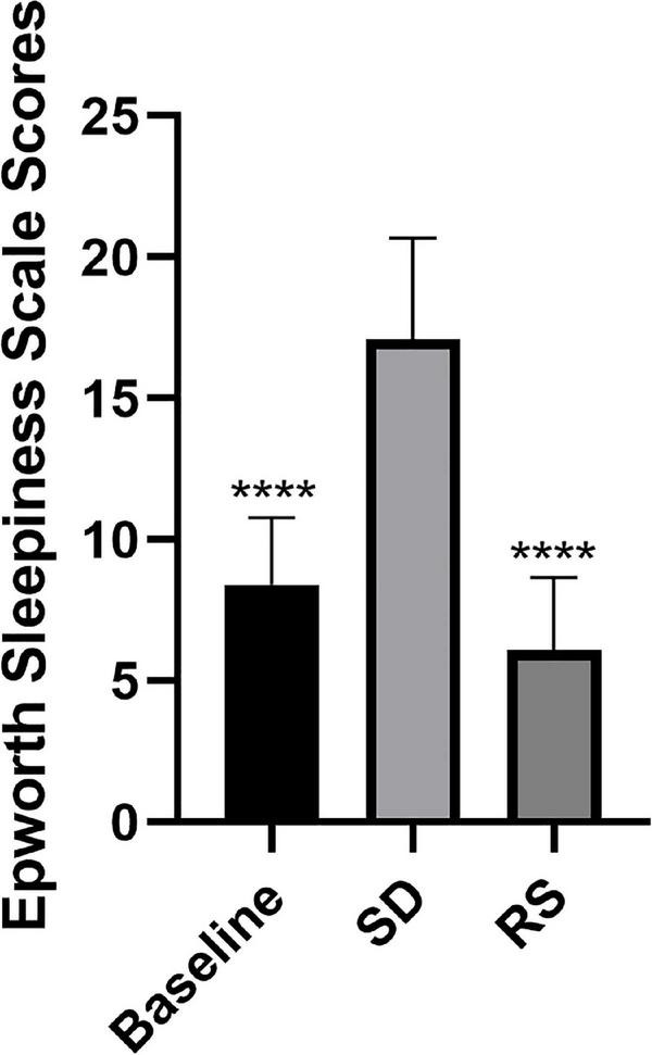
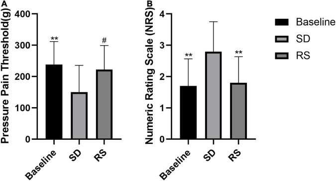
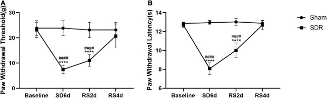
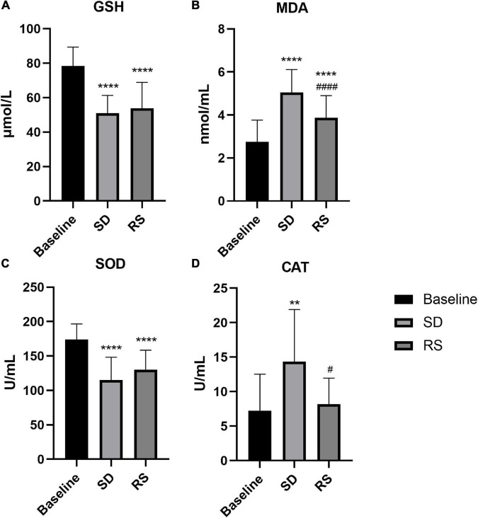
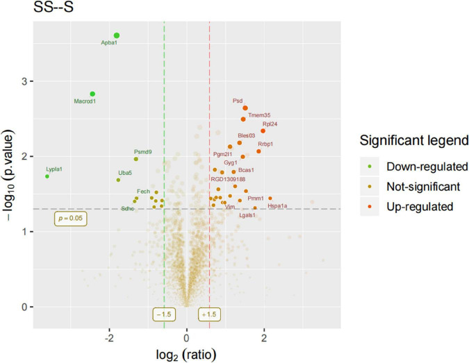
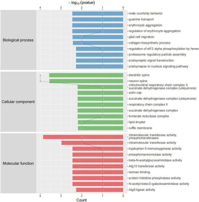
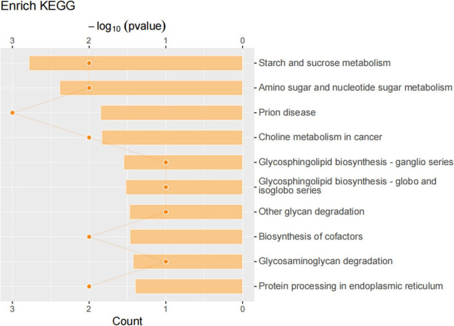
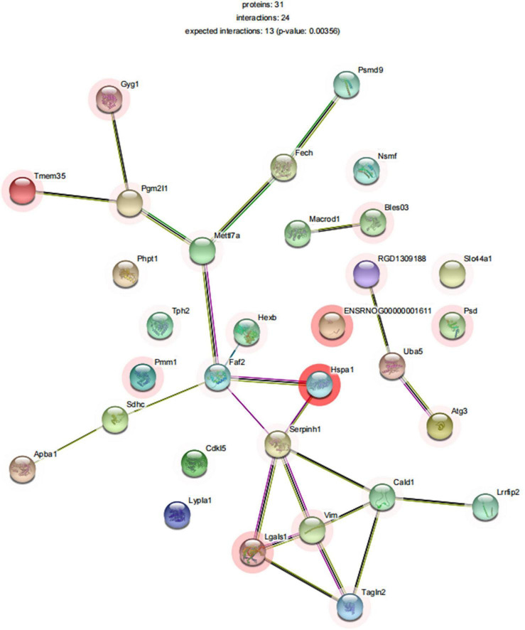
Sleep is essential for the body's repair and recovery, including supplementation with antioxidants to maintain the balance of the body's redox state. Changes in sleep patterns have been reported to alter this repair function, leading to changes in disease susceptibility or behavior. Here, we recruited healthy male physicians and measured the extent of the effect of overnight sleep deprivation (SD) and recovery sleep (RS) on nociceptive thresholds and systemic (plasma-derived) redox metabolism, namely, the major antioxidants glutathione (GSH), catalase (CAT), malondialdehyde (MDA), and superoxide dismutase (SOD). Twenty subjects underwent morning measurements before and after overnight total SD and RS. We found that one night of SD can lead to increased nociceptive hypersensitivity and the pain scores of the Numerical Rating Scale (NRS) and that one night of RS can reverse this change. Pre- and post-SD biochemical assays showed an increase in MDA levels and CAT activity and a decrease in GSH levels and SOD activity after overnight SD. Biochemical assays before and after RS showed a partial recovery of MDA levels and a basic recovery of CAT activity to baseline levels. An animal study showed that SD can cause a significant decrease in the paw withdrawal threshold and paw withdrawal latency in rats, and after 4 days of unrestricted sleep, pain thresholds can be restored to normal. We performed proteomics in the rat medial prefrontal cortex (mPFC) and showed that 37 proteins were significantly altered after 6 days of SD. Current findings showed that SD causes nociceptive hyperalgesia and oxidative stress, and RS can restore pain thresholds and repair oxidative stress damage in the body. However, one night of RS is not enough for repairing oxidative stress damage in the human body.
Keywords: night shift; oxidative stress; pain; recovery sleep; sleep deprivation.
Copyright © 2022 Chen, Xie, Li, Fan, Xing, Mao, Xing, Wang, Yang, Wang and Yuan.
Conflict of interest statement
The authors declare that the research was conducted in the absence of any commercial or financial relationships that could be construed as a potential conflict of interest.
Figures
Similar articles
-
Sleep deprivation affects pain sensitivity by increasing oxidative stress and apoptosis in the medial prefrontal cortex of rats via the HDAC2-NRF2 pathway.Biomed J. 2025 Jan 2:100826. doi: 10.1016/j.bj.2024.100826. Online ahead of print. Biomed J. 2025. PMID: 39755172
-
Melatonin ameliorates cognitive impairment induced by sleep deprivation in rats: role of oxidative stress, BDNF and CaMKII.Behav Brain Res. 2013 Nov 1;256:72-81. doi: 10.1016/j.bbr.2013.07.051. Epub 2013 Aug 6. Behav Brain Res. 2013. PMID: 23933144
-
Impact of Rapid Eye Movement Sleep Deprivation on Pain Behaviour and Oxidative Stress in the Thalamus: Role of Tualang Honey Supplementation.Malays J Med Sci. 2022 Apr;29(2):69-79. doi: 10.21315/mjms2022.29.2.7. Epub 2022 Apr 21. Malays J Med Sci. 2022. PMID: 35528807 Free PMC article.
-
Sleep Deprivation-Induced Oxidative Stress in Rat Models: A Scoping Systematic Review.Antioxidants (Basel). 2023 Aug 11;12(8):1600. doi: 10.3390/antiox12081600. Antioxidants (Basel). 2023. PMID: 37627596 Free PMC article.
-
The effects of recovery sleep on pain perception: A systematic review.Neurosci Biobehav Rev. 2020 Jun;113:408-425. doi: 10.1016/j.neubiorev.2020.03.028. Epub 2020 Apr 8. Neurosci Biobehav Rev. 2020. PMID: 32275917
Cited by
-
A Narrative Review of the Reciprocal Relationship Between Sleep Deprivation and Chronic Pain: The Role of Oxidative Stress.J Pain Res. 2024 May 20;17:1785-1792. doi: 10.2147/JPR.S455621. eCollection 2024. J Pain Res. 2024. PMID: 38799272 Free PMC article. Review.
-
Sleep and Oxidative Stress: Current Perspectives on the Role of NRF2.Cell Mol Neurobiol. 2024 Jun 25;44(1):52. doi: 10.1007/s10571-024-01487-0. Cell Mol Neurobiol. 2024. PMID: 38916679 Free PMC article. Review.
-
Thyroid-associated ophthalmopathy: the role of oxidative stress.Front Endocrinol (Lausanne). 2024 Jul 11;15:1400869. doi: 10.3389/fendo.2024.1400869. eCollection 2024. Front Endocrinol (Lausanne). 2024. PMID: 39055057 Free PMC article. Review.
-
Nanowired Delivery of Cerebrolysin Together with Antibodies to Amyloid Beta Peptide, Phosphorylated Tau, and Tumor Necrosis Factor Alpha Induces Superior Neuroprotection in Alzheimer's Disease Brain Pathology Exacerbated by Sleep Deprivation.Adv Neurobiol. 2023;32:3-53. doi: 10.1007/978-3-031-32997-5_1. Adv Neurobiol. 2023. PMID: 37480458 Review.
-
Unraveling the interplay between sleep, redox metabolism, and aging: implications for brain health and longevity.Front Aging. 2025 May 21;6:1605070. doi: 10.3389/fragi.2025.1605070. eCollection 2025. Front Aging. 2025. PMID: 40469623 Free PMC article. Review.
References
-
- Al’absi M., Petersen K. L., Wittmers L. E. (2022). Adrenocortical and hemodynamic predictors of pain perception in men and women. Pain 96 197–204. - PubMed
LinkOut - more resources
Full Text Sources
Miscellaneous