Review
. 2022 Aug 18:16:988283.
doi: 10.3389/fnins.2022.988283. eCollection 2022.

Cerebral edema after ischemic stroke: Pathophysiology and underlying mechanisms

Affiliations
Review

Cerebral edema after ischemic stroke: Pathophysiology and underlying mechanisms

Yuhang Gu et al. Front Neurosci. .

Abstract

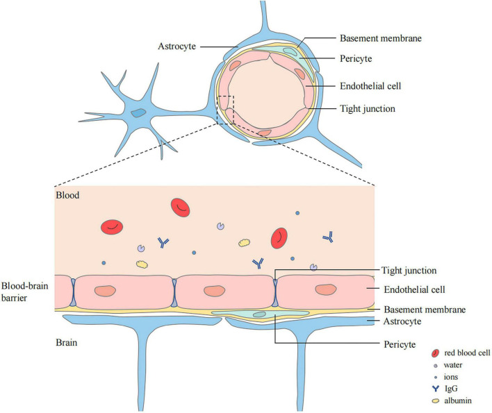
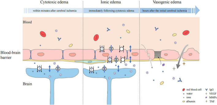
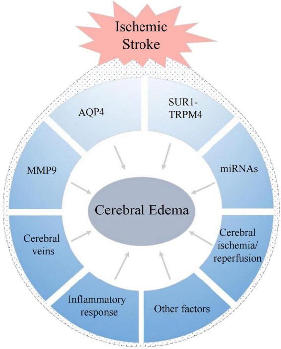
Ischemic stroke is associated with increasing morbidity and has become the main cause of death and disability worldwide. Cerebral edema is a serious complication arising from ischemic stroke. It causes an increase in intracranial pressure, rapid deterioration of neurological symptoms, and formation of cerebral hernia, and is an important risk factor for adverse outcomes after stroke. To date, the detailed mechanism of cerebral edema after stroke remains unclear. This limits advances in prevention and treatment strategies as well as drug development. This review discusses the classification and pathological characteristics of cerebral edema, the possible relationship of the development of cerebral edema after ischemic stroke with aquaporin 4, the SUR1-TRPM4 channel, matrix metalloproteinase 9, microRNA, cerebral venous reflux, inflammatory reactions, and cerebral ischemia/reperfusion injury. It also summarizes research on new therapeutic drugs for post-stroke cerebral edema. Thus, this review provides a reference for further studies and for clinical treatment of cerebral edema after ischemic stroke.

Keywords: blood-brain barrier; cerebral edema; cerebrovascular disease; ischemic stroke; pathophysiology.


Conflict of interest statement

The authors declare that the research was conducted in the absence of any commercial or financial relationships that could be construed as a potential conflict of interest.

Figures

FIGURE 1
FIGURE 1
Structure of blood–brain barrier (BBB). BBB locates between the luminal substances of the blood vasculature and the brain interstitium. The capillary lumen is surrounded with endothelial cells connected by tight junctions. Pericytes and Endothelial cells are ensheathed by a basement membrane surrounded by the end-feet of astrocytes.
FIGURE 2
FIGURE 2
Status of the blood–brain barrier at three phases of cerebral edema. Cytotoxic edema is the initial step and particularly prominent in astrocytes. Cerebral ischemia and hypoxia induced the ion influx (black arrow), which leads to osmotic gradient changes. Water may flow into astrocytes in three ways, simple diffusion (thick blue double-headed arrows), passive transport through transmembrane channels (thin blue double-headed arrows), and water co-transport (blue single-headed arrows). In ionic edema, ion, and water influx are mediated by plasmalemma channels and transporters of endothelial cells. Upregulation of transporters and ion channels also occurs in astrocytes. Vasogenic edema is characterized by destruction of the BBB. The transport of ions, water, and serum proteins such as albumin and IgG may occur directly (thick gray arrow) or via pinocytic vesicles (dashed gray arrow). Multiple factors, including VEGF, MMPs, and pro-inflammatory cytokines such as TNF are involved. They mediate neuroinflammation and tight junction degradation, aggravating cerebral edema.
FIGURE 3
FIGURE 3
Factors associated with the formation of cerebral edema after ischemic stroke.

References

    1. Aittoniemi J., Fotinou C., Craig T. J., de Wet H., Proks P., Ashcroft F. M. (2009). Review. SUR1: A unique ATP-binding cassette protein that functions as an ion channel regulator. Philos. Trans. R. Soc. Lond. B Biol. Sci. 364 257–267. 10.1098/rstb.2008.0142 - DOI - PMC - PubMed
    1. Alquisiras-Burgos I., Franco-Perez J., Rubio-Osornio M., Aguilera P. (2022). The short form of the SUR1 and its functional implications in the damaged brain. Neural Regen. Res. 17 488–496. 10.4103/1673-5374.320967 - DOI - PMC - PubMed
    1. Alquisiras-Burgos I., Ortiz-Plata A., Franco-Perez J., Millan A., Aguilera P. (2020). Resveratrol reduces cerebral edema through inhibition of de novo SUR1 expression induced after focal ischemia. Exp. Neurol. 330:113353. 10.1016/j.expneurol.2020.113353 - DOI - PubMed
    1. Battey T. W., Karki M., Singhal A. B., Wu O., Sadaghiani S., Campbell B. C., et al. (2014). Brain edema predicts outcome after nonlacunar ischemic stroke. Stroke 45 3643–3648. 10.1161/STROKEAHA.114.006884 - DOI - PMC - PubMed
    1. Beker M. C., Caglayan A. B., Altunay S., Ozbay E., Ates N., Kelestemur T., et al. (2022). Phosphodiesterase 10A is a critical target for neuroprotection in a mouse model of ischemic stroke. Mol. Neurobiol. 59 574–589. 10.1007/s12035-021-02621-5 - DOI - PubMed

LinkOut - more resources