. 2022 Aug 18;7(34):29714-29727.
doi: 10.1021/acsomega.2c02265. eCollection 2022 Aug 30.

Site-Specific Intact N-Linked Glycopeptide Characterization of Prostate-Specific Membrane Antigen from Metastatic Prostate Cancer Cells

Affiliations

Site-Specific Intact N-Linked Glycopeptide Characterization of Prostate-Specific Membrane Antigen from Metastatic Prostate Cancer Cells

Stephen Mackay et al. ACS Omega. .

Abstract

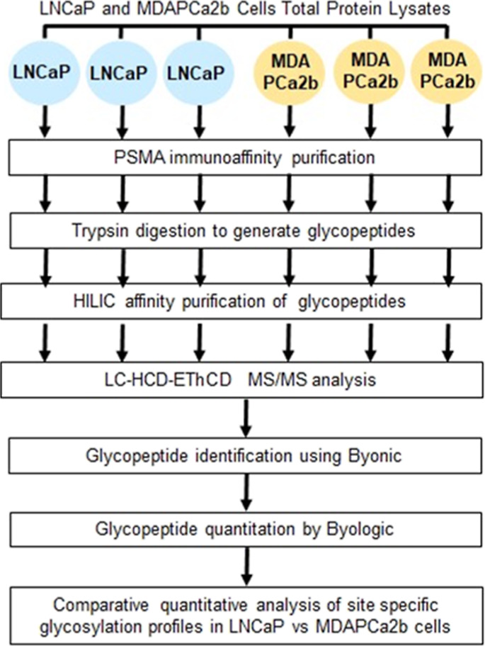
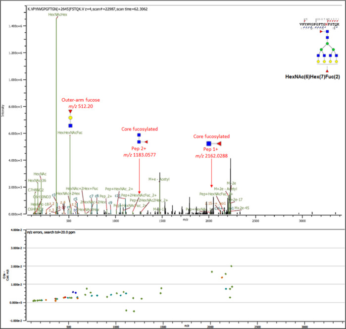
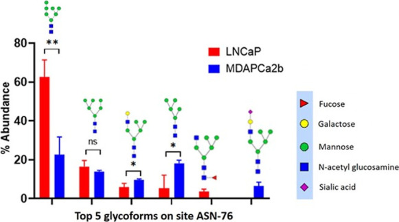
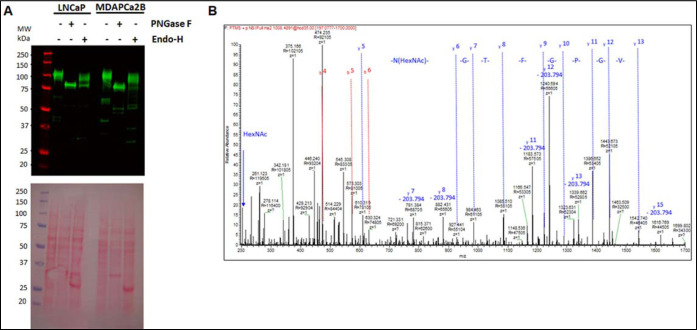
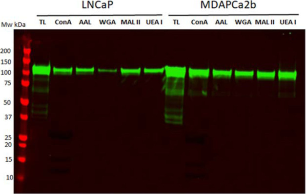
The composition of N-linked glycans that are conjugated to the prostate-specific membrane antigen (PSMA) and their functional significance in prostate cancer progression have not been fully characterized. PSMA was isolated from two metastatic prostate cancer cell lines, LNCaP and MDAPCa2b, which have different tissue tropism and localization. Isolated PSMA was trypsin-digested, and intact glycopeptides were subjected to LC-HCD-EThcD-MS/MS analysis on a Tribrid Orbitrap Fusion Lumos mass spectrometer. Differential qualitative and quantitative analysis of site-specific N-glycopeptides was performed using Byonic and Byologic software. Comparative quantitative analysis demonstrates that multiple glycopeptides at asparagine residues 51, 76, 121, 195, 336, 459, 476, and 638 were in significantly different abundance in the two cell lines (p < 0.05). Biochemical analysis using endoglycosidase treatment and lectin capture confirm the MS and site occupancy data. The data demonstrate the effectiveness of the strategy for comprehensive analysis of PSMA glycopeptides. This approach will form the basis of ongoing experiments to identify site-specific glycan changes in PSMA isolated from disease-stratified clinical samples to uncover targets that may be associated with disease progression and metastatic phenotypes.


Conflict of interest statement

The authors declare no competing financial interest.

Figures

Figure 1
Figure 1
Schematic diagram of the experimental workflow. Summary of the workflow for qualitative and quantitative analysis of intact PSMA N-linked glycopeptides in PCa cells with different malignant phenotypes.
Figure 2
Figure 2
Western blot detection of PSMA expression in four PCa cell lines with different malignancy phenotypes. (A) PSMA was detected in LNCaP and MDAPCa2b but not in the RWPE-1 control lines and PC3-ML2 cell lines.
Figure 3
Figure 3
MS/MS Fragmentation spectrum for ASN-336 PSMA glycopeptides after PNGase-F cleavage of the glycans in the presence of light and heavy water. Spectrum showing b and y ions from the PSMA glycopeptide with sequence VPYNVGPGFTGN*FSTQK in the presence of 18O heavy water; the deamidation results in an increase in mass of the aspartic acid by 2.98 Da in the upper panel. A similar spectrum showing b and y ions after ASN-336 deamidation in the presence of normal 16O water with a mass increase of 0.98 Da of the aspartic acid is shown in the lower panel.
Figure 4
Figure 4
MS/MS spectra of an intact PSMA N-glycopeptide with sequence VPNYVGPGFTGN*FSTQK and conjugated with the glycan FA4G4F [depicted as HexNAc(6)Hex(7)Fuc(2)] at ASN-336. Both b and y peptide fragment ions and oxonium ions include outer-arm terminal fucose fragment ions HexNAc(1)Hex(1)Fuc(1) with m/z 512.20 and composite fragment ions comprise the intact peptide and core fucosylated [HexNAc(2)Fuc(1) sugar moiety with m/z 1183.0577 (+2) and peptide + HexNAc + Fuc m/z 2162.0288 (+1)].
Figure 5
Figure 5
Site-specific comparative quantitative glycan analysis for the relative abundance of the five most abundant glycopeptides at the ASN-76 glycosylation site in LNCaP (red bars) and MAPCa2b cells (blue bars). Error bars show the standard deviations of the means across triplicate analyses from each cell line. Multiple unpaired t-tests were used to determine differences in abundance. There are glycans that are differentially expressed between the different cell lines. Man7: LNCaP versus MDAPCa2b, P = 0.0053**. M5G1: LNCaP versus MDAPCa2b, P = 0.034593*; Man5: LNCaP versus MDAPCa2b, P = 0.032044*; FM5G1 was exclusively detected in LNCaP cells whereas M5G1S1 was exclusively detected in MDAPCa2b cells.
Figure 6
Figure 6
(A,B) Detection of the PSMA total protein lysates from LNCaP and MDAPCa2b cells treated with endoglycosidases. (A). Western blot analysis of PSMA in LNCaP and MDAPCa2b cells after treatment with PNGase-F and Endo-H. (B). MS/MS analysis of PSMA glycopeptides after cleavage with Endo-H glycosidase identifies HexNAc-modified ASN-336 in the VPYNVGPGFTGN*FSTQK glycopeptide.
Figure 7
Figure 7
PSMA detection after lectin affinity purification. Total protein lysates from the two cell lines were incubated with agarose beads conjugated with ConA, AAL, WGA, MAA/MAL II, and UAE1 lectins overnight. Proteins bound to the lectins were eluted and separated by SDS-gel and used for western blot to detect PSMA.

References

    1. https://seer.cancer.gov/statfacts/html/prost.html#content (accessed on March 10, 2022).
    1. Siegel R. L.; Miller K. D.; Fuchs H. E.; Jemal A. Cancer statistics, 2022. Ca-Cancer J. Clin. 2022, 72, 7–33. 10.3322/caac.21708. - DOI - PubMed
    1. Bray F.; Ferlay J.; Soerjomataram I.; Siegel R. L.; Torre L. A.; Jemal A. Global cancer statistics 2018: GLOBOCAN estimates of incidence and mortality worldwide for 36 cancers in 185 countries. Ca—Cancer J. Clin. 2018, 68, 394–424. 10.3322/caac.21492. - DOI - PubMed
    1. Berman D. M.; Epstein J. I. When is prostate cancer really cancer?. Urol. Clin. 2014, 41, 339.10.1016/j.ucl.2014.01.006. - DOI - PubMed
    1. Schröder F. H.; Hugosson J.; Roobol M. J.; Tammela F. H.; Zappa J.; Nelen M. J.; Kwiatkowski T. L.; Lujan M.; Määttänen V.; Lilja M.; et al. Screening and prostate cancer mortality: results of the European Randomised Study of Screening for Prostate Cancer (ERSPC) at 13 years of follow-up. Lancet 2014, 384, 2027.10.1016/s0140-6736(14)60525-0. - DOI - PMC - PubMed