

Subclavian Effort Thrombosis: Pathophysiology, Diagnosis, and Management
- PMID: 36062232
- PMCID: PMC9433153
- DOI: 10.1055/s-0042-1753481
Subclavian Effort Thrombosis: Pathophysiology, Diagnosis, and Management
Erratum in
-
Erratum: Subclavian Effort Thrombosis: Pathophysiology, Diagnosis, and Management.Semin Intervent Radiol. 2022 Sep 20;39(3):e1. doi: 10.1055/s-0042-1756698. eCollection 2022 Jun. Semin Intervent Radiol. 2022. PMID: 36147191 Free PMC article.
Abstract
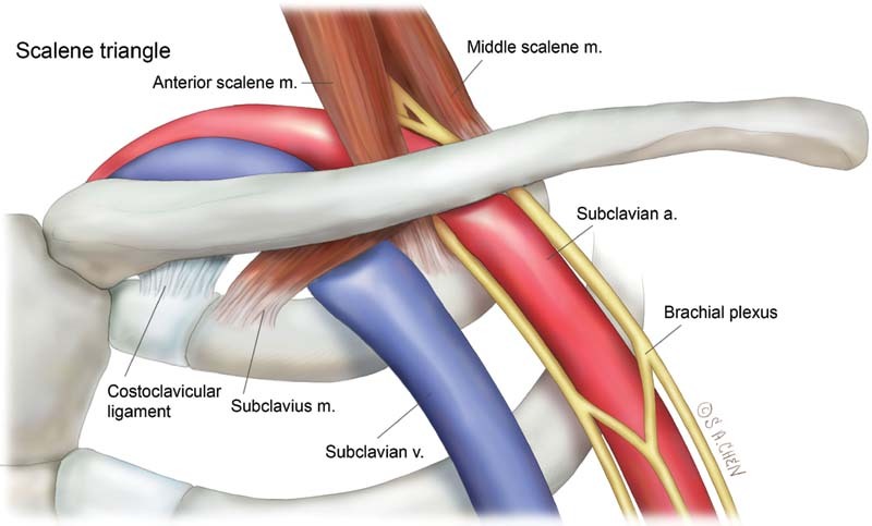
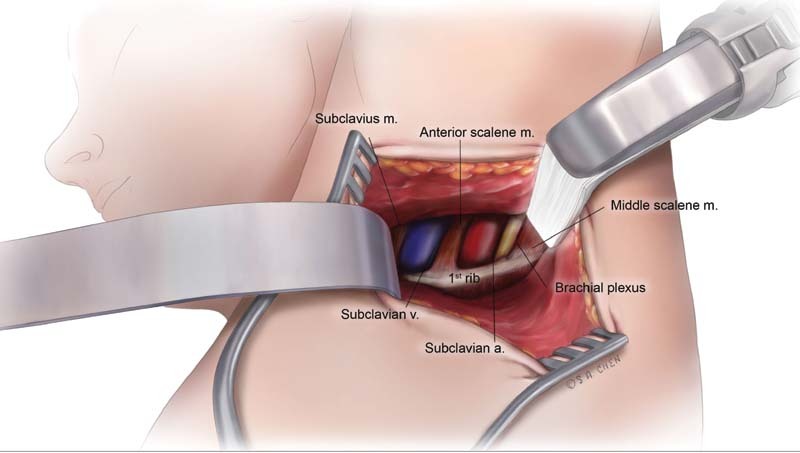
Subclavian vein (SCV) effort thrombosis, also known as Paget-Schroetter syndrome or venous thoracic outlet syndrome, is an uncommon condition that affects individuals with an irregularly narrow thoracic outlet who engage in repetitive overhead motions of the affected arm. Venous injury arises from microtraumas that occur from the repetitive compression of the SCV between the first rib and the overlying clavicle. Additional sources of extrinsic compression can be due to the anterior scalene muscle, subclavius muscle, and costoclavicular ligament. SCV effort thrombosis is a distinct entity from other forms of deep venous thrombosis and requires unique diagnostic and treatment considerations. Early catheter-directed therapy in the form of pharmacomechanical or catheter-directed thrombolysis combined with prompt surgical thoracic outlet decompression offers patients the best chances for early and durable symptom relief.
Keywords: Paget-Schroetter; angioplasty; chest; effort thrombosis; interventional radiology; thoracic outlet syndrome; thrombolysis; venous thrombosis.
Thieme. All rights reserved.
Conflict of interest statement
Conflict of Interest None declared.
Figures

References
-
- Karaolanis G, Antonopoulos C N, Koutsias S G. A systematic review and meta-analysis for the management of Paget-Schroetter syndrome. J Vasc Surg Venous Lymphat Disord. 2021;9(03):801–8.1E7. - PubMed
-
- Kucher N. Clinical practice. Deep-vein thrombosis of the upper extremities. N Engl J Med. 2011;364(09):861–869. - PubMed
-
- Illig K A, Doyle A J. A comprehensive review of Paget-Schroetter syndrome. J Vasc Surg. 2010;51(06):1538–1547. - PubMed
