

Sex-specific Cutoff Values of Visceral Fat Area for Lean vs. Overweight/Obese Nonalcoholic Fatty Liver Disease in Asians
- PMID: 36062272
- PMCID: PMC9396328
- DOI: 10.14218/JCTH.2021.00379
Sex-specific Cutoff Values of Visceral Fat Area for Lean vs. Overweight/Obese Nonalcoholic Fatty Liver Disease in Asians
Abstract
Background and aims: Visceral obesity is a risk factor for nonalcoholic fatty liver disease (NAFLD). We investigated sex-specific optimal cutoff values for visceral fat area (VFA) associated with lean and overweight/obese NAFLD in an Asian population.
Methods: This retrospective study included 678 potential living liver donors (mean age, 30.8±9.4 years; 434 men and 244 women) who had undergone abdominal computed tomography (CT) imaging and liver biopsy between November 2016 and October 2017. VFA was measured using single-slice abdominal CT. NAFLD was evaluated by liver biopsy (≥5% hepatic steatosis). Receiver operating characteristic curve analysis was used to determine cutoff values for VFA associated with lean (body mass index [BMI] <23 kg/m2) and overweight/obese (BMI ≥23 kg/m2) NAFLD.
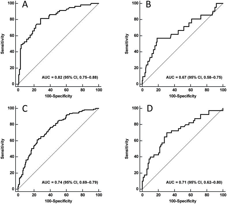
Results: Area under the curve (AUC) values with 95% confidence intervals (CI) for VFA were 0.82 (95% CI, 0.75-0.88) for lean and 0.74 (95% CI, 0.69-0.79) for overweight/obese men with NAFLD. The AUC values were 0.67 (95% CI, 0.58-0.75) for lean and 0.71 (95% CI, 0.62-0.80) for overweight/obese women with NAFLD. The cutoff values for VFA associated with lean NAFLD were 50.2 cm2 in men and 40.5 cm2 in women. The optimal cutoff values for VFA associated with overweight/obese NAFLD were 100.6 cm2 in men and 68.0 cm2 in women.
Conclusions: Sex-specific cutoff values for VFA may be useful for identifying subjects at risk of lean and overweight/obese NAFLD.
Keywords: Adipose tissue; Computed tomography.; Hepatic steatosis; Liver biopsy.
© 2022 Authors.
Conflict of interest statement
The authors have no conflict of interests related to this publication.
Figures
References
LinkOut - more resources
Full Text Sources