. 2022 Nov;12(11):2025-2041.
doi: 10.1002/2211-5463.13479. Epub 2022 Sep 22.

PSMB5 overexpression is correlated with tumor proliferation and poor prognosis in hepatocellular carcinoma

Affiliations

PSMB5 overexpression is correlated with tumor proliferation and poor prognosis in hepatocellular carcinoma

Jun Liu et al. FEBS Open Bio. 2022 Nov.

Abstract

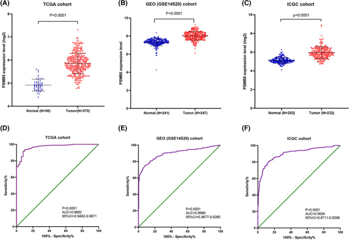
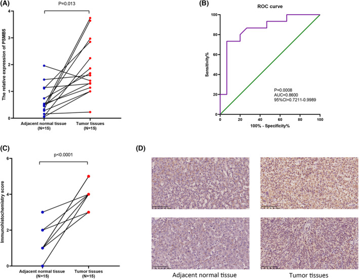
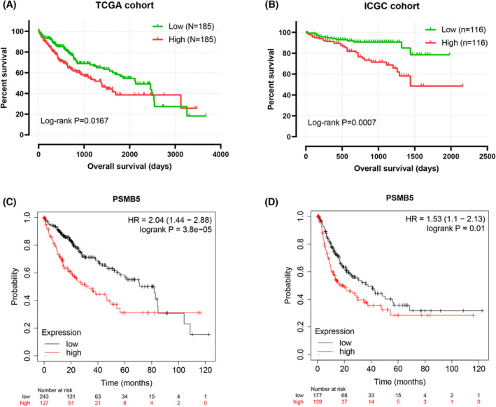
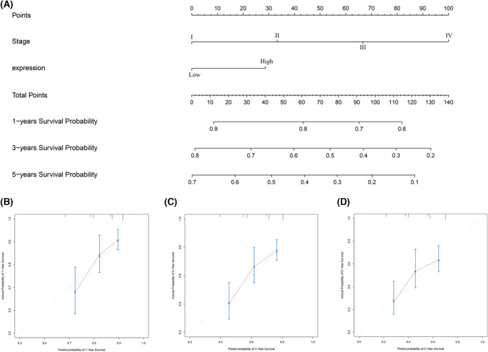
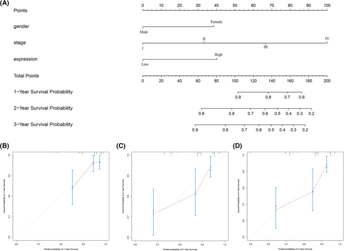
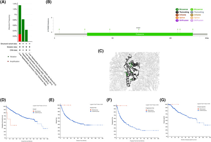
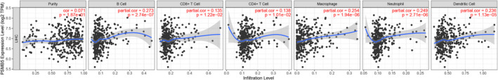
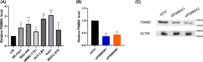
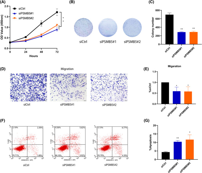
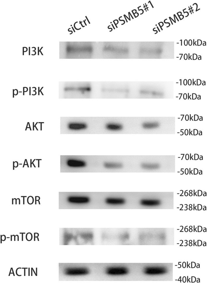
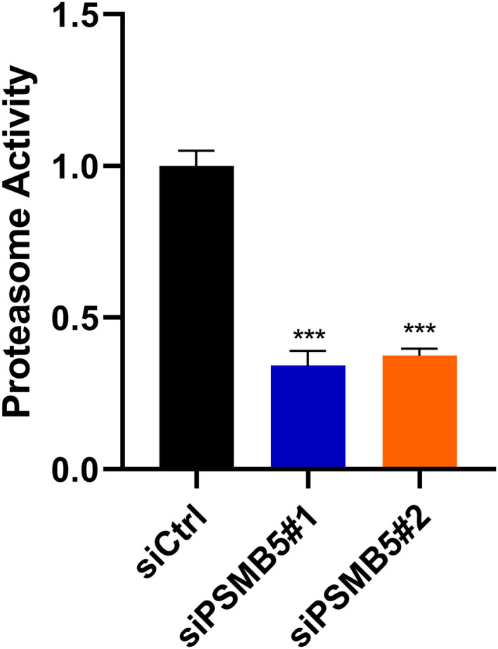
Aberrant expression of members of the proteasome subunit beta (PSMB) family (including PSMB2, PSMB4, PSMB7 and PSMB8) has been reported in hepatocellular carcinoma (HCC). However the role of PSMB5 in HCC is unclear. To address this issue, we examined the expression of PSMB5 in HCC tissues using the The Cancer Genome Atlas, International Cancer Genome Consortium and Gene Expression Omnibus databases. A quantitative real-time PCR and immunohistochemistry were performed to validate the expression of PSMB5 in HCC. The survival mutation status and immune cell infiltration of PSMB5 were also evaluated in HCC. We then examined the effect of knocking down PSMB5 expression through RNA interference in the HCC cell line Huh7. High expression of PSMB5 was observed in HCC tissues and was associated with poor prognosis. PSMB5 expression and clinical characteristics were then incorporated to build a prognostic nomogram. We observed that PSMB5 expression was closely related to the abundance of B cells, CD4+ T cells, CD8+ T cells, dendritic cell macrophages and neutrophils. Moreover silencing of PSMB5 in Huh7 significantly suppressed cell proliferation and migration at the same time as increasing apoptosis. Inhibition of the phosphatidylinositol-3-kinase/Akt/mechanistic target of rapamycin pathway was observed after PSMB5 downregulation in Huh7 cells. Our findings suggest that PSMB5 may promote the proliferation of HCC cells by inactivating the phosphatidylinositol-3-kinase/Akt/mechanistic target of rapamycin signaling pathway and thus PSMB5 may have potential as a biomarker for diagnosis and prognosis of HCC.

Keywords: PSMB5; diagnosis; prognosis; proliferation and hepatocellular carcinoma.


Conflict of interest statement

The authors declare that they have no conflicts of interest.

Figures

Fig. 1
Fig. 1
The expression of PSMB5 in HCC. (A–C) The expression of PSMB5 was signifcantly higher in HCC than normal tissue based on TCGA, GEO (GSE14520) and ICGC cohorts. (D–F) The diagnostic ROC curves of PSMB5 based on TCGA, GEO (GSE14520) and ICGC cohorts.
Fig. 2
Fig. 2
mRNA and protein expression of PSMB5 was confirmed by qRT‐PCR and immunohistochemistry. (A) mRNA expression level of PSMB5 between HCC and adjacent normal tissues. (B) The diagnostic ROC curves of PSMB5. (C, D) Immunohistochemistry score and protein expression of PSMB5 between HCC and adjacent normal tissues. Scale bars = 100 μm.
Fig. 3
Fig. 3
Kaplan–Meier survival curves for PSMB5 in HCC. (A) Overall survival curves of PSMB5 in HCC from the TCGA cohort. (B) Overall survival curves of PSMB5 in HCC from the ICGC cohort. (C, D) Overall survival and recurrence‐free survival curves of PSBM5 in HCC based on the Kaplan–Meier plotter database.
Fig. 4
Fig. 4
Construction of the nomogram consisted of PSMB5 expression and clinical features based on the TCGA cohort. (A) Nomogram. (B–D) Calibration curves of the nomogram to predict 1‐, 3‐ and 5‐year overall survival.
Fig. 5
Fig. 5
Construction of the nomogram consisted of PSMB5 expression and clinical features based on ICGC cohort. (A) Nomogram. (B–D) Calibration curves of the nomogram to predict 1‐, 2‐ and 3‐year overall survival.
Fig. 6
Fig. 6
Genetic alteration of PSMB5 in HCC. (A) The alteration frequency with mutation type of PSMB5. (B) Mutation sites of PSMB5. (C) The mutation site in the 3D structure of PSMB5. (D–G) Survival analysis of overall survival, disease‐free survival, progression‐free survival and disease‐specific survival with or without PSMB5 alteration.
Fig. 7
Fig. 7
Association of PSMB5 expression with immune infltration levels in HCC from the TCGA.
Fig. 8
Fig. 8
PSMB5 expression level in HCC cell lines and construction of HCC cell models with PSMB5 knockdown. (A) Levels of psmb5 mRNA in difference HCC cell lines. (B, C) qPCR and western blotting were applied to examine the knockdown efficiency of PSMB5 in huh7. Unpaired two‐tailed Student's t‐tests were used to determine significance. Error bars indicate the SD. The precise n value (number of biologically‐independent replicates) is 3, except for western blotting where n = 1. *P < 0.05 **P < 0.01.
Fig. 9
Fig. 9
PSMB5 modulated proliferation, migration and apoptosis in vitro. (A) A CCK‐8 assay was conducted to detect the proliferation of PSMB5 knockdown in huh7. (B, C) The effect of PSMB5 knockdown on the colony forming capacity of huh7. (D, E) The effect of PSMB5 knockdown on the migration capacity of huh7. Scale bars = 100 μm. (F, G) The effect of PSMB5 knockdown on the cell apoptosis in huh7. Unpaired two‐tailed Student's t‐tests and one‐way or two‐way analysis of variance were used to determine significance. Error bars indicate the SD. The precise n value (number of biologically‐independent replicates) is 3. *P < 0.05 **P < 0.01.
Fig. 10
Fig. 10
PSMB5 regulates the PI3K/Akt/mTOR signaling pathway. Western blotting was performed against the indicated proteins in huh7 cells treated with siCtrl siPSMB5#1 or siPSMB5#2.
Fig. 11
Fig. 11
Proteasome activity of huh7 cells treated with siCtrl, siPSMB5#1 or siPSMB5#2 were detected using a Proteasome 20S Activity Assay Kit (Abcam) with a fluorescence microplate reader. Unpaired two‐tailed Student's t‐tests were used to determine significance. Error bars indicate the SD. The precise n value (number of biologically‐independent replicates) is 3. ***P < 0.001.

Similar articles

Cited by

References

    1. Llovet JM, Zucman‐Rossi J, Pikarsky E, Sangro B, Schwartz M, Sherman M, et al. Hepatocellular carcinoma. Nat Rev Dis Primers. 2016;2:16018. - PubMed
    1. McGlynn KA, Petrick JL, El‐Serag HB. Epidemiology of hepatocellular carcinoma. Hepatology. 2021;73(Suppl 1):4–13. - PMC - PubMed
    1. Sung H, Ferlay J, Siegel RL. Global cancer statistics 2020: GLOBOCAN estimates of incidence and mortality worldwide for 36 cancers in 185 countries. CA Cancer J Clin. 2021;71:209–49. - PubMed
    1. Zhou J, Yu L, Gao X, Hu J, Wang J, Dai Z, et al. Plasma microRNA panel to diagnose hepatitis B virus‐related hepatocellular carcinoma. J Clin Oncol. 2011;29:4781–8. - PubMed
    1. Nie Y, Li J, Wu W, Guo D, Lei X, Zhang T, et al. A novel nine‐lncRNA risk signature correlates with immunotherapy in hepatocellular carcinoma. Front Oncol. 2021;11:706915. - PMC - PubMed

Publication types

MeSH terms