. 2022 Jan-Dec;14(1):2119054.
doi: 10.1080/19490976.2022.2119054.

Microbiota manipulation to increase macrophage IL-10 improves colitis and limits colitis-associated colorectal cancer

Affiliations

Microbiota manipulation to increase macrophage IL-10 improves colitis and limits colitis-associated colorectal cancer

Daniel F Zegarra Ruiz et al. Gut Microbes. 2022 Jan-Dec.

Abstract

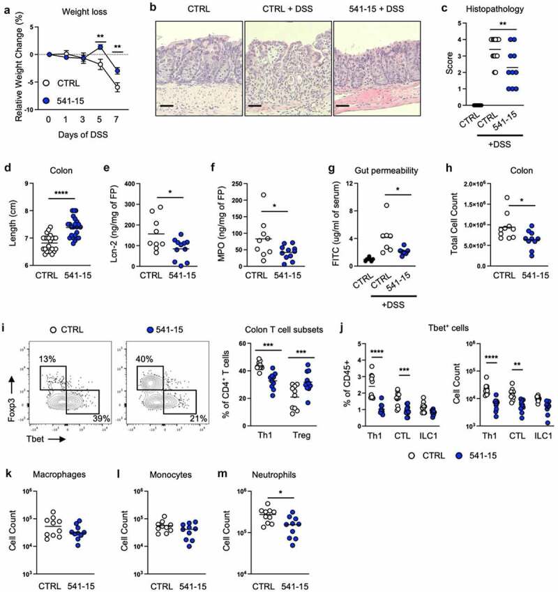
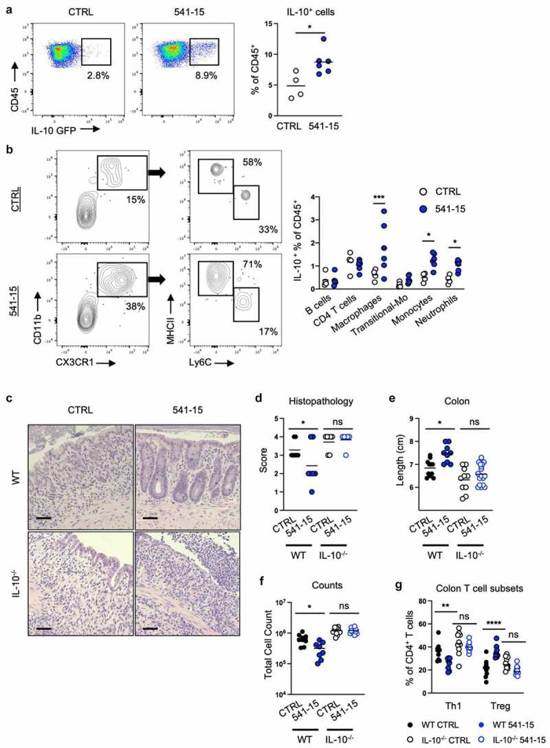
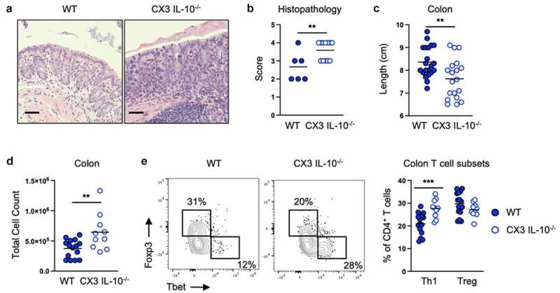
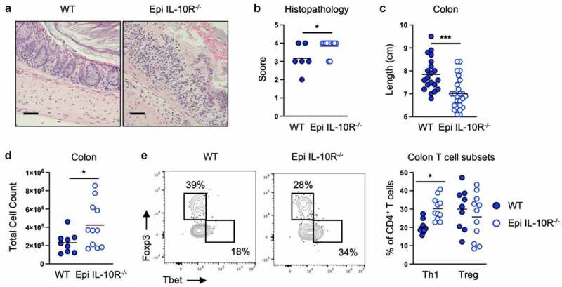
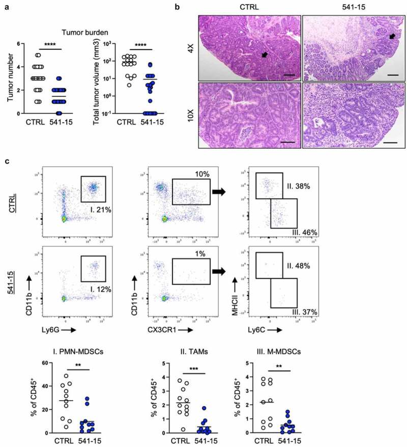
Inflammatory bowel disease (IBD) is a chronic life-long inflammatory disease affecting almost 2 million Americans. Although new biologic therapies have been developed, the standard medical treatment fails to selectively control the dysregulated immune pathways involved in chronic colonic inflammation. Further, IBD patients with uncontrolled colonic inflammation are at a higher risk for developing colorectal cancer (CRC). Intestinal microbes can impact many immune functions, and here we asked if they could be used to improve intestinal inflammation. By utilizing an intestinal adherent E. coli that we find increases IL-10 producing macrophages, we were able to limit intestinal inflammation and restrict tumor formation. Macrophage IL-10 along with IL-10 signaling to the intestinal epithelium were required for protection in both inflammation and tumor development. Our work highlights that administration of immune modulating microbes can improve intestinal outcomes by altering tissue inflammation.

Keywords: E. coli; IL-10; Microbiota; colitis-associated cancer; colorectal cancer; intestinal epithelium; intestinal inflammation; intestinal macrophages.


Conflict of interest statement

The authors declare no conflict of interests.

Figures

Figure 1.
Figure 1.
Intestinal colonization with E. coli 541–15 protects from colitis. Wildtype mice were colonized with E. coli 541–15 or gavaged with LB broth (CTRL) and 3 days later 2% DSS was provided in drinking water. At day 7 of DSS treatment, samples were collected. (a) Relative weight change. (b) Representative H&E staining of distal colons. Scale bars represent 50 μm. (c) Histopathology scores. (d) Colon lengths. (e) Lipocalin-2 (Lcn-2) and (f) Myeloperoxidase (MPO) concentration in fecal pellets. At day 7 mice were gavaged with FITC-dextran and 3 hours later blood was collected. (g) FITC concentration in serum. Colon lamina propria cells were isolated. (h) Number of immune cells. (i) Flow cytometry and frequencies of Th1 and Tregs. (j) Frequencies and counts of total Th1 cells, CTLs, and ILC1s. Counts of (k) Macrophages, (l) Monocytes, and (m) Neutrophils. Each replicate is a biologically independent sample. Individual dots represent samples from individual mice. Data are shown as individual values and mean, compared by two-tailed unpaired t-test or two-way ANOVA with Fisher’s LSD post hoc test. The results are representative of at least two independent experiments. *P < .05 was considered statistically significant; **P <.01; ***P < .001; ****P < .0001. See also Figure S1.
Figure 2.
Figure 2.
Intestinal colonization with E. coli 541–15 prevents colitis through IL-10 induction. Vert-X mice were colonized with E. coli 541–15 or gavaged with LB broth (CTRL) and 3 days later 2% DSS was provided. At day 7 of DSS treatment, samples were collected. Flow cytometry and frequencies of (a) total IL-10+ immune cells from the colonic lamina propria and (b) identification of IL-10+ immune cells. IL-10 deficient (IL-10-/-) and wildtype (WT) littermate mice were colonized with E. coli 541–15 or gavaged with LB broth (CTRL) and 3 days later 2% DSS was provided. At day 7 of DSS treatment, samples were collected. (c) Representative H&E staining of distal colons. Scale bars represent 50 μm. (d) Histopathology scores. (e) Colon lengths. Colon lamina propria cells were isolated. (f) Number of immune cells. (g) Frequencies of Th1 and Tregs. Each replicate is a biologically independent sample. Individual dots represent samples from individual mice. Data are shown as individual values and mean, compared by two-tailed unpaired t-test or two-way ANOVA with Fisher’s LSD post hoc test. The results are representative of at least two independent experiments. *P < .05 was considered statistically significant; **P <.01; ***P < .001; ****P < .0001. See also Figure S2.
Figure 3.
Figure 3.
Intestinal colonization with E. coli 541–15 prevents colitis by inducing IL-10 production by CX3CR1+ macrophages. IL-10flox/- (CX3 IL-10-/-) and IL-10flox/+ (WT) littermate CX3CR1-CreERT2 mice were colonized with E. coli 541–15 and 3 days later 2% DSS was provided. At day 7 of DSS treatment, samples were collected. 3 days before colonization and every 2 days throughout the experiment 4OHT was injected to both groups. (a) Representative H&E staining of distal colons. Scale bars represent 50 μm. (b) Histopathology scores. (c) Colon lengths. Colon lamina propria cells were isolated. (d) Number of immune cells. (e) Flow cytometry and frequencies of Th1 and Tregs. Each replicate is a biologically independent sample. Individual dots represent samples from individual mice. Data are shown as individual values and mean, compared by two-tailed unpaired t-test or two-way ANOVA with Fisher’s LSD post hoc test. The results are representative of at least two independent experiments. *P < .05 was considered statistically significant; **P <.01; ***P < .001. See also Figure S3.
Figure 4.
Figure 4.
Intestinal colonization with E. coli 541–15 prevents colitis by modulating IL-10 signaling to intestinal epithelium. LGR5-CreERT2+ (Epi IL-10R-/-) and LGR5- CreERT2- (WT) littermate IL-10Raflox/flox mice were colonized with E. coli 541–15 and 3 days later 2% DSS was provided. At day 7 of DSS treatment, samples were collected. 7 days before colonization 4OHT was injected on two consecutive days to both groups. (a) Representative H&E staining of distal colons. Scale bars represent 50 μm. (b) Histopathology scores. (c) Colon lengths. Colon lamina propria cells were isolated. (d) Number of immune cells. (e) Flow cytometry and frequencies of Th1 and Tregs. Each replicate is a biologically independent sample. Individual dots represent samples from individual mice. Data are shown as individual values and mean, compared by two-tailed unpaired t-test or two-way ANOVA with Fisher’s LSD post hoc test. The results are representative of at least two independent experiments. *P < .05 was considered statistically significant; ***P < .001. See also Figure S4.
Figure 5.
Figure 5.
Intestinal colonization with E. coli 541–15 protects from colitis-associated colorectal cancer. Wildtype mice were administered AOM followed by 3 cycles of 2% DSS. Before administration of DSS mice were colonized with E. coli 541–15 or gavaged with LB broth (CTRL) and samples were collected as indicated in Figure S1A. (a) Number and size of colonic tumors. (b) Representative H&E staining of distal colons. Black arrow shows a tubular adenoma. Scale bars represent 4x = 200 μm and 10x = 100 μm. Tumors were individually collected, digested, and the tumor microenvironment was analyzed by flow cytometry. (c) Flow cytometry and frequencies of PMN-MDSCs, TAMs, and MMDSCs. Each replicate is a biologically independent sample. Individual dots represent samples from individual mice. Data are shown as individual values and mean, compared by two-tailed unpaired t-test. The results are representative of at least two independent experiments. *P < .05 was considered statistically significant; **P <.01; ***P < .001; ****P < .0001. See also Figures S5 and S6.

References

    1. Baumgart DC, Carding SR.. Inflammatory bowel disease: cause and immunobiology. Lancet. 2007;369(9573):1627–15. doi:10.1016/S0140-6736(07)60750-8. - DOI - PubMed
    1. Alatab S, Sepanlou SG, Ikuta K, Vahedi H, Bisignano C, Safiri S, Sadeghi A, Nixon MR, Abdoli A, Abolhassani H, et al. The global, regional, and national burden of inflammatory bowel disease in 195 countries and territories, 1990–2017: a systematic analysis for the global burden of disease study 2017. Lancet Gastroenterology Hepatology. 2020;5:17–30. doi:10.1016/S2468-1253(19)30333-4. - DOI - PMC - PubMed
    1. Caruso R, Lo BC, Núñez G. Host–microbiota interactions in inflammatory bowel disease. Nat Rev Immunol. 2020;20:411–426. doi:10.1038/s41577-019-0268-7. - DOI - PubMed
    1. Pithadia AB, Jain S. Treatment of inflammatory bowel disease (IBD). Pharmacol Rep. 2011;63:629–642. doi:10.1016/S1734-1140(11)70575-8. - DOI - PubMed
    1. Dalal SR, Chang EB. The microbial basis of inflammatory bowel diseases. J Clin Investigation. 2014;124:4190–4196. doi:10.1172/JCI72330. - DOI - PMC - PubMed

Publication types

LinkOut - more resources