. 2022 Nov;188(11):3191-3228.
doi: 10.1002/ajmg.a.62965. Epub 2022 Sep 5.

Elements of morphology: Standard terminology for the trunk and limbs

Affiliations

Elements of morphology: Standard terminology for the trunk and limbs

Leslie G Biesecker et al. Am J Med Genet A. 2022 Nov.

Abstract

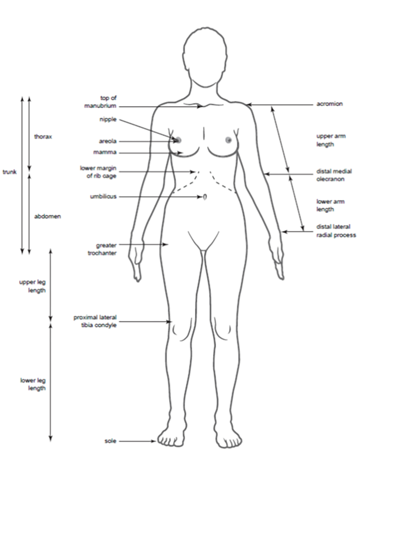
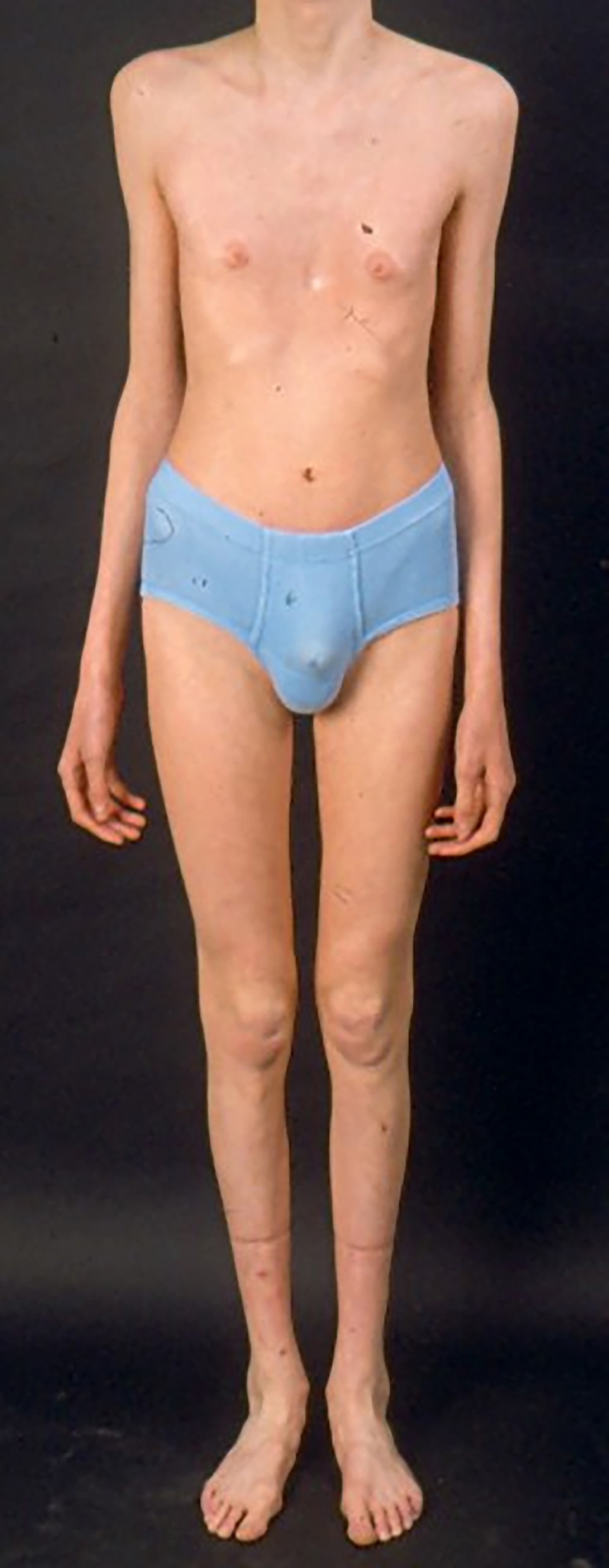
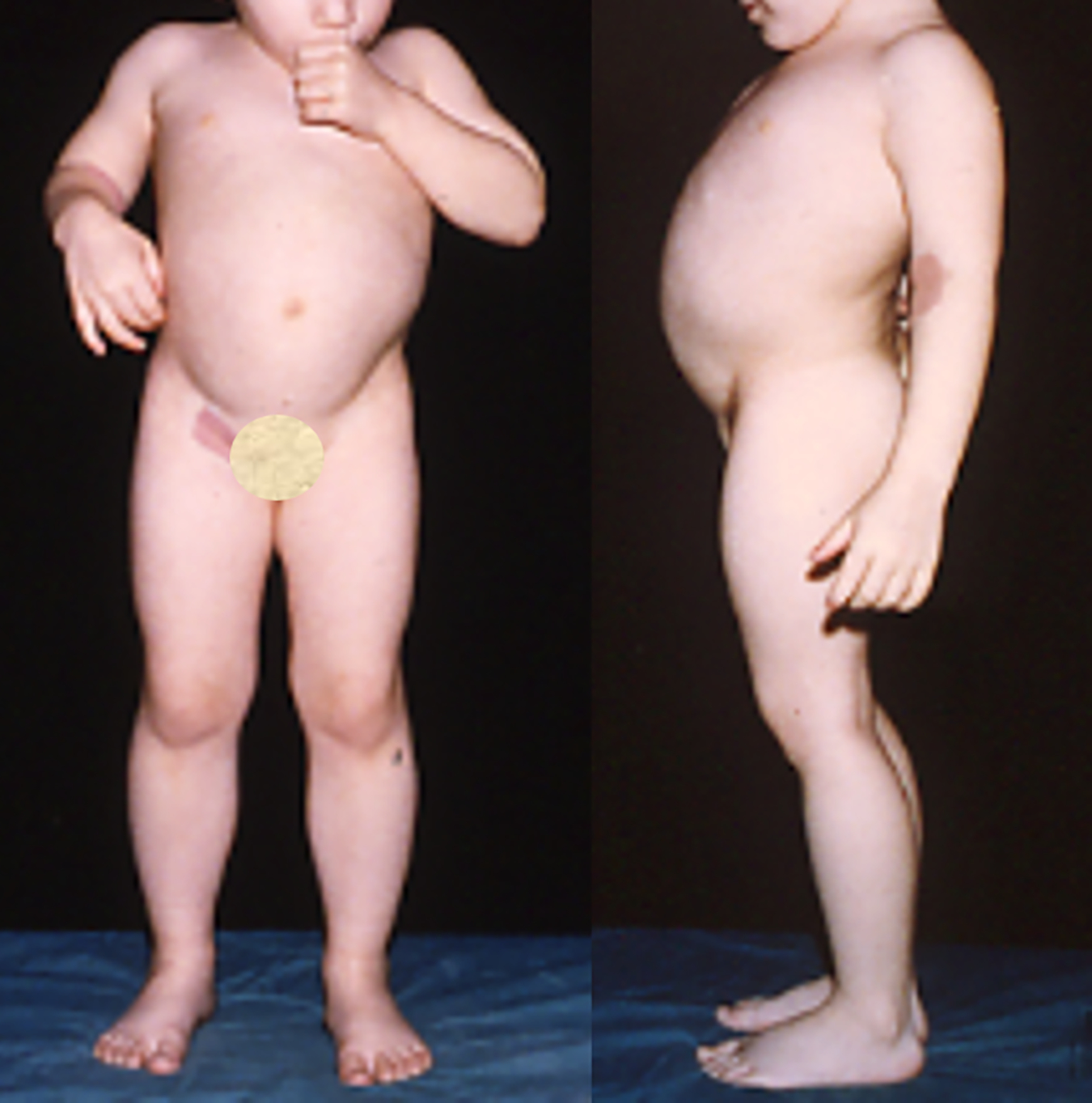
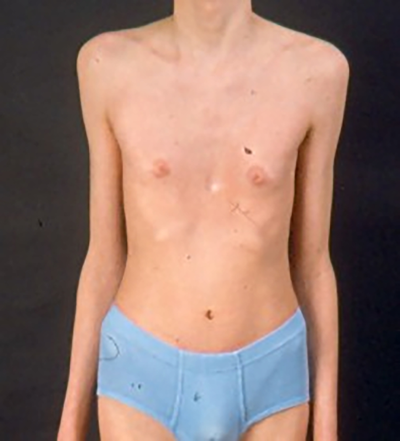
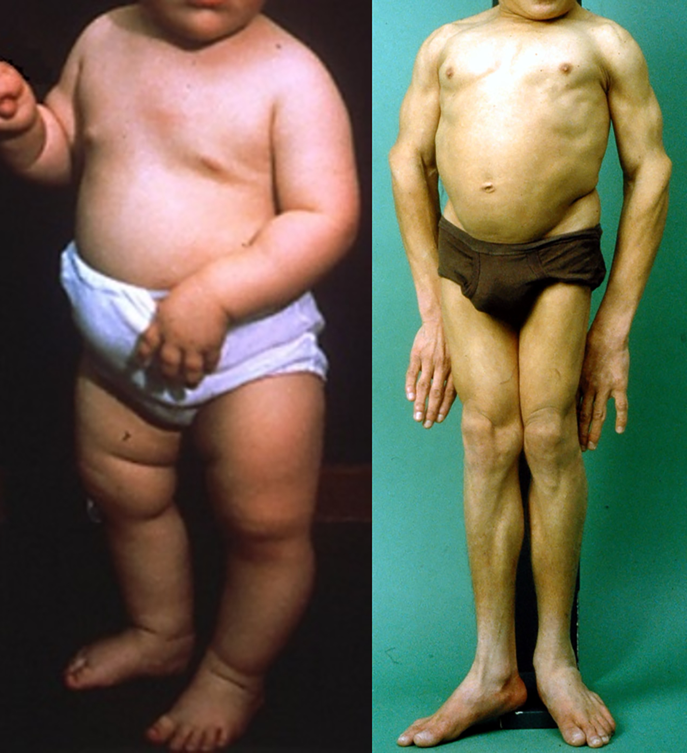
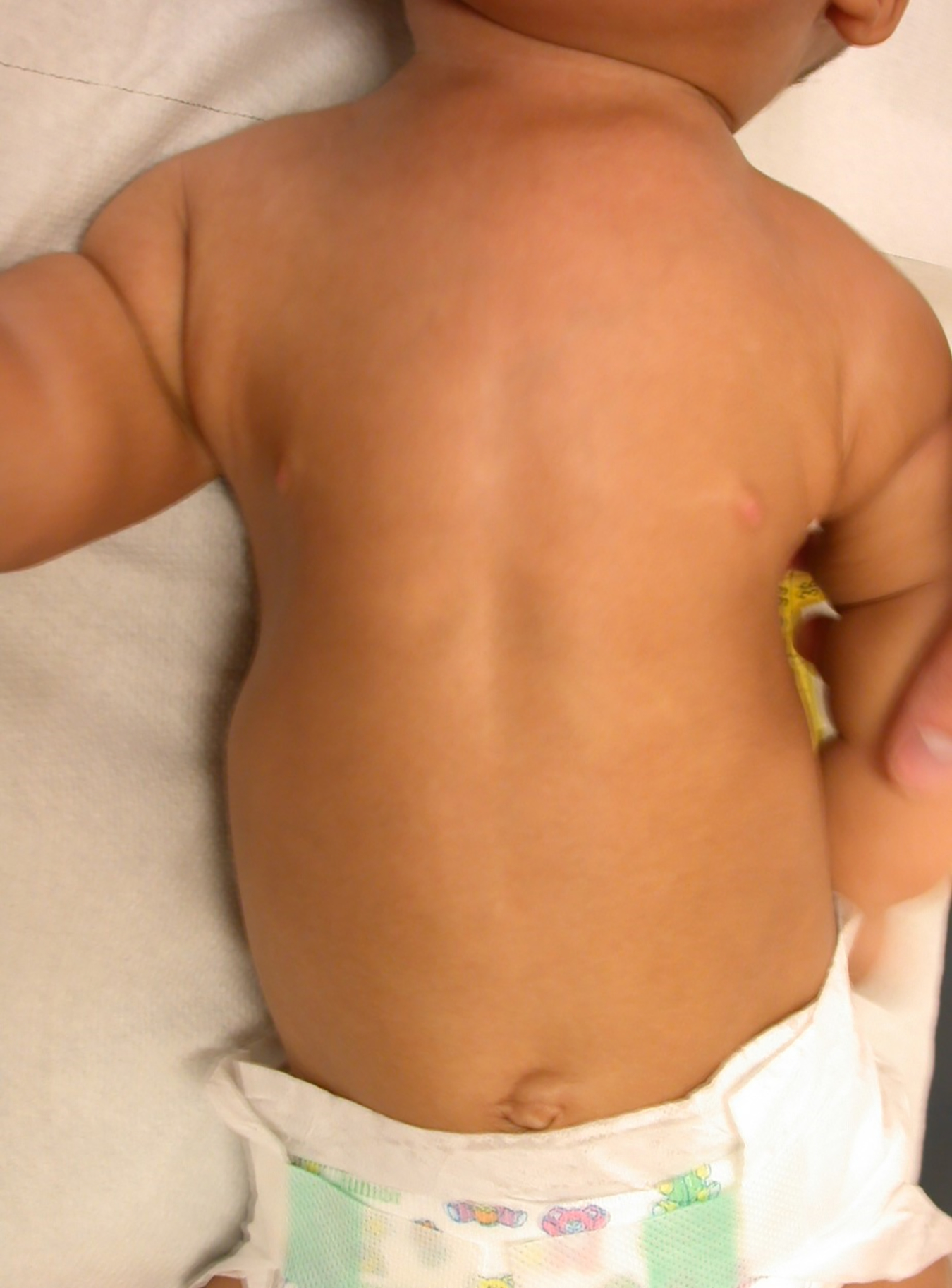
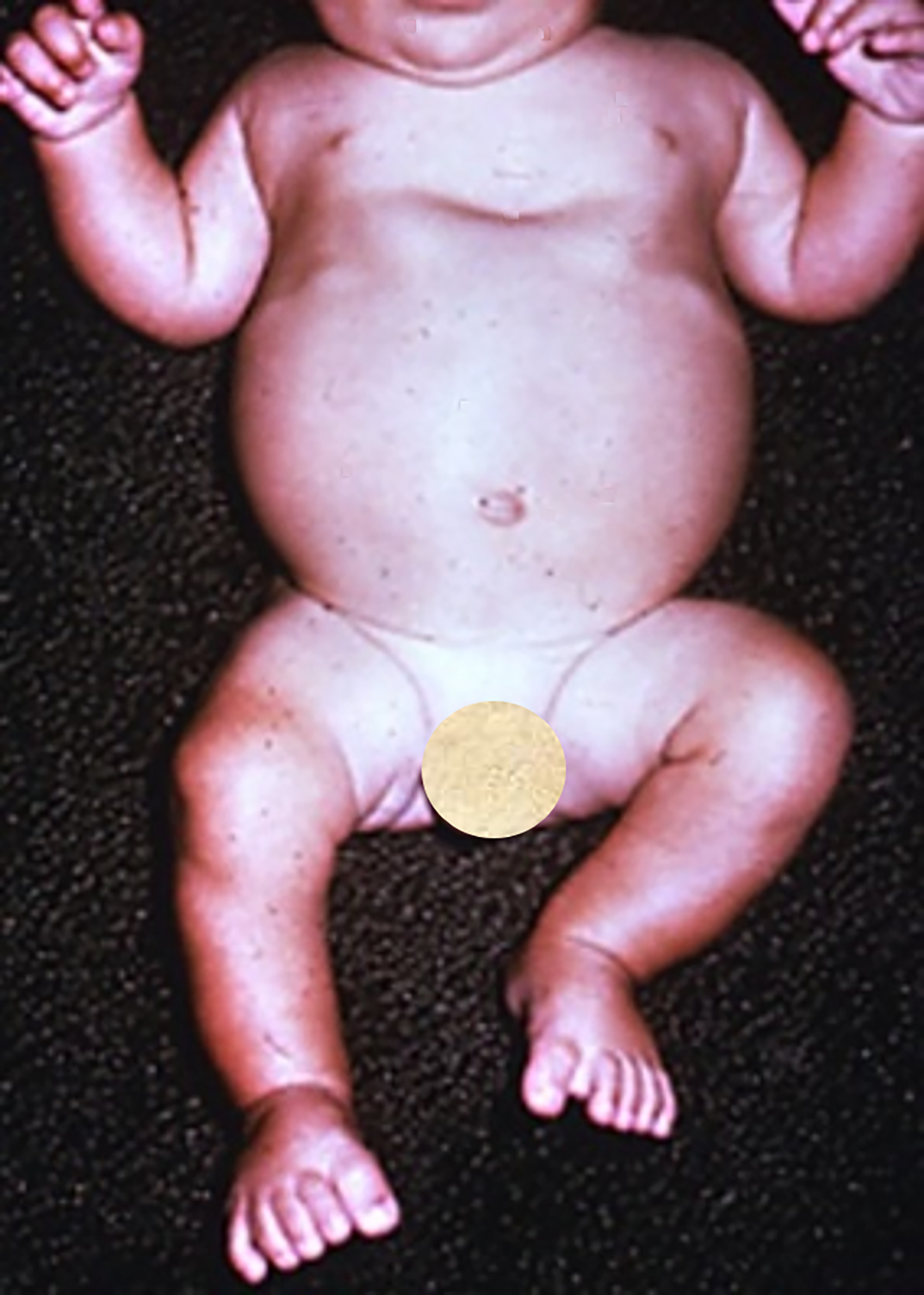
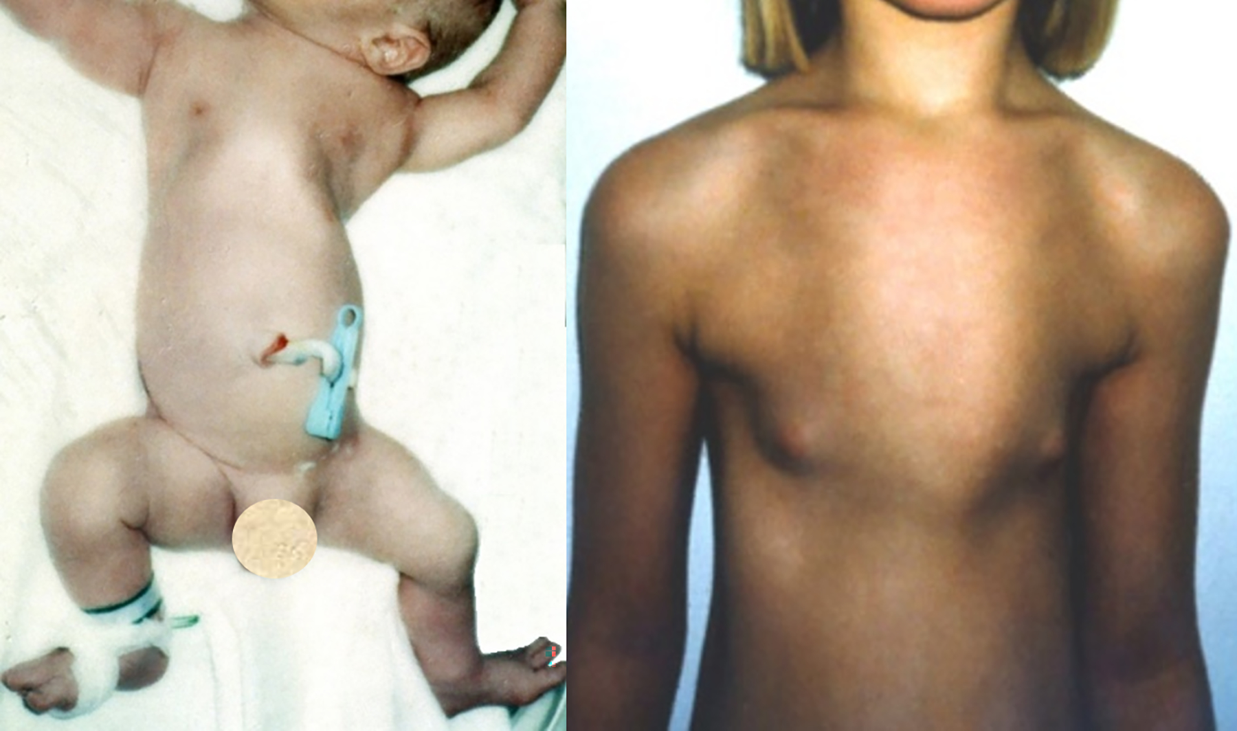
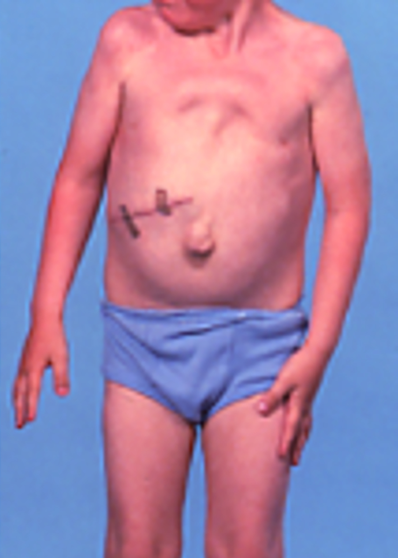
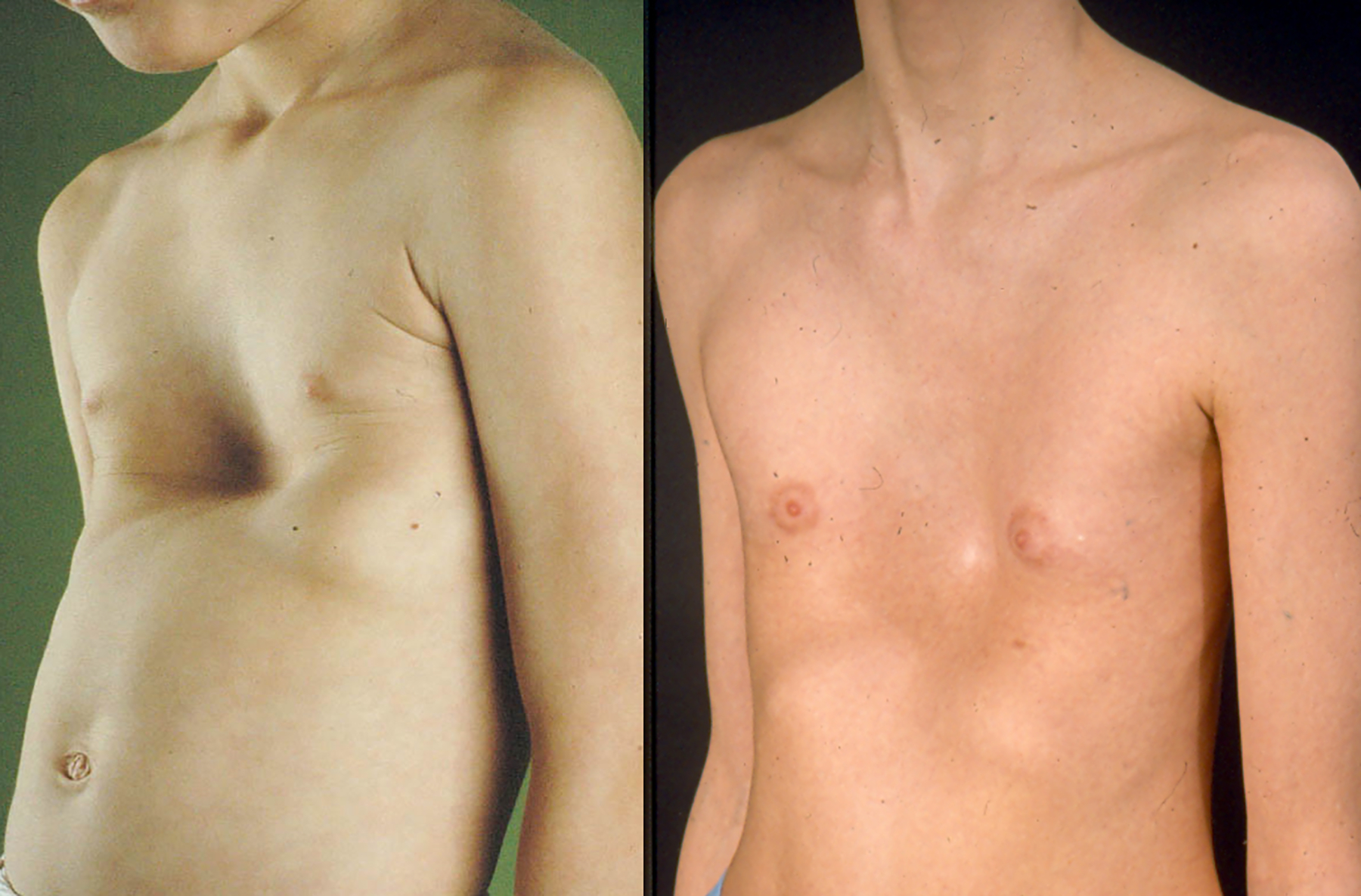
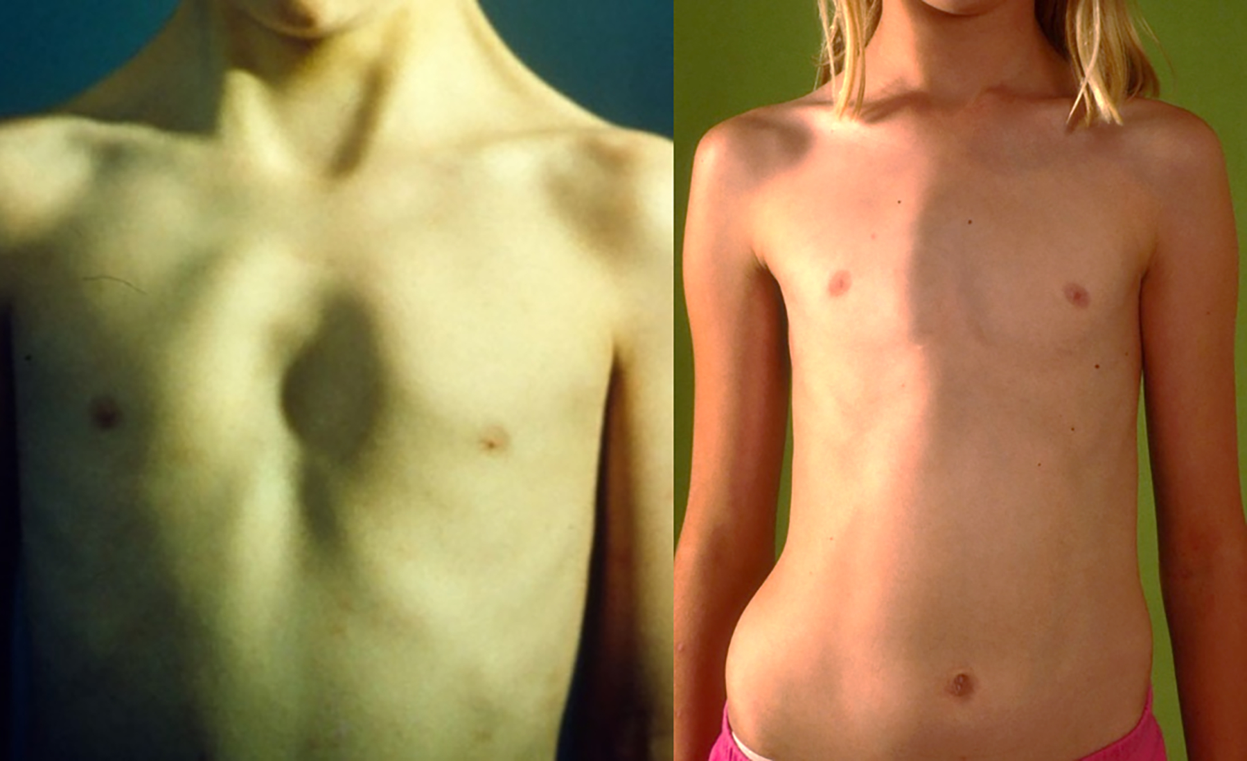
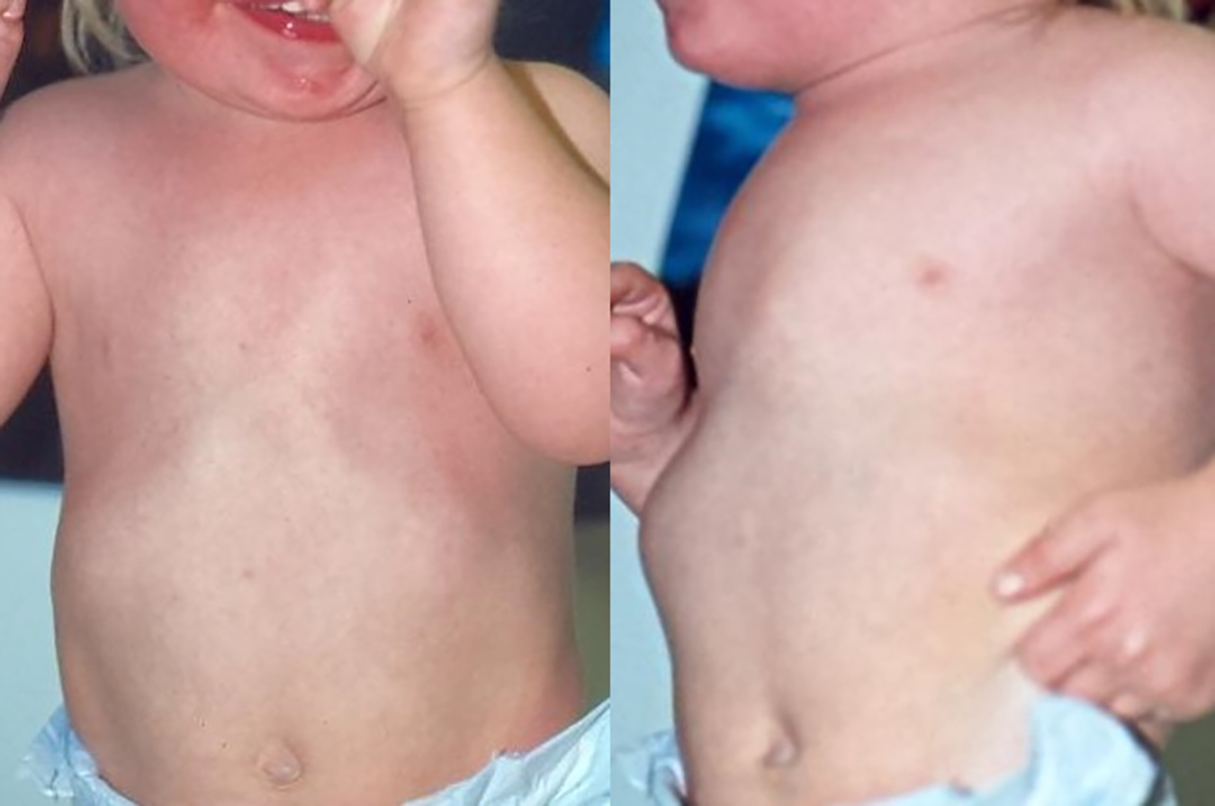
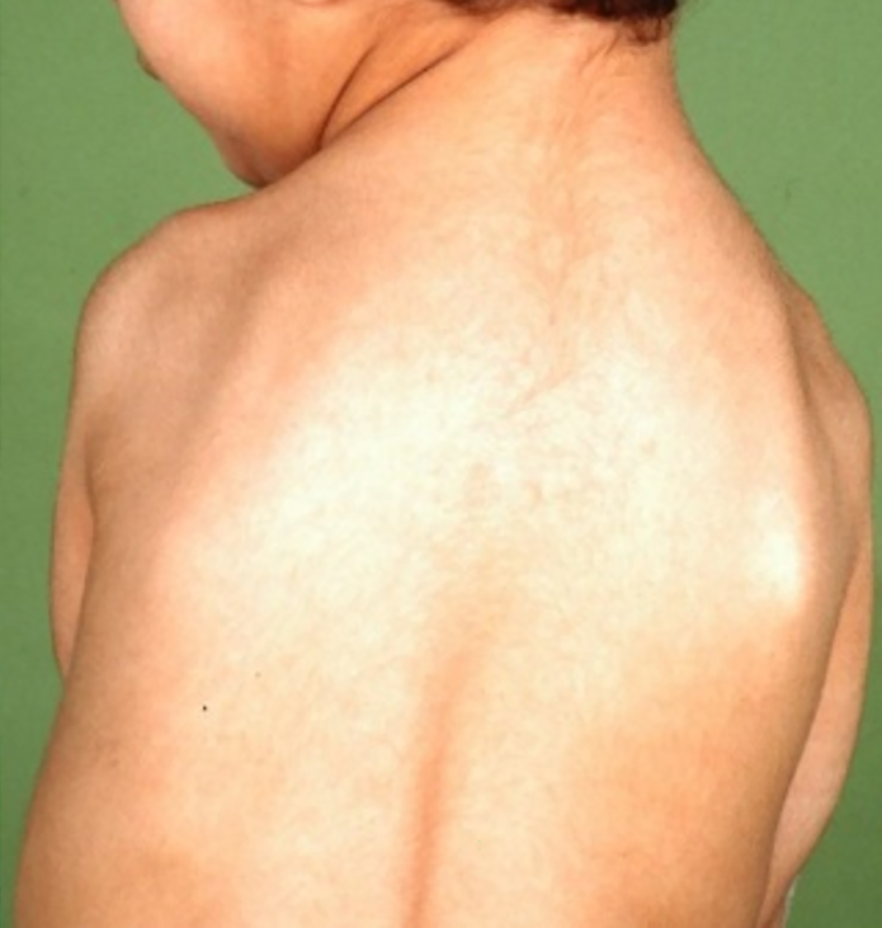
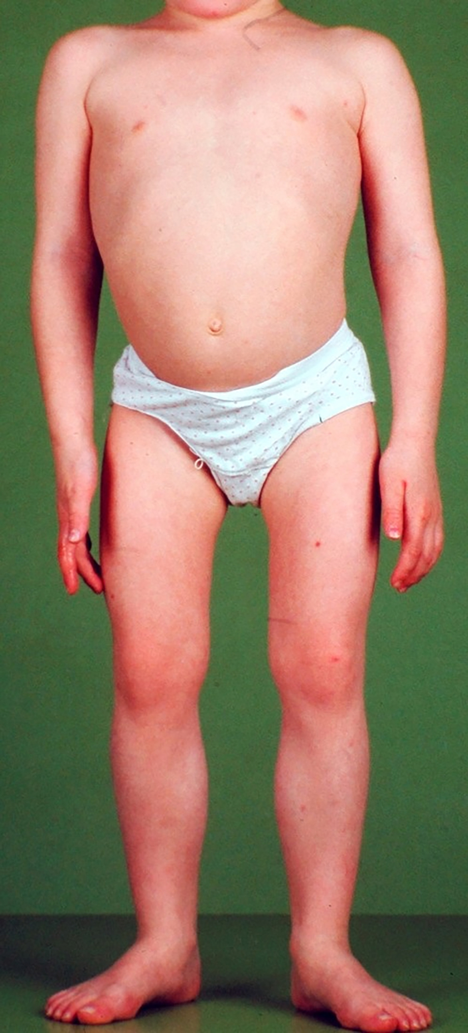
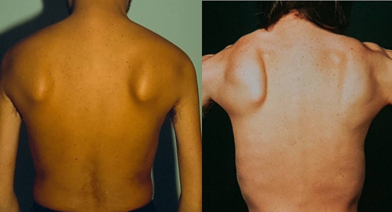
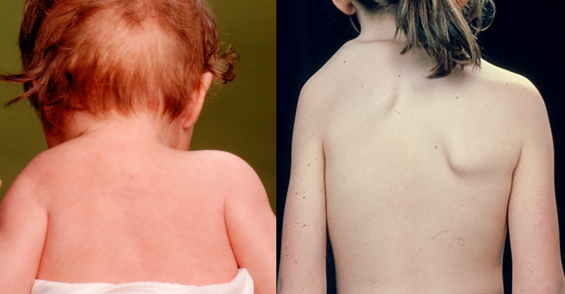
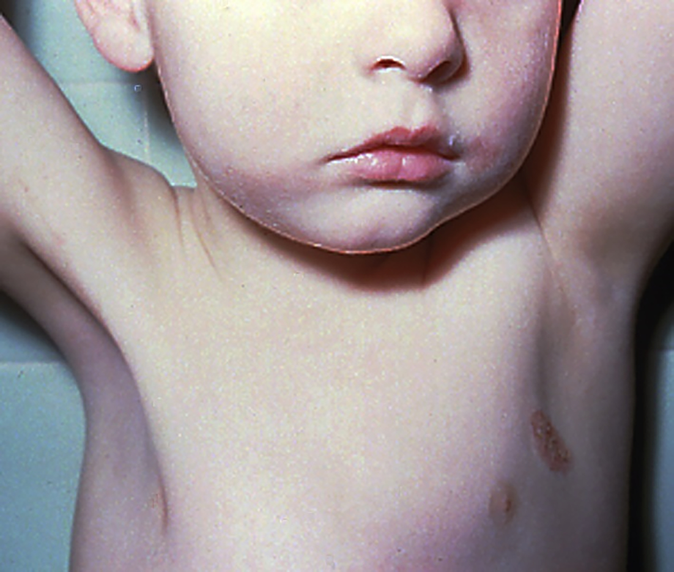
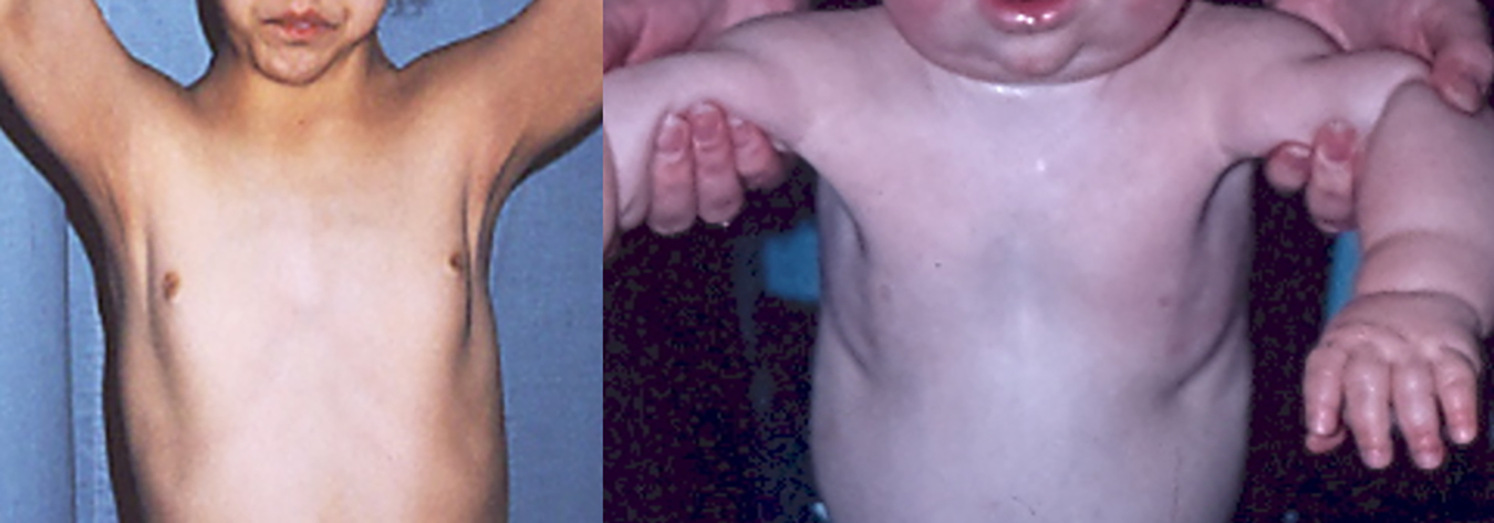
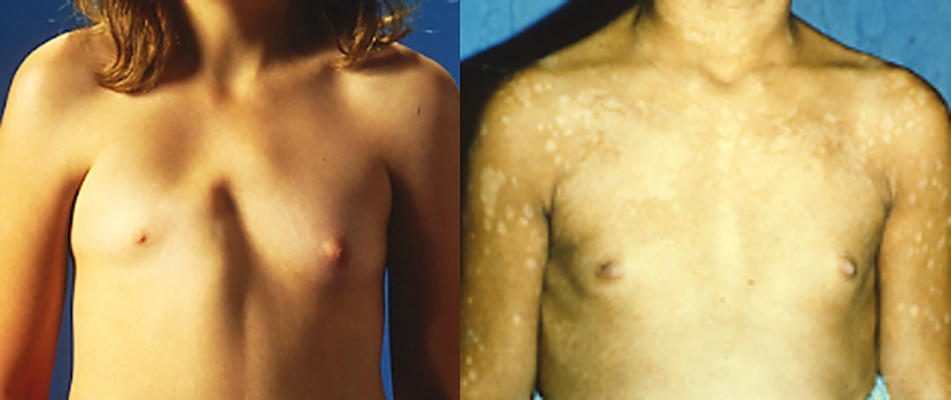
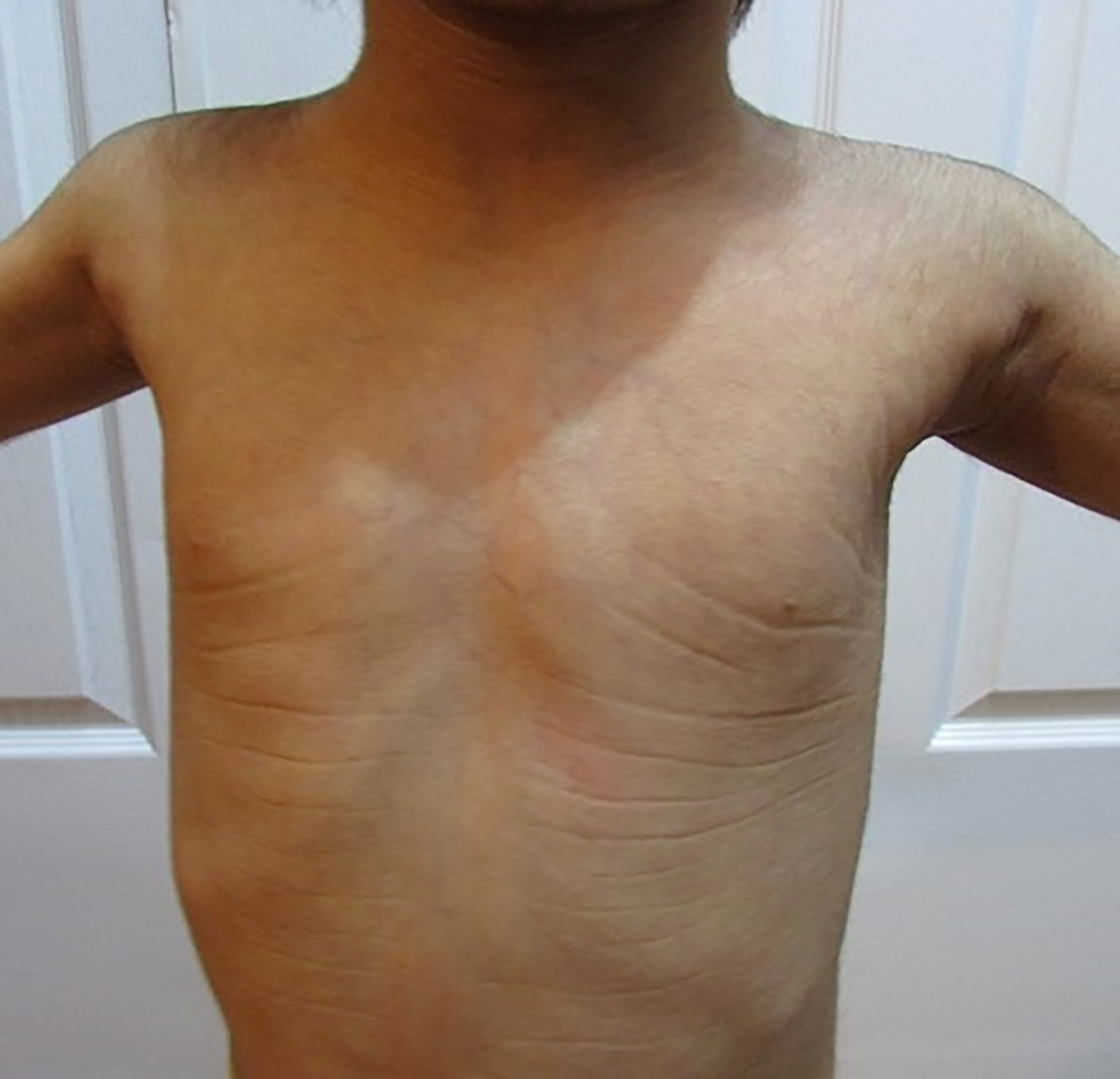
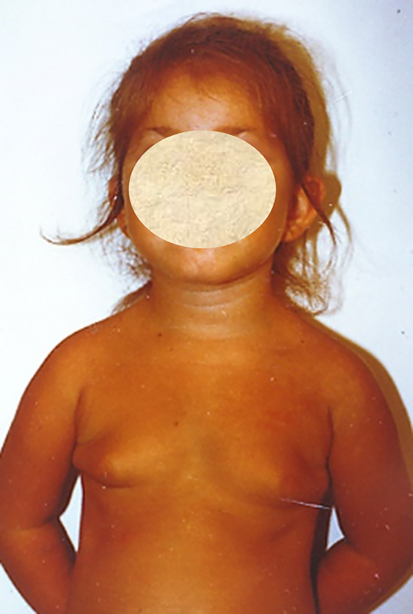
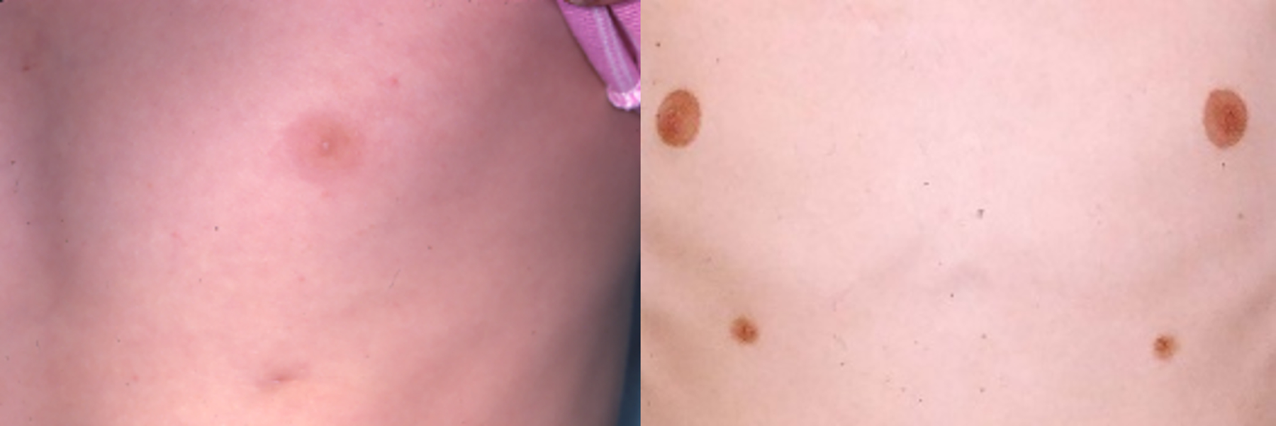
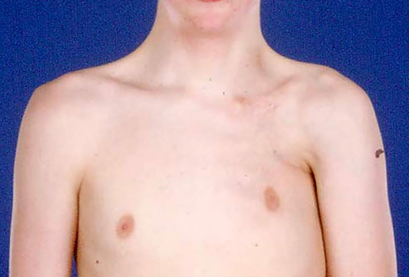
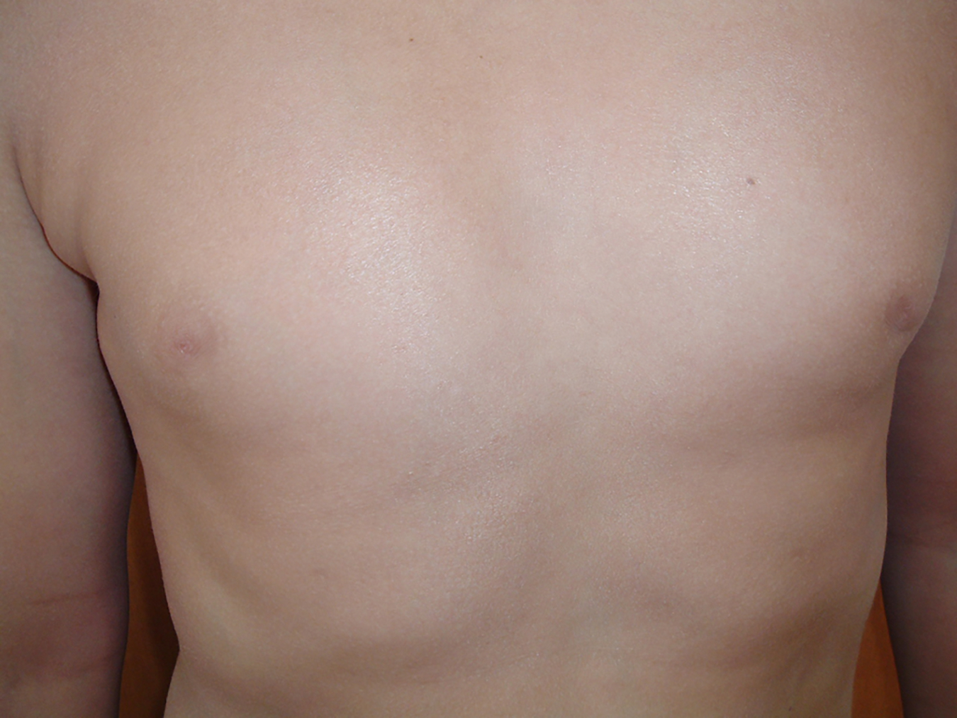
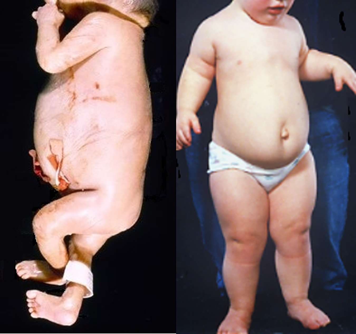
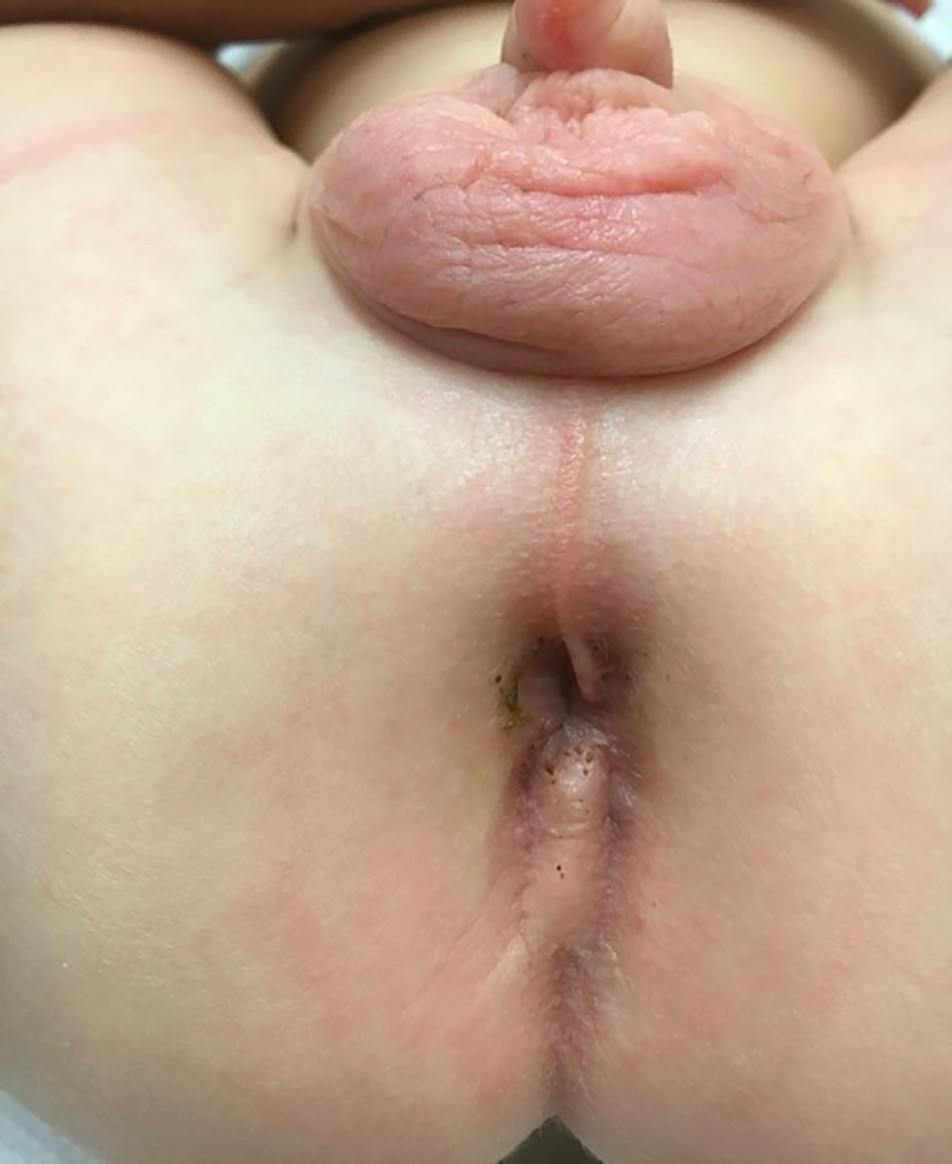
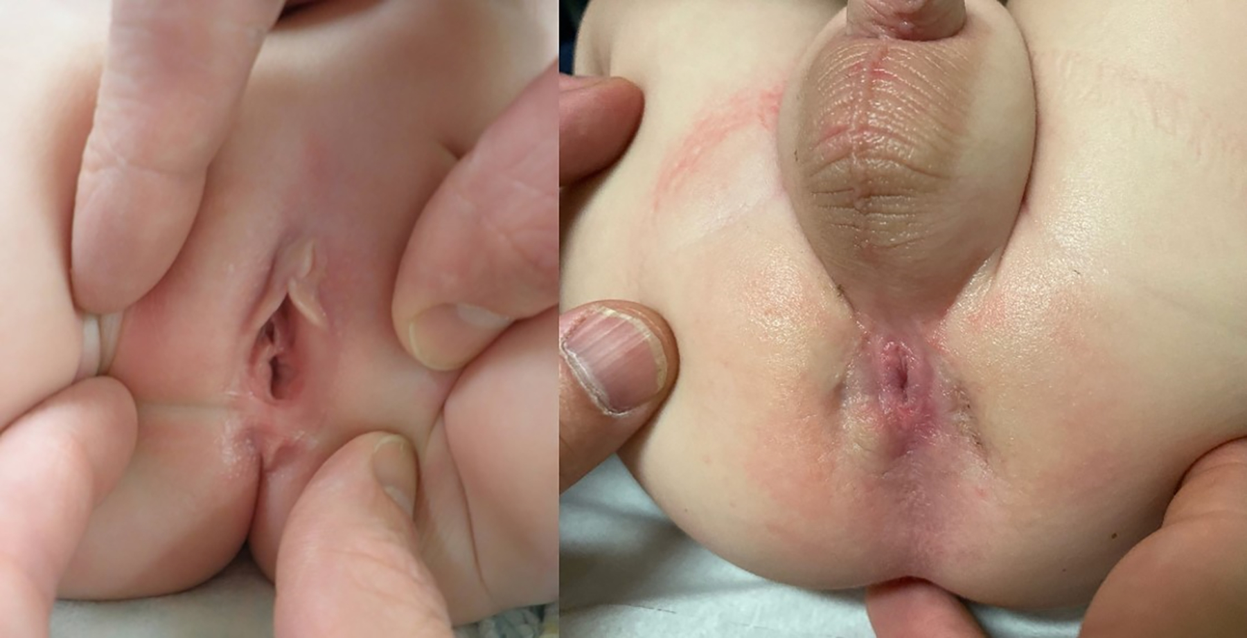
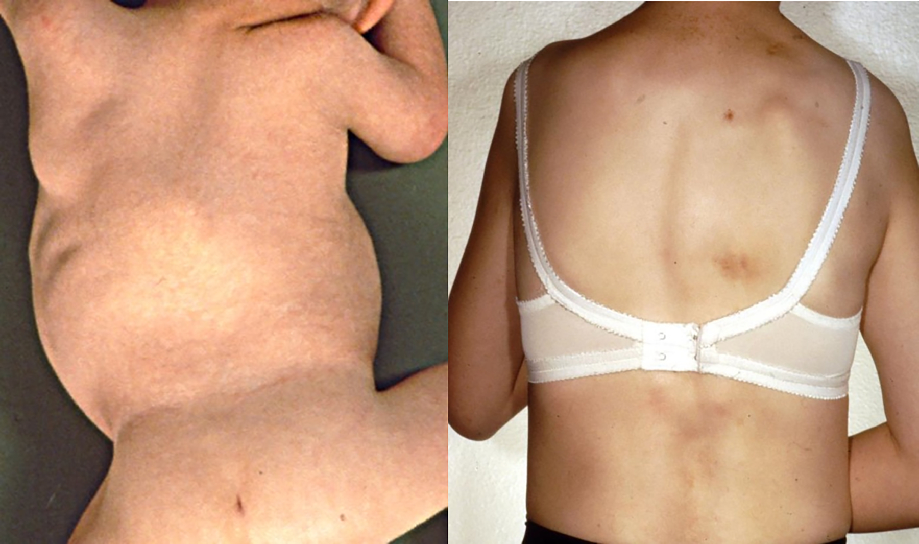
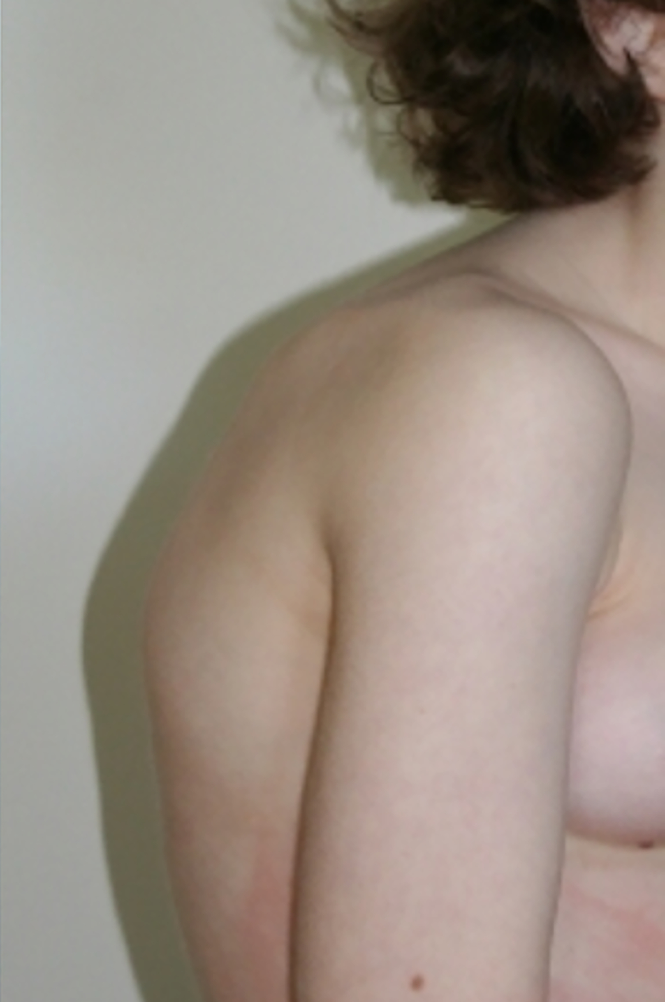
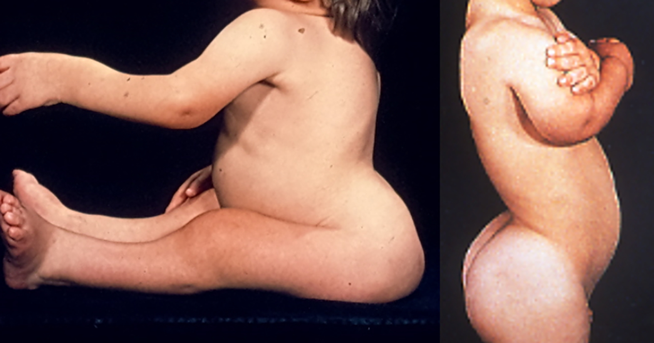
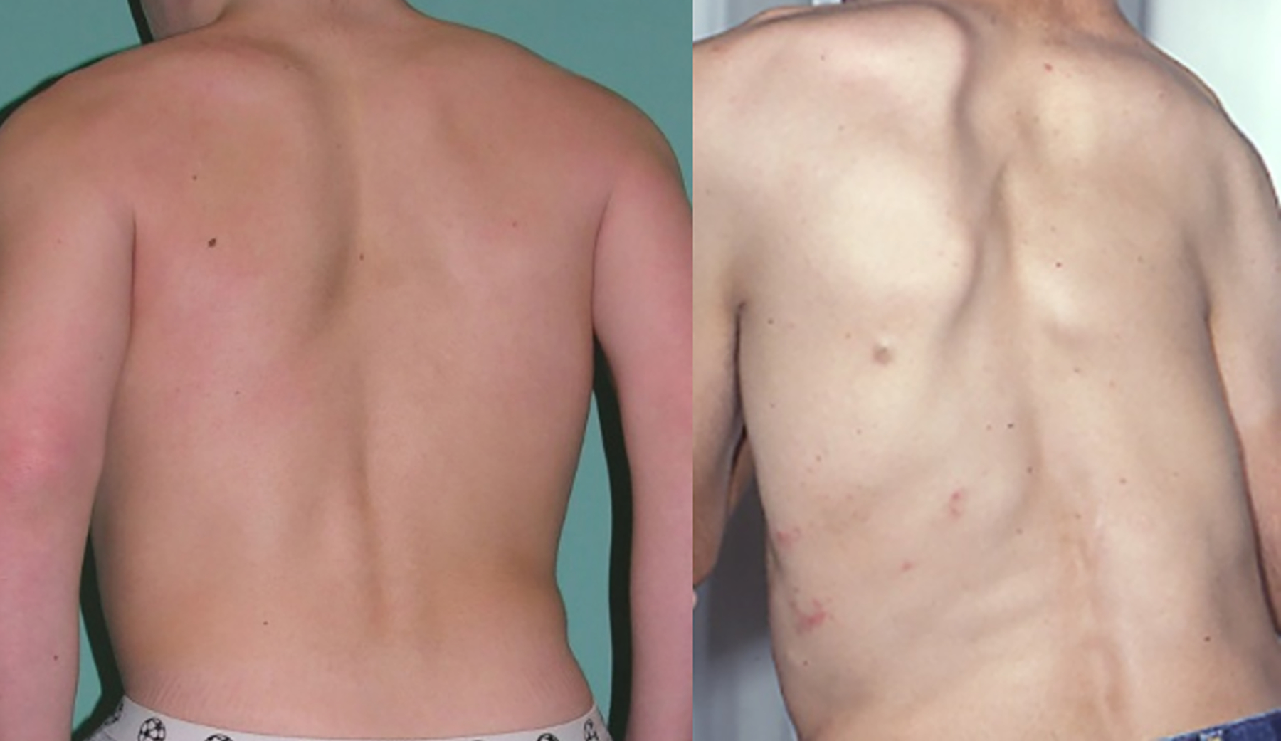
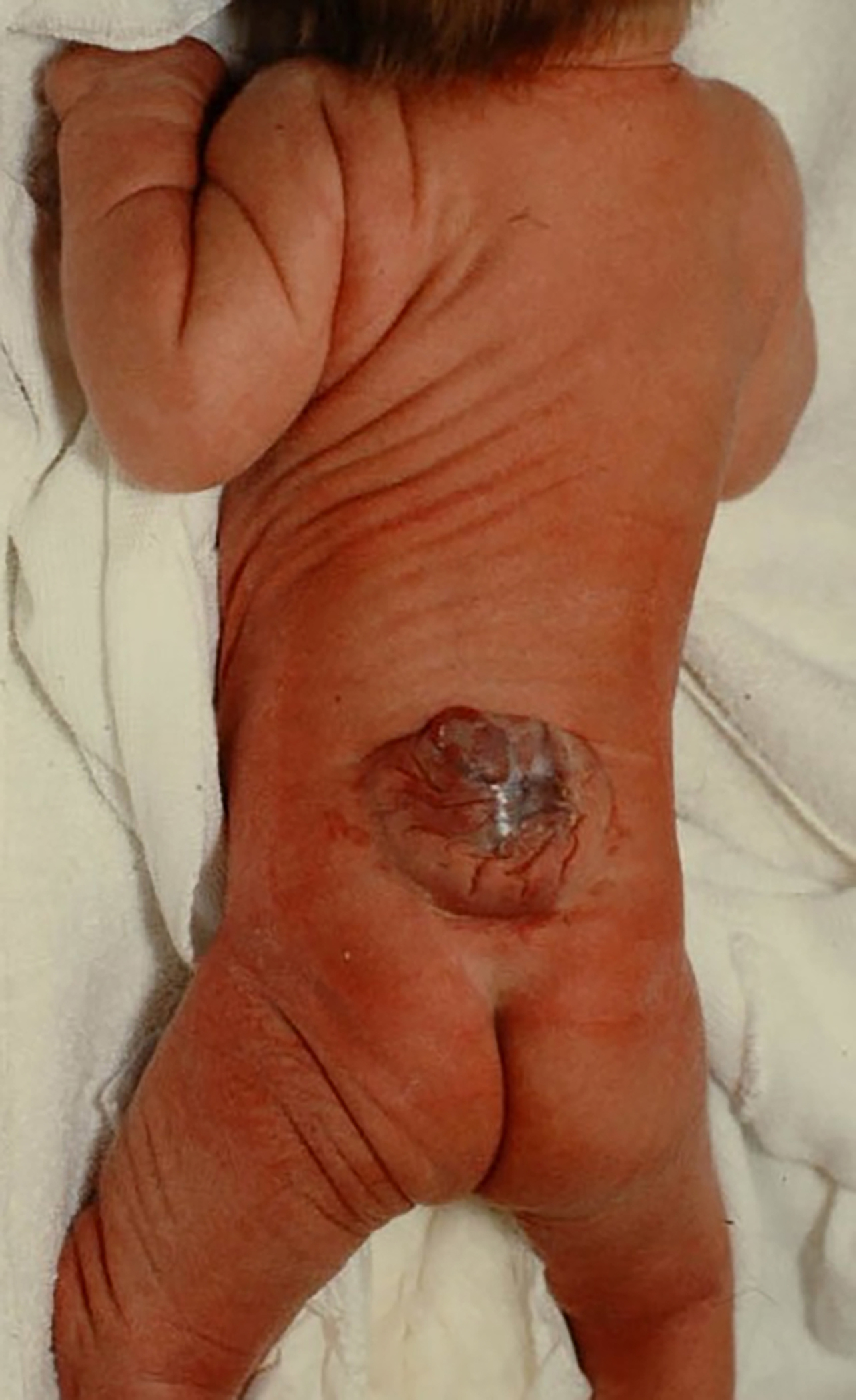
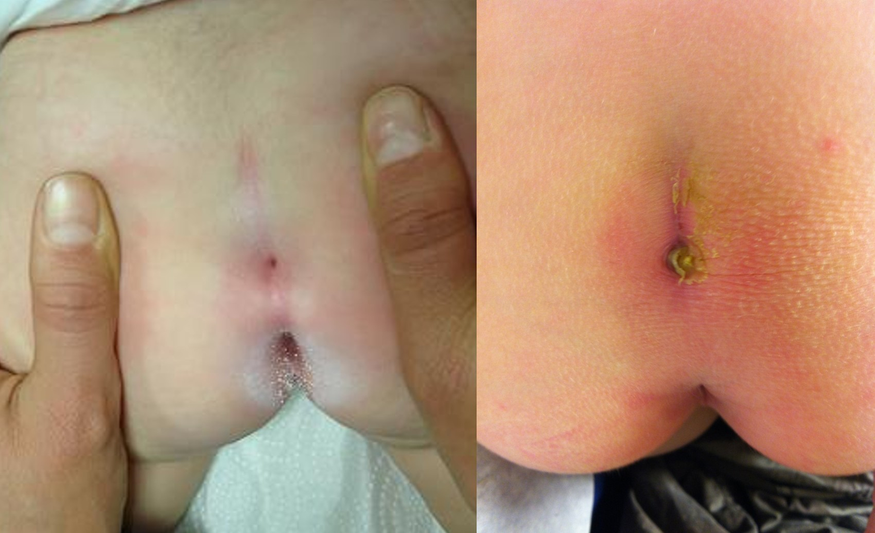
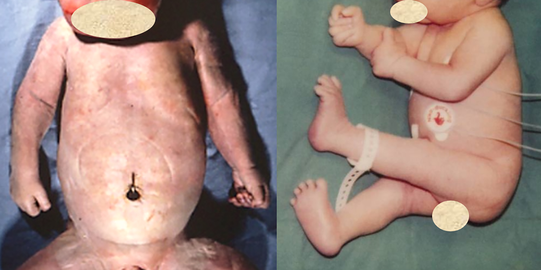
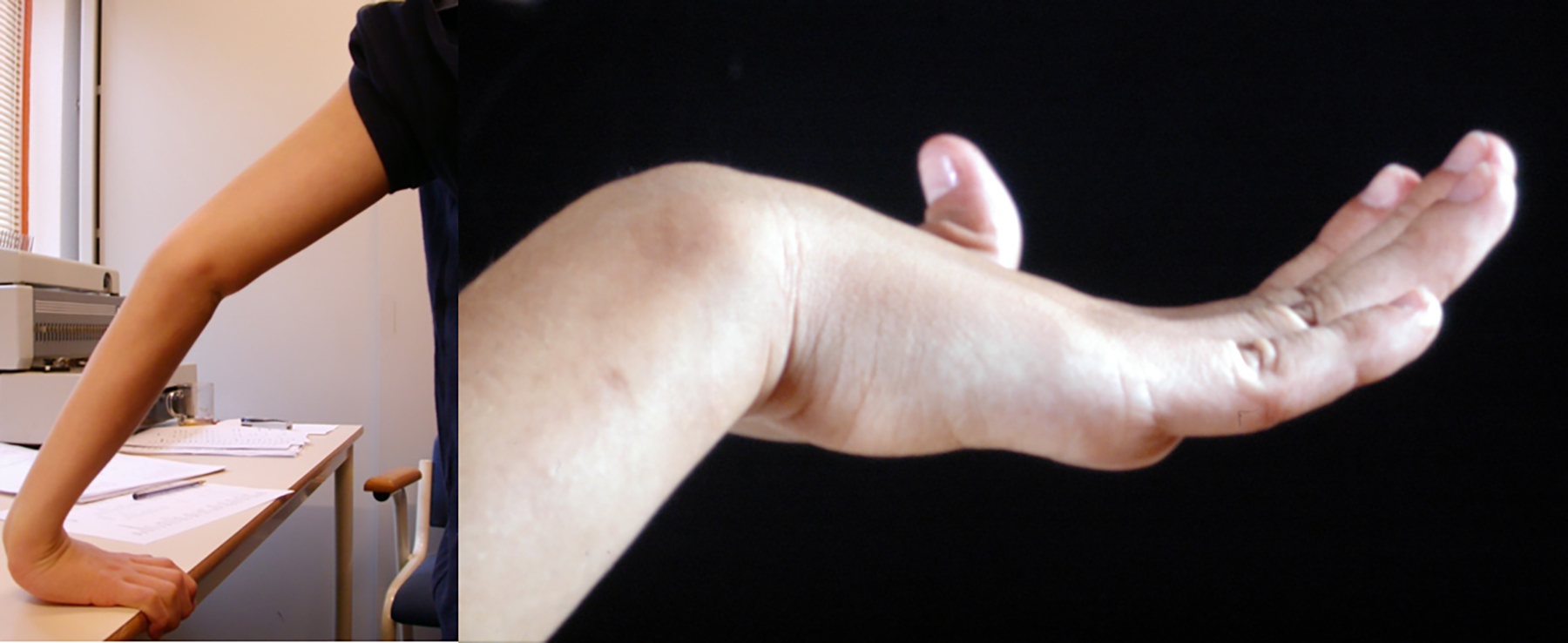
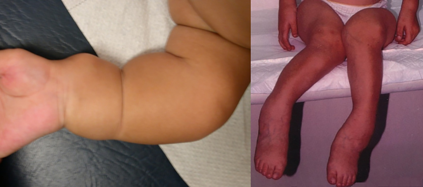
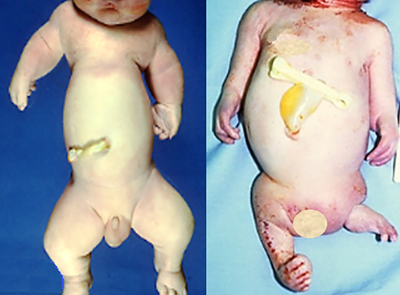
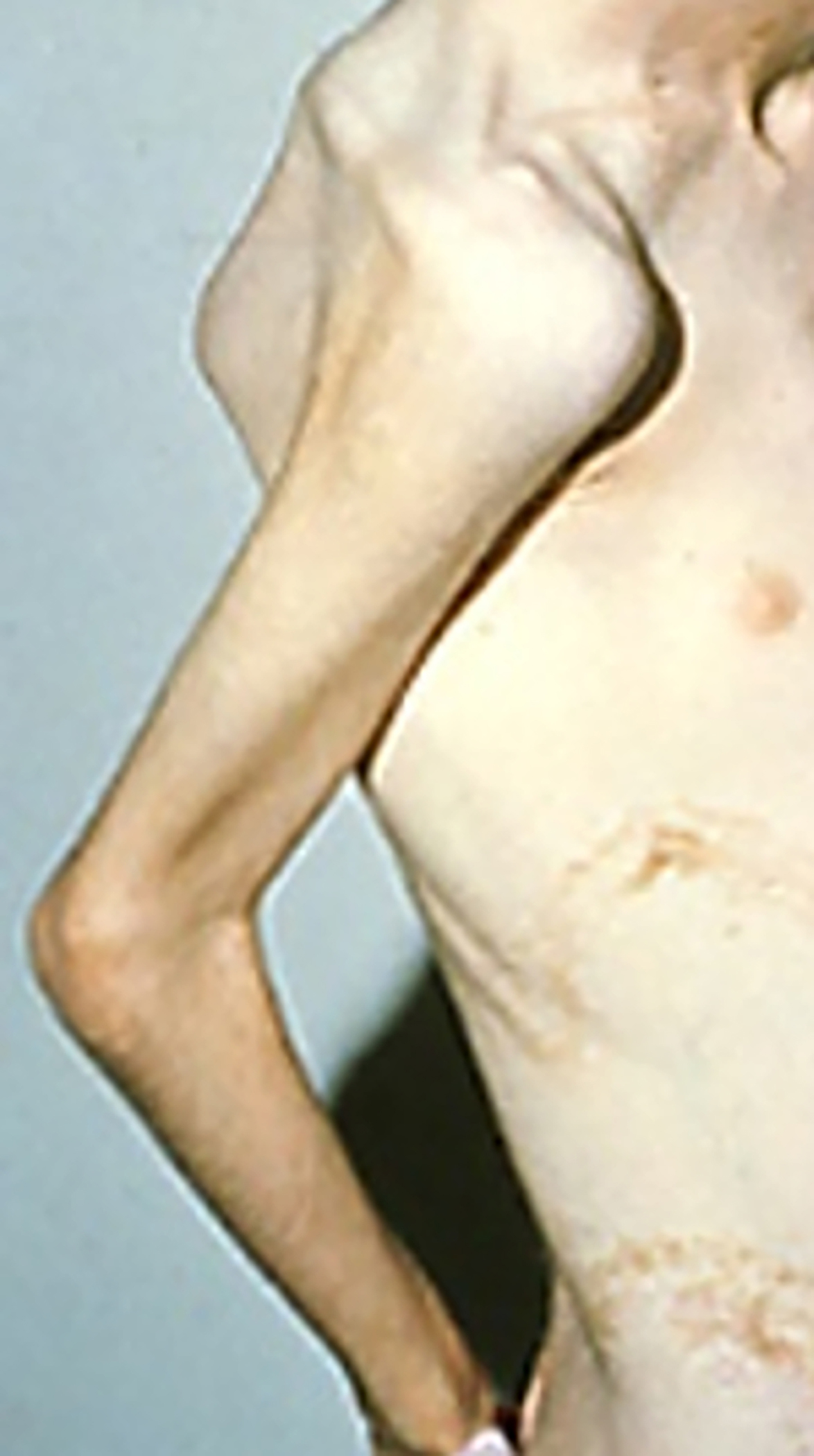
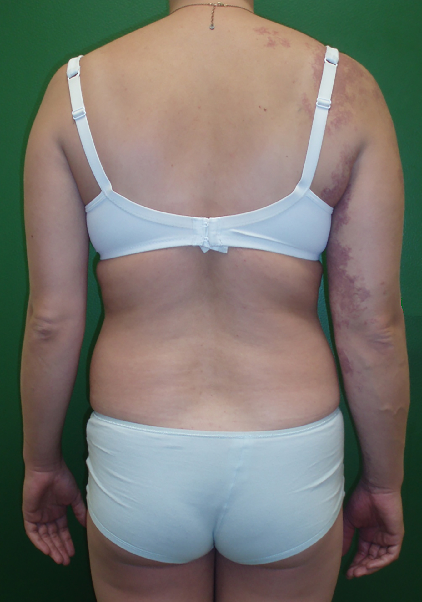
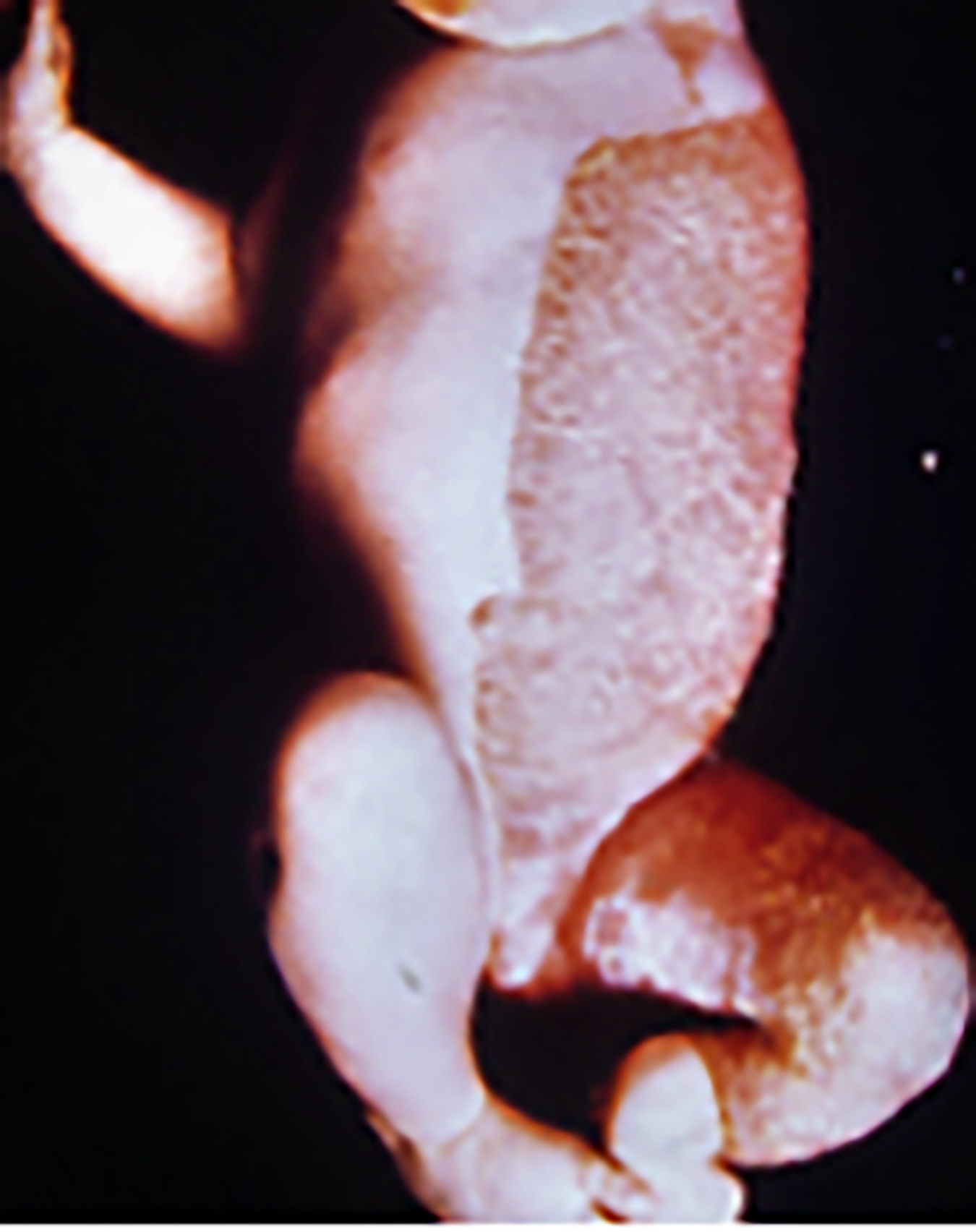
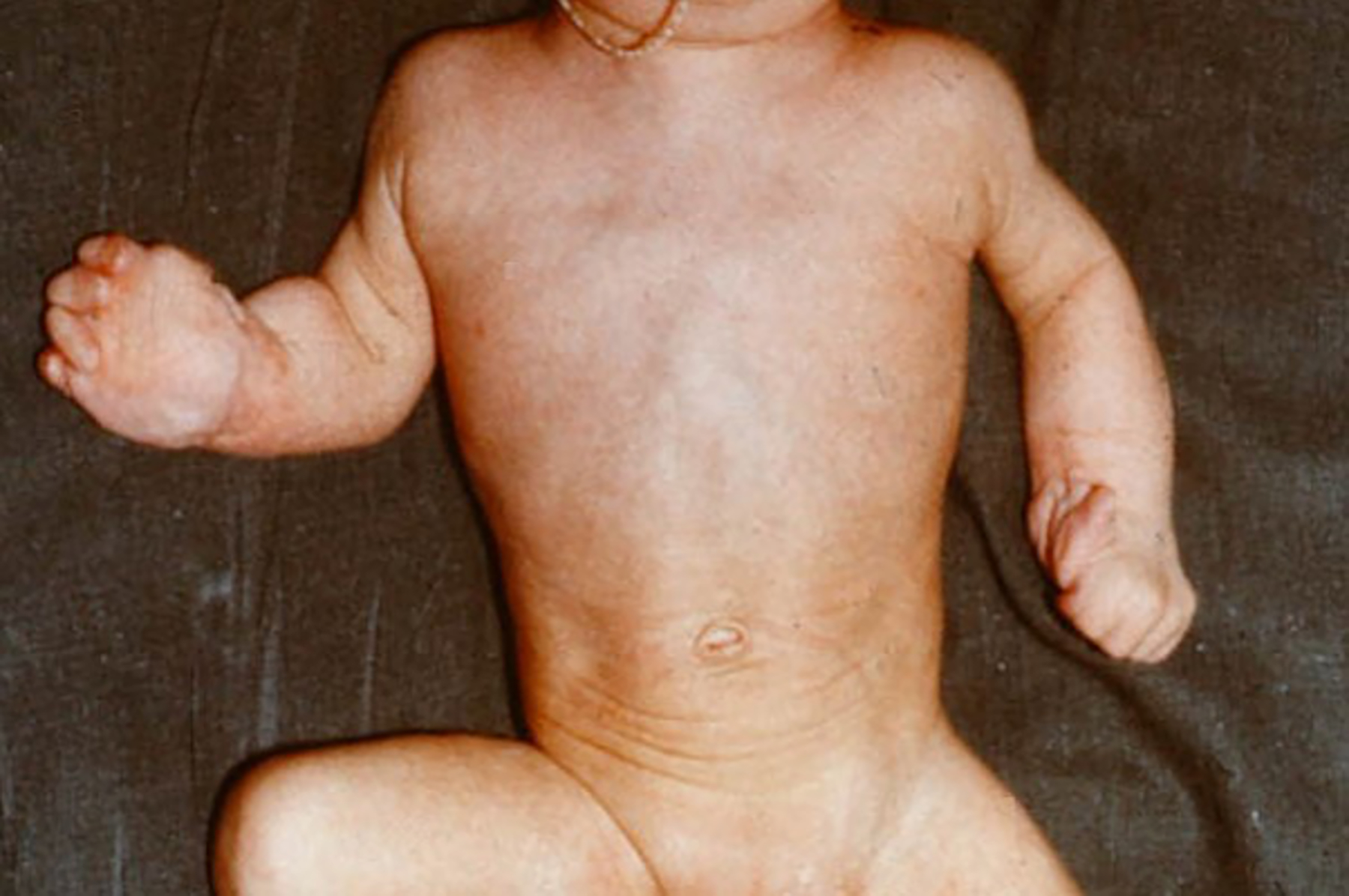
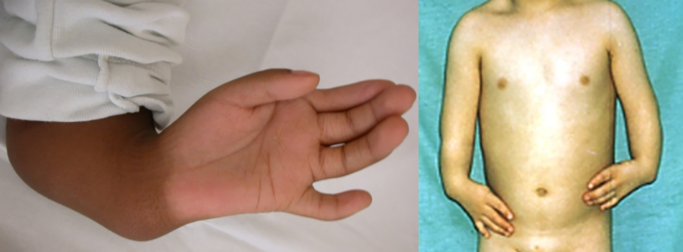
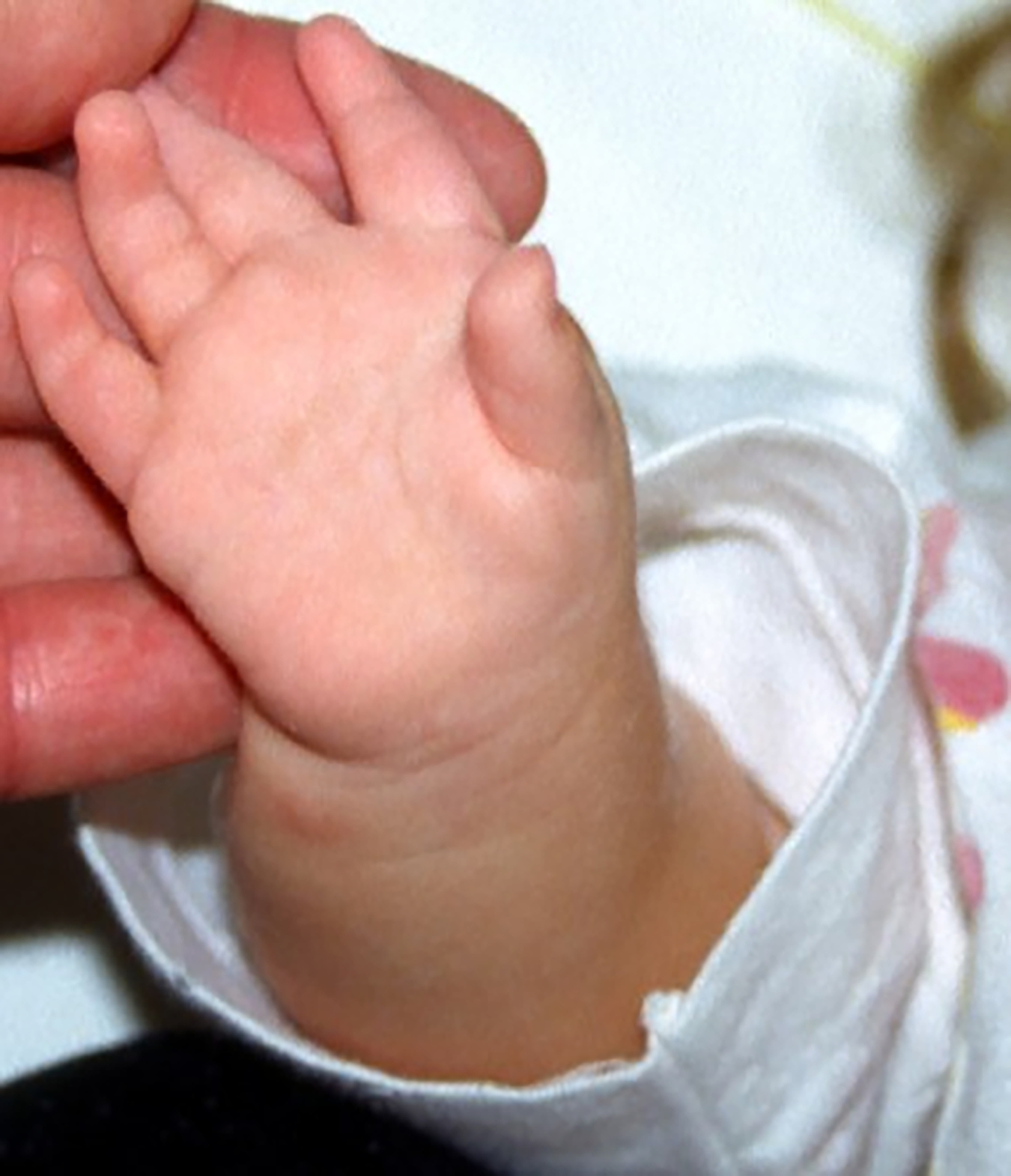
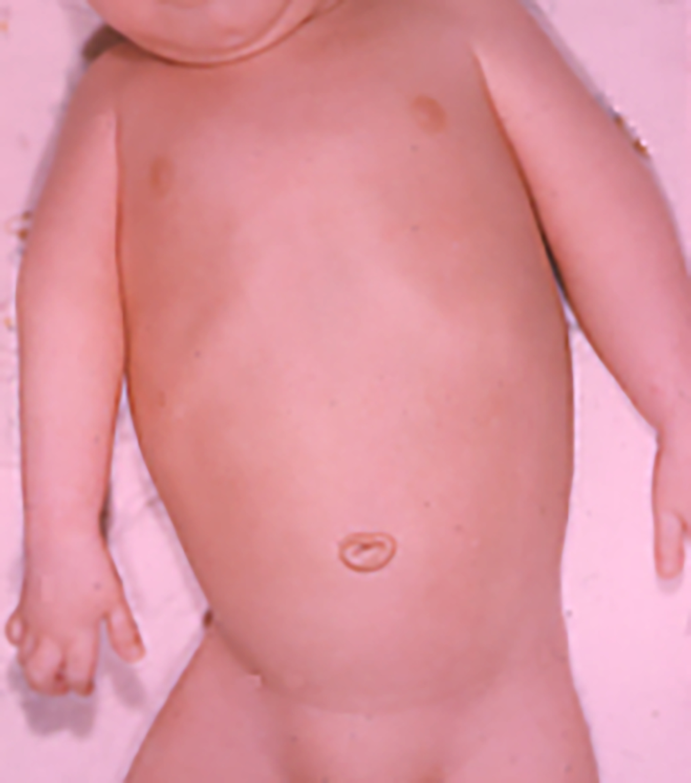
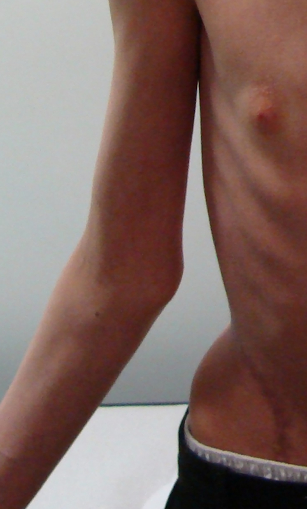
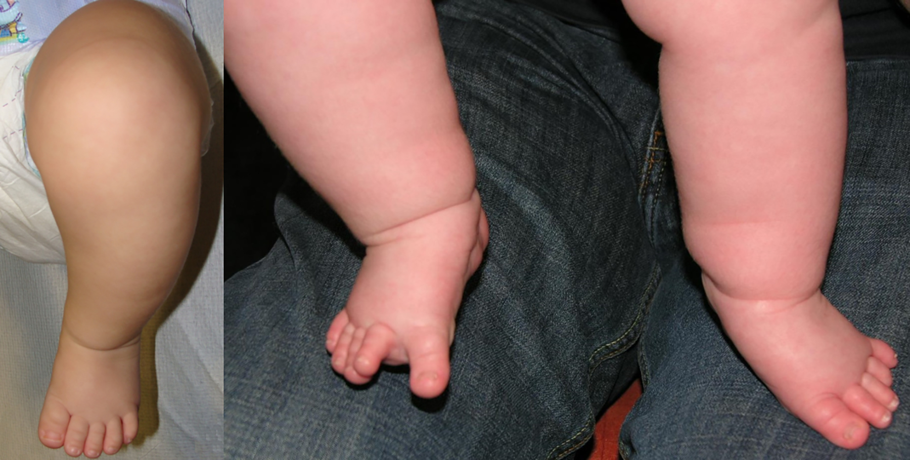
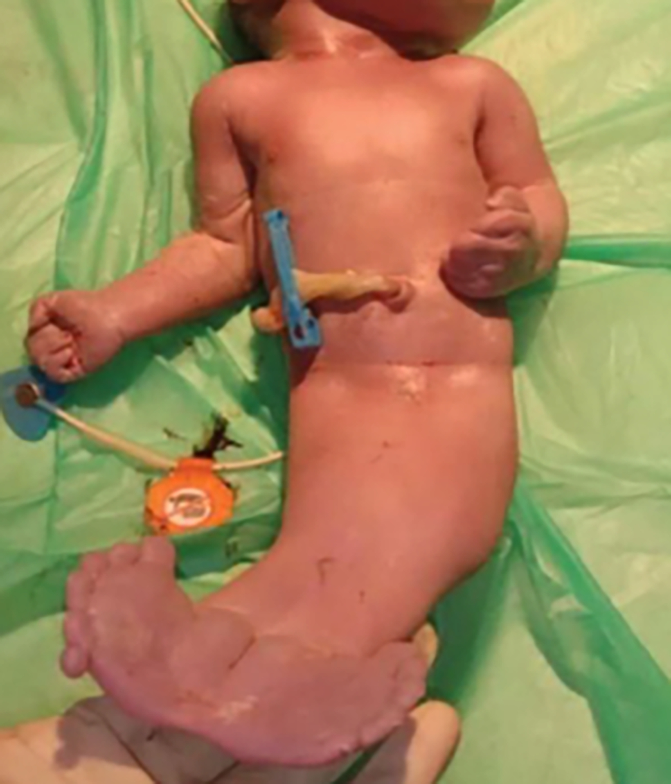
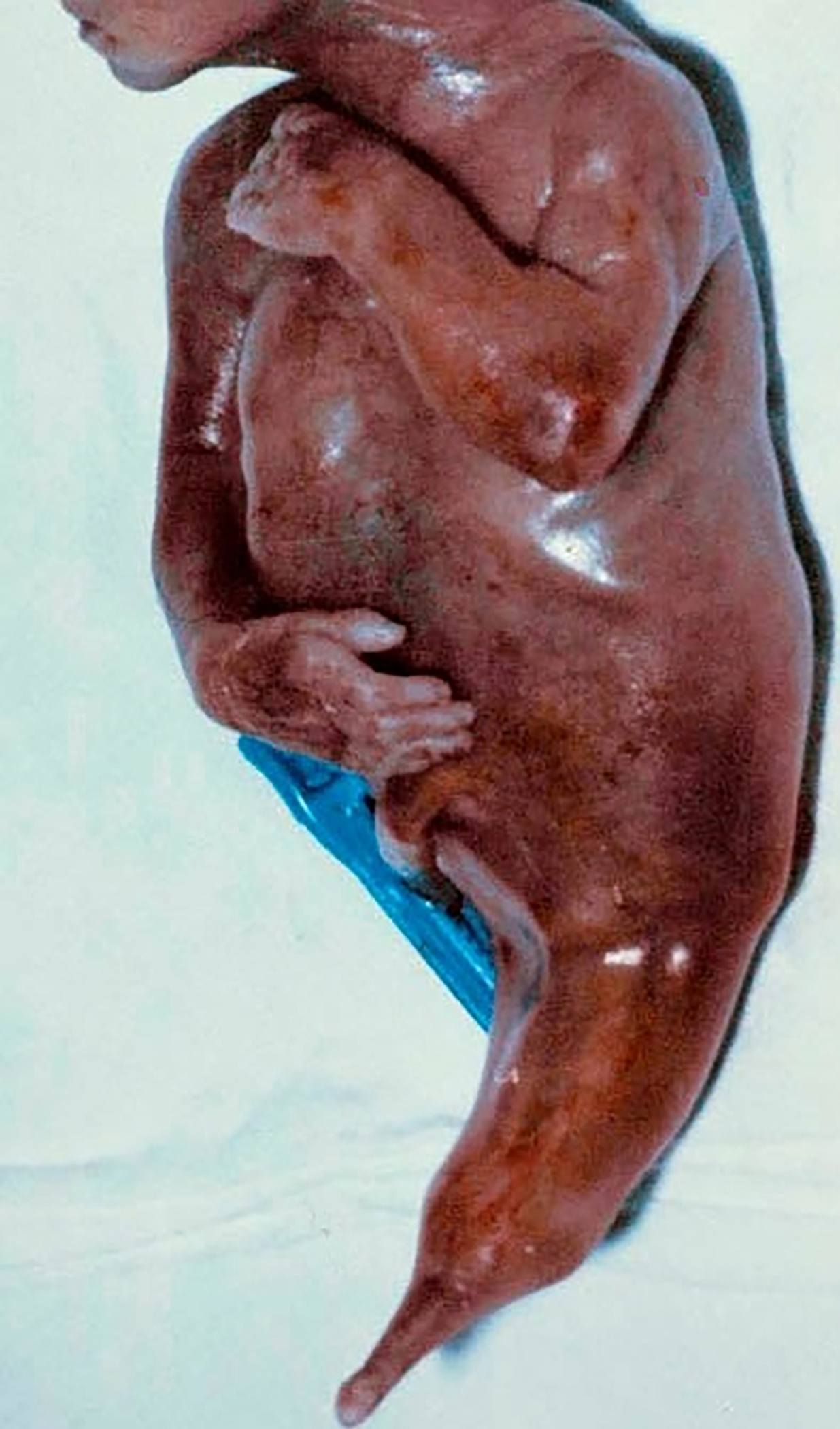
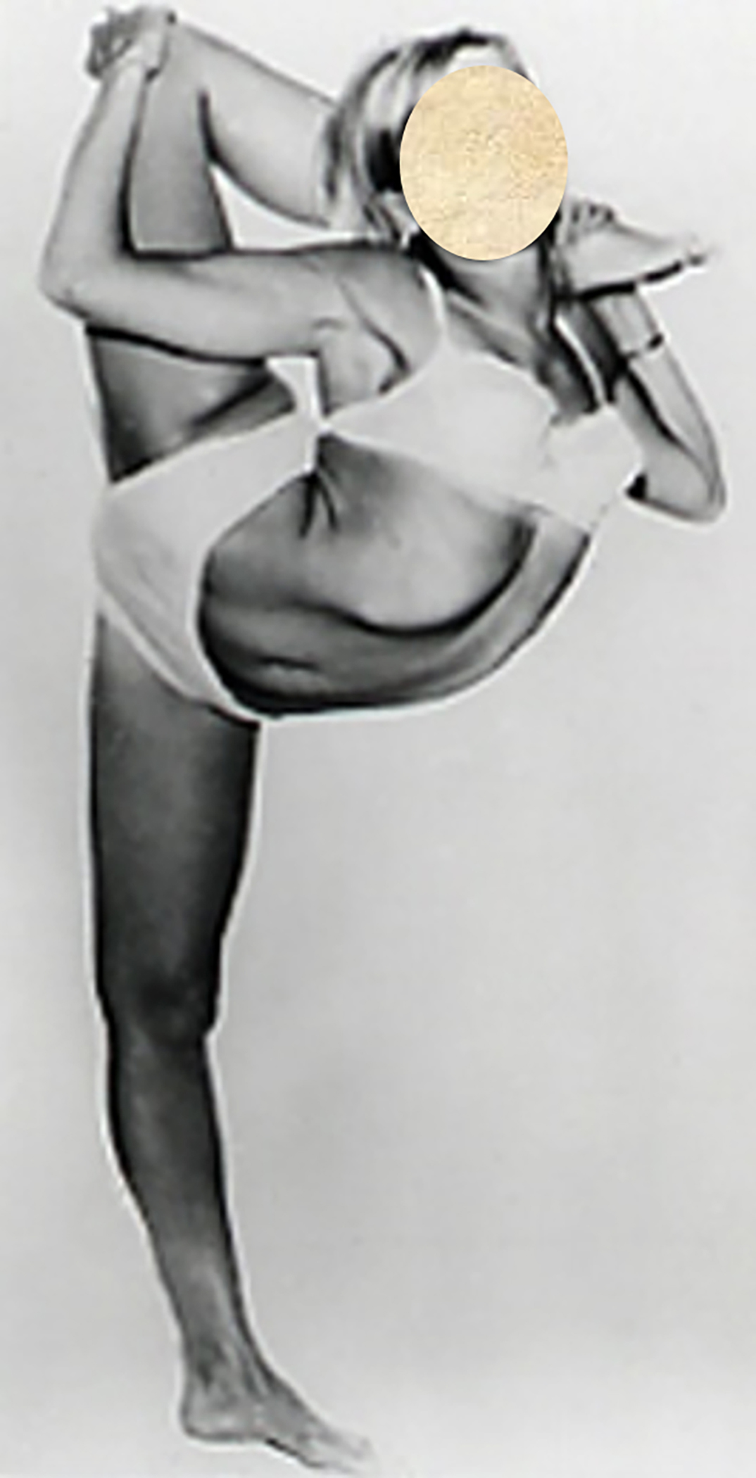
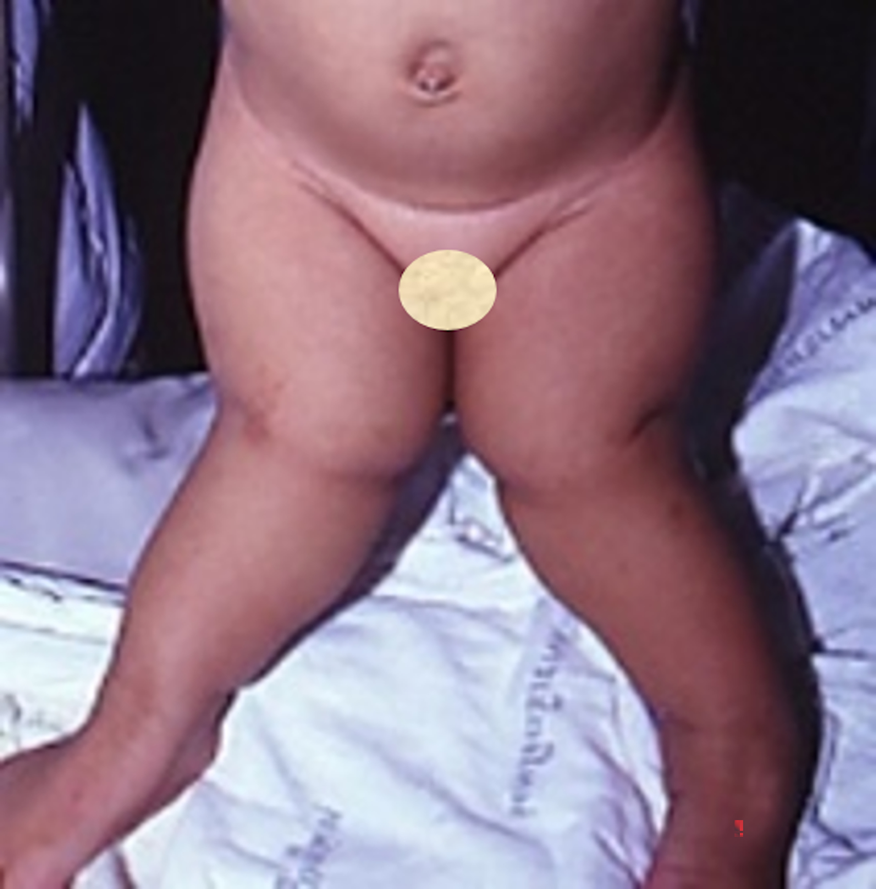
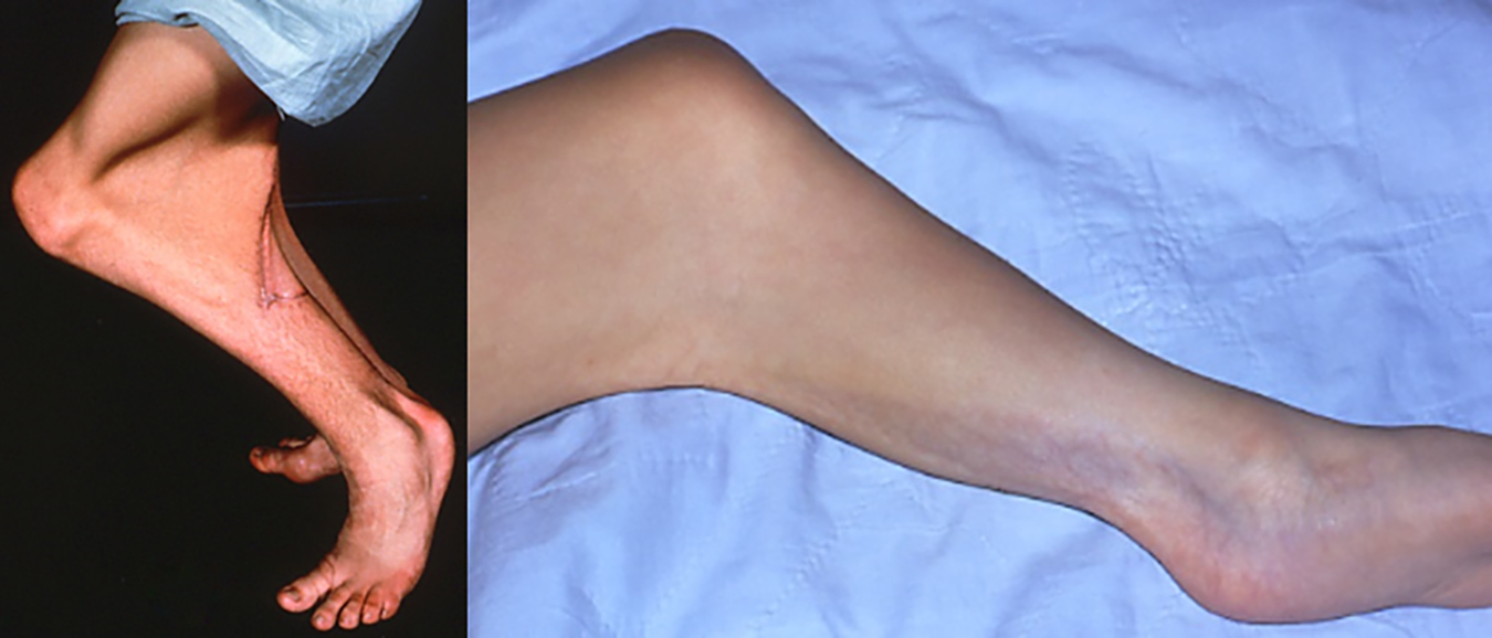
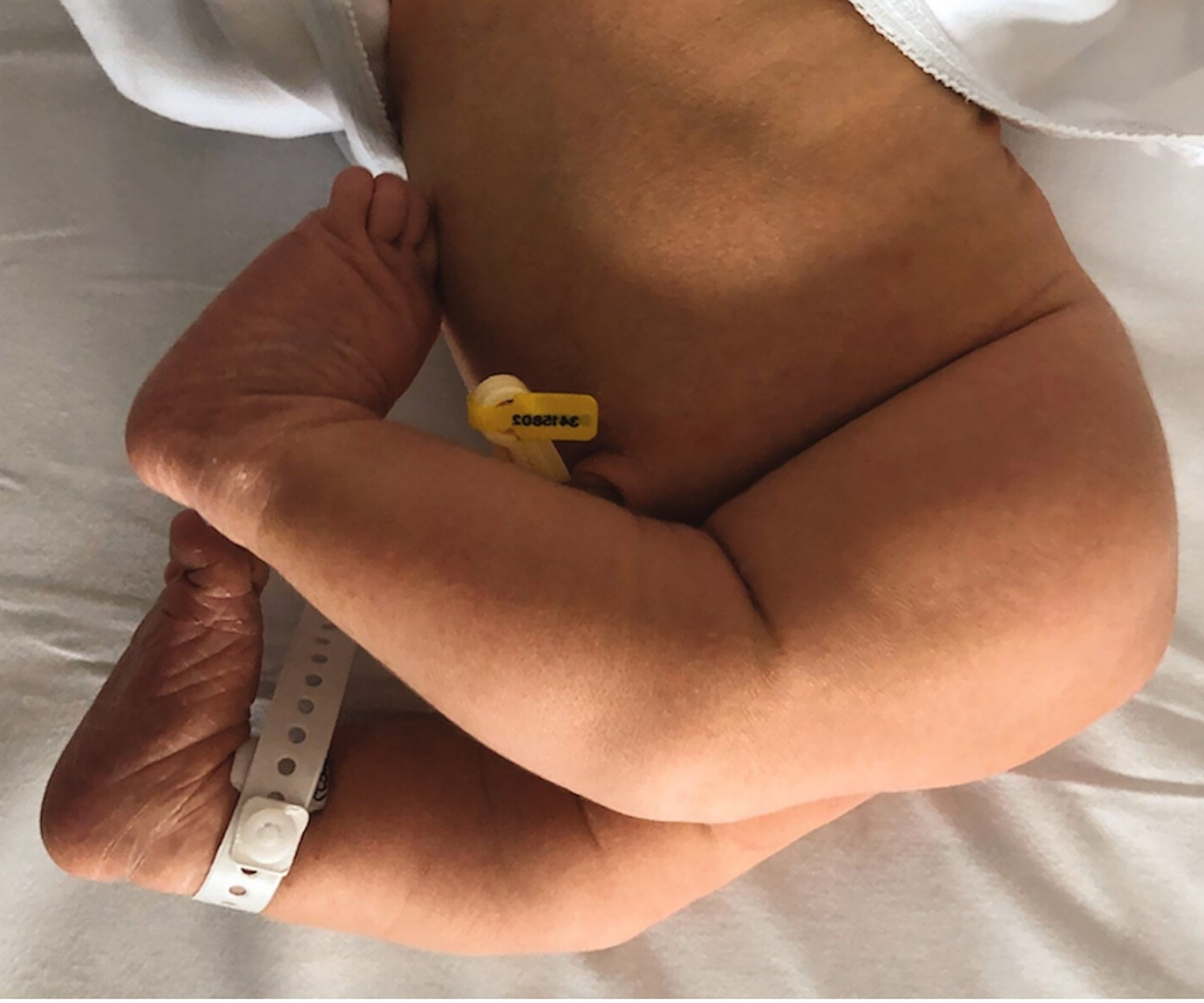
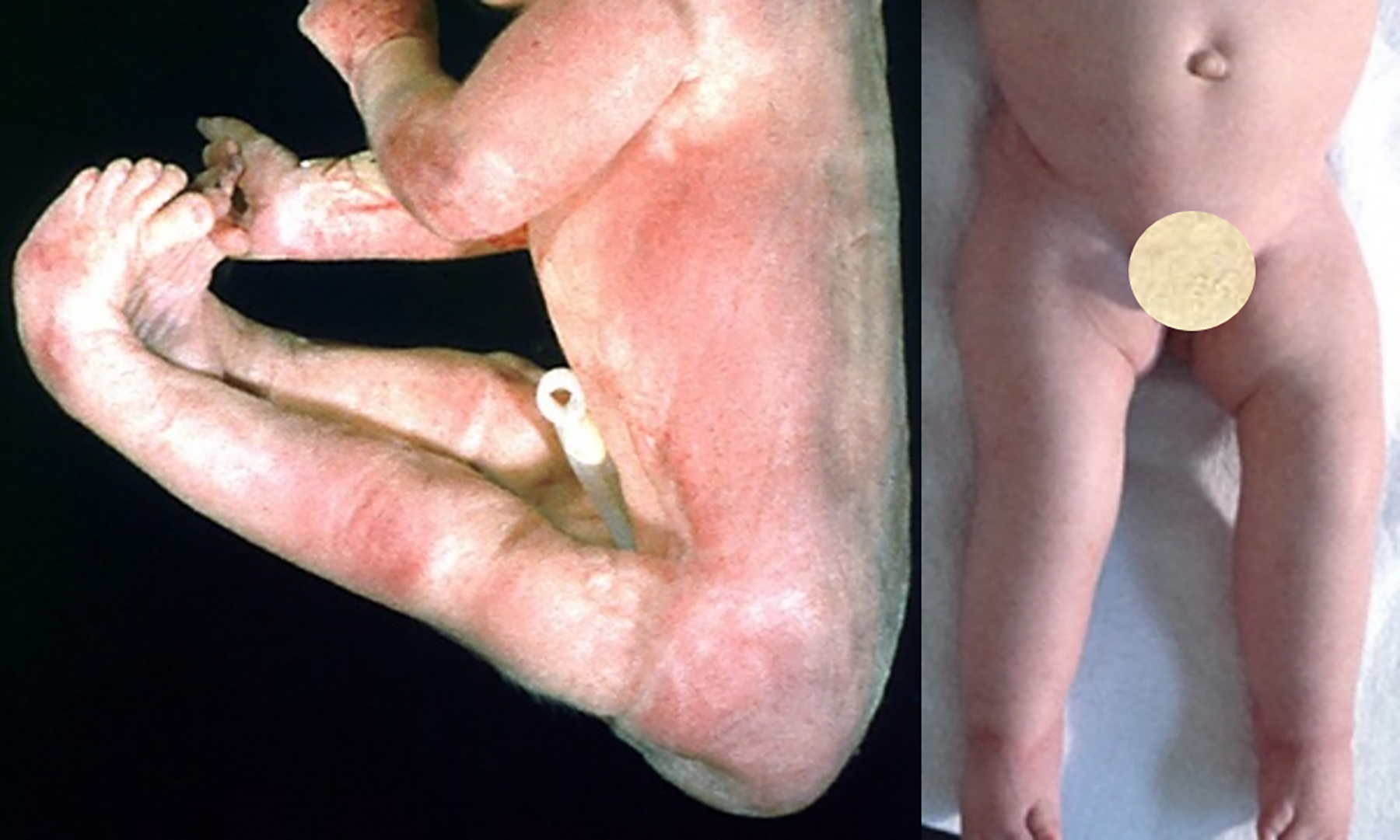
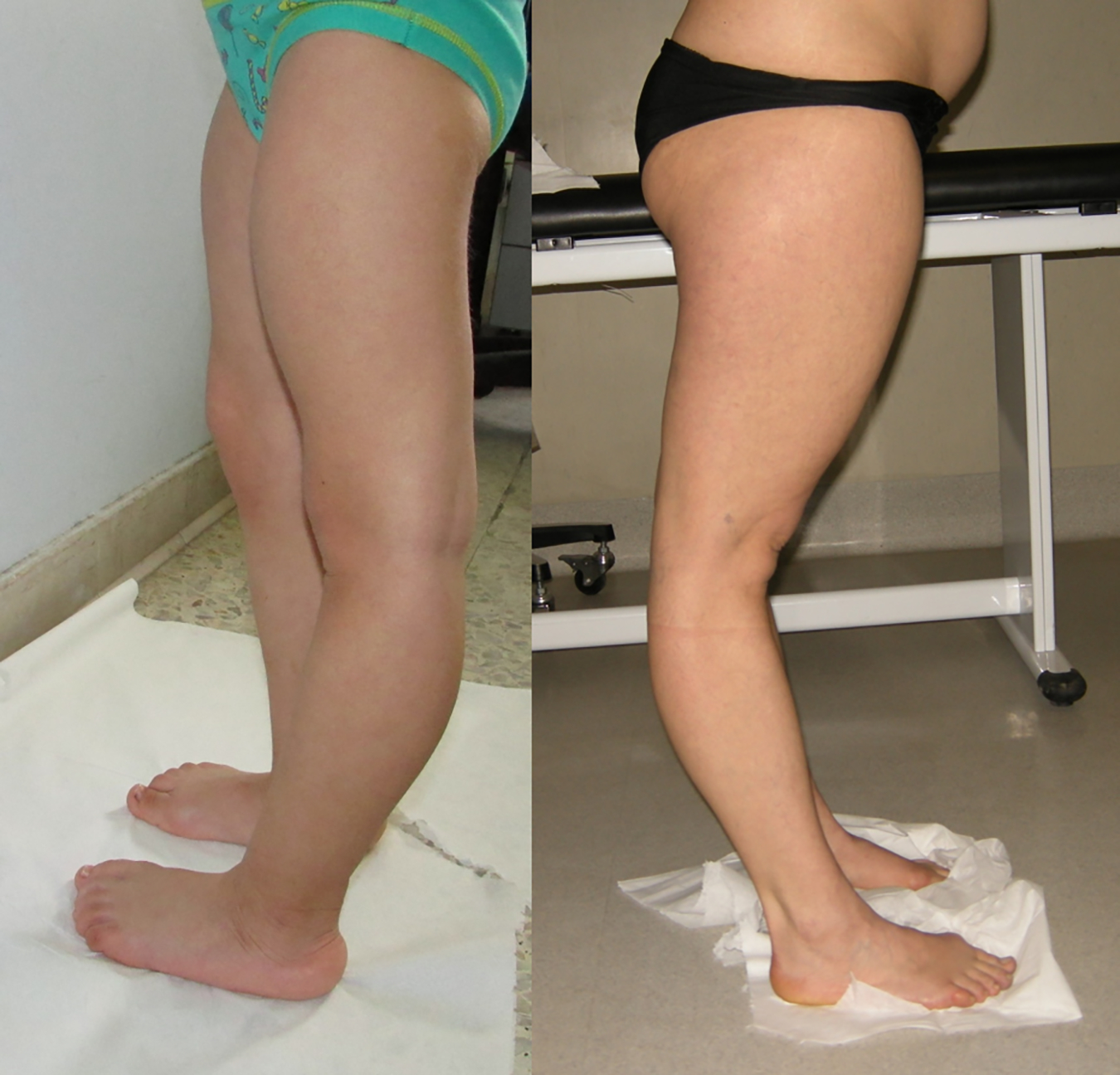
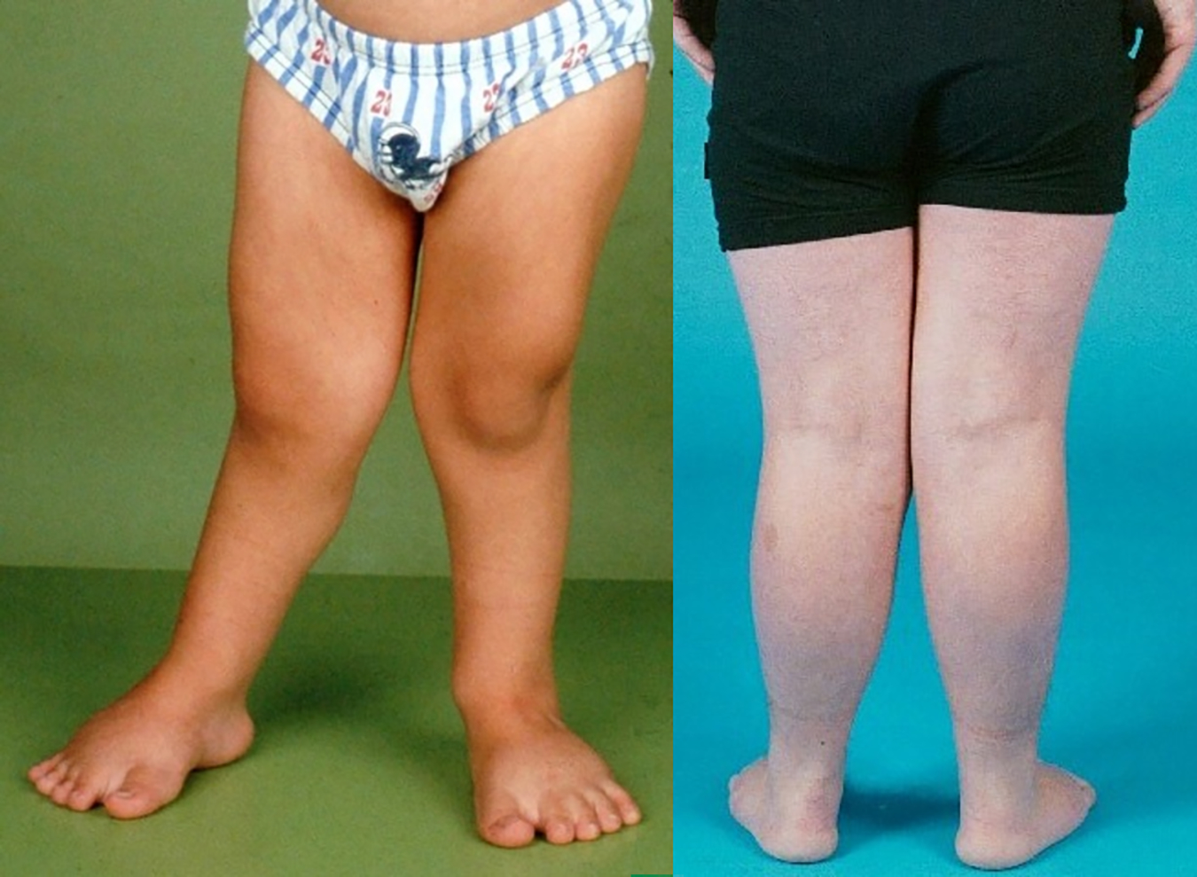
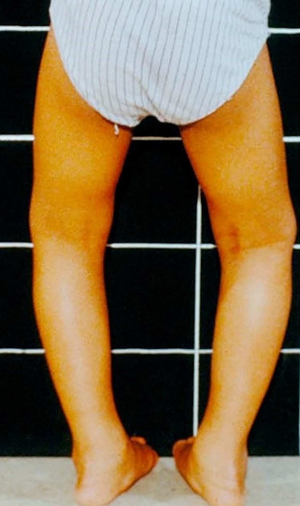
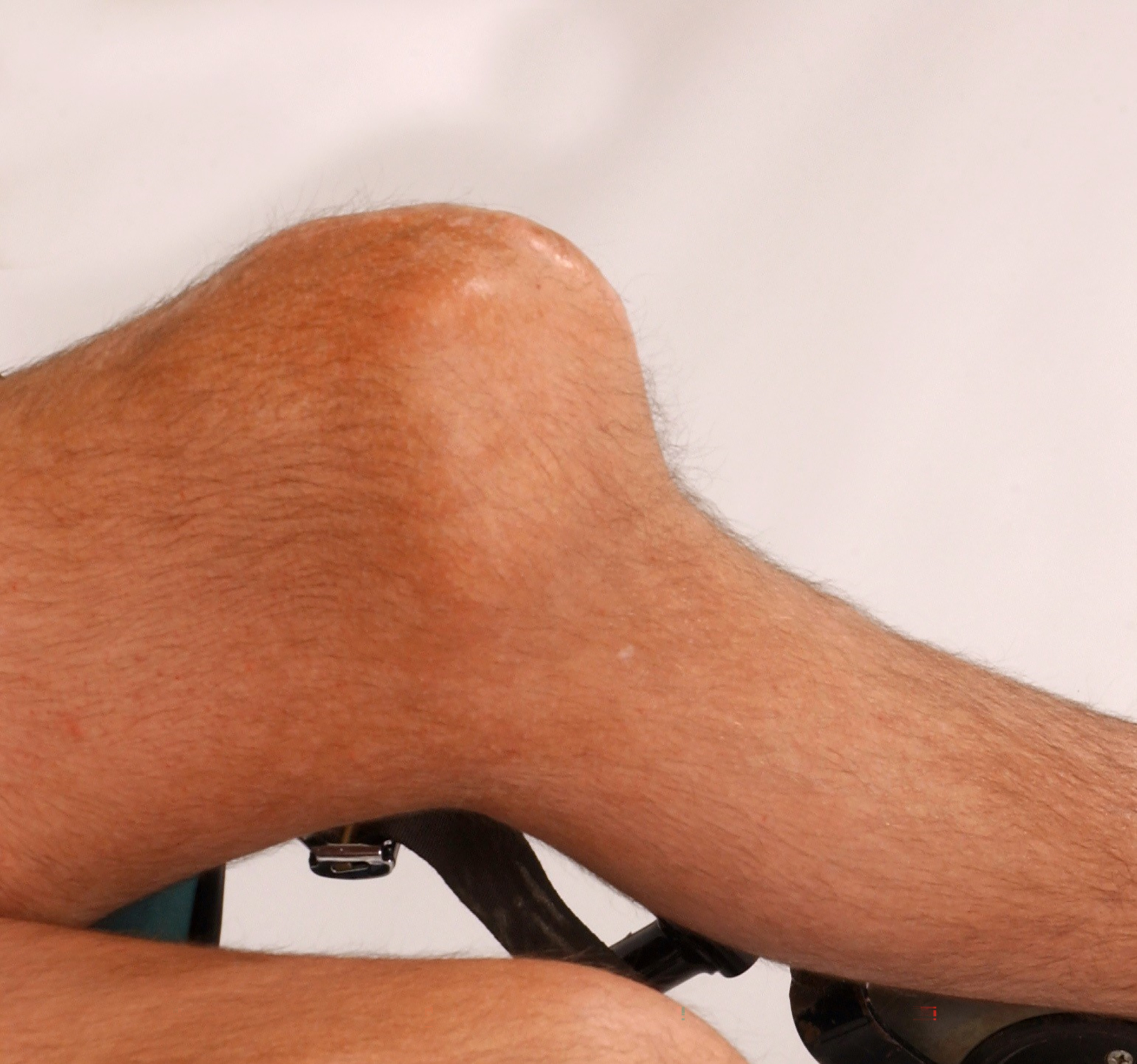
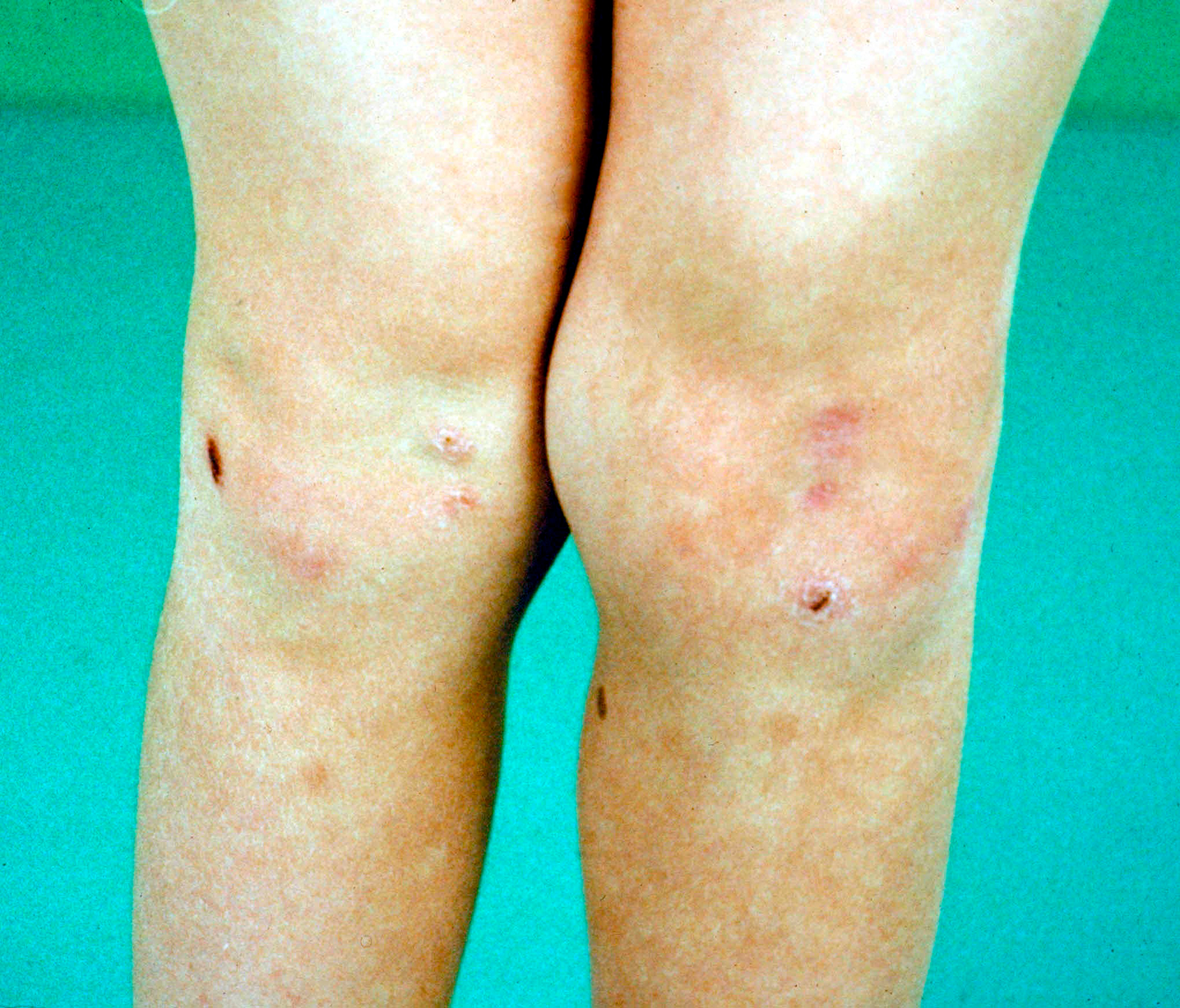
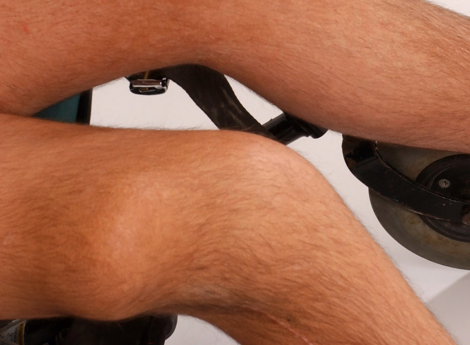
An international group of clinicians working in the field of dysmorphology has initiated the standardization of terms used to describe human morphology. The goals are to standardize these terms and reach consensus regarding their definitions. In this way, we will increase the utility of descriptions of the human phenotype and facilitate reliable comparisons of findings among patients. Additional discussions with other workers in dysmorphology and related fields, such as developmental biology and molecular genetics, will become more precise. Here we introduce the anatomy of the trunk and limbs and define and illustrate the terms that describe the major characteristics of these body regions.

Keywords: anatomy; anthropometry; classification; dysmorphology; elements of morphology; nomenclature.


Conflict of interest statement

CONFLICT OF INTEREST: LGB is a member of the Illumina Medical Ethics Committee, receives editing honoraria from Cold Spring Harbor Laboratory Press, and receives in-kind research support from Merck, Inc. All other authors declare no conflicts of interest.

Figures

Fig. 1.
Fig. 1.
Anatomy of the trunk and limbs.
Fig. 2.
Fig. 2.
Muscular body build.
Fig. 3.
Fig. 3.
Thin body build.
Fig. 4.
Fig. 4.
Generalized obesity.
Fig. 5.
Fig. 5.
Truncal obesity.
Fig. 6.
Fig. 6.
Proprotionate short stature.
Fig. 7.
Fig. 7.
Proprotionate tall stature.
Fig. 8.
Fig. 8.
Short trunk.
Fig. 9.
Fig. 9.
Long trunk.
Fig. 10.
Fig. 10.
Short thorax.
Fig. 11.
Fig. 11.
Long thorax.
Fig. 12.
Fig. 12.
Broad thorax.
Fig. 13.
Fig. 13.
Narrow thorax.
Fig. 14.
Fig. 14.
Short sternum.
Fig. 15.
Fig. 15.
Pectus excavatum.
Fig. 16.
Fig. 16.
Pectus carinatum.
Fig. 17.
Fig. 17.
Flaring ribs.
Fig. 18.
Fig. 18.
Narrow shoulders.
Fig. 19.
Fig. 19.
Broad shoulders.
Fig. 20.
Fig. 20.
Sloping shoulders.
Fig. 21.
Fig. 21.
Winging of the scapulae.
Fig. 22.
Fig. 22.
Sprengel anomaly.
Fig. 23.
Fig. 23.
Underdeveloped pectoralis muscle on the left.
Fig. 24.
Fig. 24.
Webbing of the axilla.
Fig. 25.
Fig. 25.
Small breast (note: unilater on left panel, bilateral in an adult female on right panel).
Fig. 26.
Fig. 26.
Large breast.
Fig. 27.
Fig. 27.
Impalpable breast.
Fig. 28.
Fig. 28.
Premature development of the breast.
Fig. 29.
Fig. 29.
Gynecomastia (note: left side only).
Fig. 30.
Fig. 30.
Small nipple.
Fig. 31.
Fig. 31.
Large nipple (note: in a newborn child).
Fig. 32.
Fig. 32.
Absent nipple.
Fig. 33.
Fig. 33.
Supernumerary nipple.
Fig. 34.
Fig. 34.
Ectopic nipple (note: acquired).
Fig. 35.
Fig. 35.
Widely spaced nipple.
Fig. 36.
Fig. 36.
Protuberant abdomen.
Fig.37.
Fig.37.
Diastasis recti (note: also depression of the umbilicus visible).
Fig. 38.
Fig. 38.
Underdeveloped abdominal muscles.
Fig. 39.
Fig. 39.
Prune belly.
Fig. 40.
Fig. 40.
Umbilical hernia.
Fig. 41.
Fig. 41.
Gastroschisis (Courtesy of Dr. Justin R De Jong).
Fig. 42.
Fig. 42.
Omphalocele.
Fig. 43.
Fig. 43.
Low-positioned umbilicus (courtesy of Dr Sumru Kavurt [Akman et al., 2013]).
Fig. 44.
Fig. 44.
Protruding umbilicus.
Fig. 45.
Fig. 45.
Short umbilical cord.
Fig. 46.
Fig. 46.
Inguinal hernia(Courtesy of Dr. Justin R De Jong).
Fig. 47.
Fig. 47.
Anal stenosis (Courtesy of Dr. Justin R De Jong)
Fig. 48.
Fig. 48.
Anal atresia.
Fig. 49.
Fig. 49.
Ectopic anus (Courtesy of Dr. Justin R De Jong).
Fig. 50.
Fig. 50.
Anal fistula. Left panel into vestibulum, right panel into perineum (Courtesy of Dr. Justin R De Jong).
Fig. 51.
Fig. 51.
Scoliosis.
Fig. 52.
Fig. 52.
Increased kyphosis.
Fig. 53.
Fig. 53.
Increased lordosis.
Fig. 54.
Fig. 54.
Straight spine.
Fig. 55.
Fig. 55.
Spina bifida aperta.
Fig. 56.
Fig. 56.
Sacral sinus (Courtesy of Dr. Justin R De Jong).
Fig. 57.
Fig. 57.
Sacral dimple.
Fig. 58.
Fig. 58.
Caudal appendage.
Fig. 59.
Fig. 59.
Limb constriction ring. Caused by amniotic bands (a and b). Congenital and symmetrical (c and d). (Courtesy of Dr. Ariana Kariminejad and Dr. Nima Jafari-Rastegar).
Fig. 60.
Fig. 60.
Enlarged limb muscles.
Fig. 61.
Fig. 61.
Small limb muscles.
Fig, 62.
Fig, 62.
Arthrogryposis.
Fig. 63.
Fig. 63.
Hypermobile joints.
Fig. 64.
Fig. 64.
Enlarged joint.
Fig. 65.
Fig. 65.
Proportionately short arm.
Fig. 66.
Fig. 66.
Underdeveloped arm.
Fig. 67.
Fig. 67.
Overdeveloped arm on the rigth compared to the left.
Fig. 68.
Fig. 68.
Absent arm.
Fig. 69.
Fig. 69.
Rhizomelic shortening of the arms.
Fig. 70.
Fig. 70.
Mesomelic shortening of the arms.
Fig. 71.
Fig. 71.
Acromelic shortening of the arms.
Fig. 72
Fig. 72
Webbing of the elbow.
Fig. 73.
Fig. 73.
Decreased mobility of the elbow. Note: The individual’s elbows are in maximum flexion.
Fig. 74.
Fig. 74.
Increased mobility of the elbow.
Fig. 75.
Fig. 75.
Cubitus valgus.
Fig. 76.
Fig. 76.
Limited pronation-supination of the forearm. Note: The individual’s hands are in maximal supination position.
Fig. 77.
Fig. 77.
Absent mobility of the wrist.
Fig. 78.
Fig. 78.
Increased mobility of the wrist.
Fig. 79.
Fig. 79.
Proportionately short legs.
Fig. 80.
Fig. 80.
Underdeveloped leg.
Fig. 81.
Fig. 81.
Overdeloped leg.
Fig. 82.
Fig. 82.
Absent leg.
Fig. 83.
Fig. 83.
Rhizomelic shortening of the leg.
Fig. 84.
Fig. 84.
Mesomelic shortening of the leg.
Fig. 85.
Fig. 85.
Acromelic shortening of the leg.
Fig. 86.
Fig. 86.
Sirenomelia (courtesy of Dr Rakesh Kotha [Kotha et al., 2021]).
Fig. 87.
Fig. 87.
Sympodia.
Fig. 88.
Fig. 88.
Increased mobility of the hip.
Fig. 89.
Fig. 89.
Dislocated knee.
Fig. 90.
Fig. 90.
Webbing of the knee.
Fig. 91.
Fig. 91.
Increased mobility of the knee (courtesy of Dr Jose Morales-Roselló [Morales-Roselló et al., 2020]).
Fig. 92.
Fig. 92.
Decreased mobility of the knee. Note: For both photographs, the knees are in maximum flexion.
Fig. 93.
Fig. 93.
Genu revurvatum (courtesy of Dr Marco Castori).
Fig. 94.
Fig. 94.
Genu valgum.
Fig. 95.
Fig. 95.
Genu varum.
Fig. 96.
Fig. 96.
Overdeveloped patella.
Fig. 97.
Fig. 97.
Underdeveloped patella
Fig. 98.
Fig. 98.
Dislocated patella.
Fig. 99.
Fig. 99.
Increased mobility of the ankle.

Similar articles

Cited by

References

    1. Akman AO, Ezturk N, Kavurt S, Celik U, Aydemir O, Yesiltepe C, Bas AY, Demirel N. (2013). An unusual localization of the umbilicus in a neonate. Eur J Pediatr Surg Ped 1:41–42. - PMC - PubMed
    1. Allanson JE, Biesecker LG, Carey JC, Hennekam RC. (2009a). Elements of morphology: Introduction. Am J Med Genet 149A:2–5. - PMC - PubMed
    1. Allanson JE, Cunniff C, Hoyme HE, McGaughran J, Muenke M, Neri G. (2009b). Elements of morphology: Standard terminology for the head and face. Am J Med Genet 149A:6–28. - PMC - PubMed
    1. Ambardar S, Cabot J, Cekic V, Baxter K, Arnell TD, Forde KA, Nihalani A, Whelan RL. (2009). Abdominal wall dimensions and umbilical position vary widely with BMI and should be taken into account when choosing port locations. Surg Endosc 23(9):1995–2000. - PubMed
    1. Biesecker LG, Aase JM, Clericuzio C, Gurrieri F, Temple K, Toriello H. (2009). Elements of morphology: Standard terminology for the hands and feet. Am J Med Genet 149A:93–127. - PMC - PubMed

Publication types