. 2022 Sep;11(9):e12261.
doi: 10.1002/jev2.12261.

Necroptosis is associated with Rab27-independent expulsion of extracellular vesicles containing RIPK3 and MLKL

Affiliations

Necroptosis is associated with Rab27-independent expulsion of extracellular vesicles containing RIPK3 and MLKL

Kartik Gupta et al. J Extracell Vesicles. 2022 Sep.

Abstract

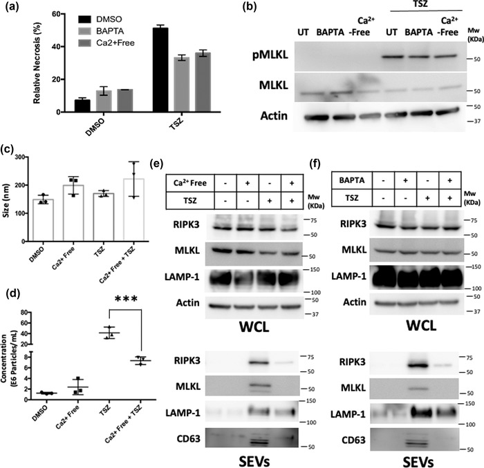
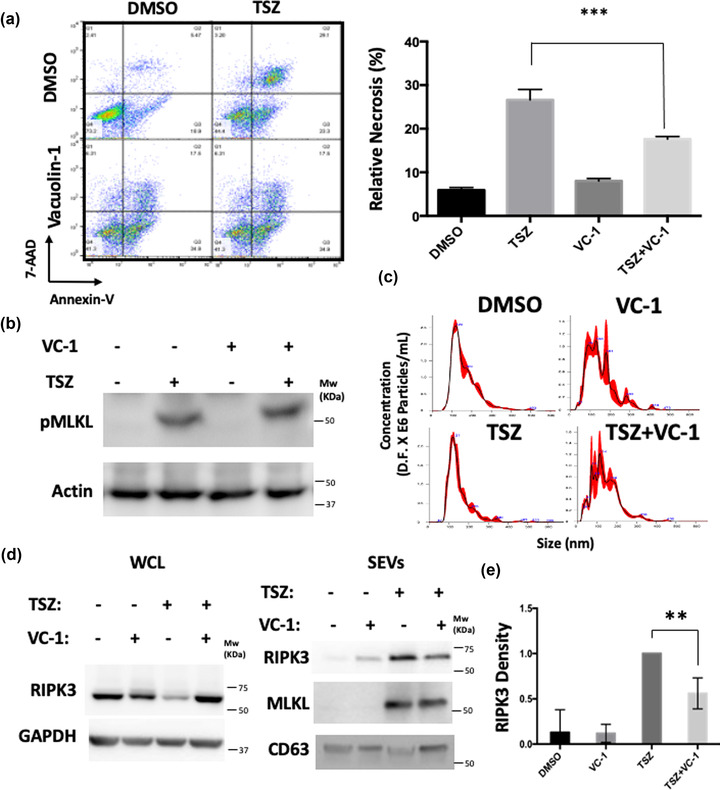
Extracellular vesicle (EV) secretion is an important mechanism used by cells to release biomolecules. A common necroptosis effector-mixed lineage kinase domain like (MLKL)-was recently found to participate in the biogenesis of small and large EVs independent of its function in necroptosis. The objective of the current study is to gain mechanistic insights into EV biogenesis during necroptosis. Assessing EV number by nanoparticle tracking analysis revealed an increased number of EVs released during necroptosis. To evaluate the nature of such vesicles, we performed a newly adapted, highly sensitive mass spectrometry-based proteomics on EVs released by healthy or necroptotic cells. Compared to EVs released by healthy cells, EVs released during necroptosis contained a markedly higher number of unique proteins. Receptor interacting protein kinase-3 (RIPK3) and MLKL were among the proteins enriched in EVs released during necroptosis. Further, mouse embryonic fibroblasts (MEFs) derived from mice deficient of Rab27a and Rab27b showed diminished basal EV release but responded to necroptosis with enhanced EV biogenesis as the wildtype MEFs. In contrast, necroptosis-associated EVs were sensitive to Ca2+ depletion or lysosomal disruption. Neither treatment affected the RIPK3-mediated MLKL phosphorylation. An unbiased screen using RIPK3 immunoprecipitation-mass spectrometry on necroptotic EVs led to the identification of Rab11b in RIPK3 immune-complexes. Our data suggests that necroptosis switches EV biogenesis from a Rab27a/b dependent mechanism to a lysosomal mediated mechanism.

Keywords: MLKL; RIPK3; SEVs; lysosomal exocytosis; necroptosis; proteomics.


Conflict of interest statement

KAB and YG are co‐inventors on a patent that covers the detergent Azo. Other authors have no conflict of interest to declare.

Figures

FIGURE 1
FIGURE 1
Necroptotic cells release extracellular vesicles. (a) Ripk3+/+ and Ripk3–/– MEFs were treated with DMSO or TSZ for 6 h. Cell death was analyzed by flow cytometry. Representative scatter plot is depicted. (b) Schematic outline of SEV isolation. Cell culture media from cultured MEFs was centrifuged to remove large cellular debris and SEVs were then isolated using ultracentrifugation or ExoQuick reagent. (c and d) Representative traces from nanoparticle tracking analysis (NTA) are presented. SEVs isolated from DMSO‐ or TSZ‐treated Ripk3+/+ or Ripk3–/– MEFs were isolated by ultracentrifugation (c) or ExoQuick (d) and analyzed by NTA. (e and f) Mean particle size obtained by NTA analyses is depicted for SEVs isolated from DMSO‐ or TSZ‐treated Ripk3+/+ or Ripk3–/– MEFs by ultracentrifugation (e) or ExoQuick (f). (g and h) Mean particle number measured by NTA analysis is depicted for SEVs isolated from DMSO‐ or TSZ‐treated Ripk3+/+ or Ripk3–/– MEFs by ultracentrifugation (g) or ExoQuick (h). All data shown are mean ± SD of three or more independent experiments. ***p < 0.001 using one‐way ANOVA (g, h)
FIGURE 2
FIGURE 2
Mass spectrometry (MS) analysis of NEEs. (a) Schematic representation of MS‐based proteomics workflow for the analysis of extracellular vesicles. Proteins were extracted from isolated vesicles using a photocleavable surfactant, Azo, reduced with tris(2‐carboxyethyl)phosphine (TCEP), alkylated with 2‐chloroacetamide (CA), digested with trypsin, and analyzed by using reversed‐phase liquid chromatography‐mass spectrometry (RPLC‐MS) after surfactant removal. (b) Venn diagram comparing protein identified in the control (DMSO) and TSZ treated samples. (c and d) Overview of GO enrichment analysis of proteins uniquely found in the TSZ treated sample sorted by molecular function (MF), biological process, cellular component, KEGG, and reactome. (e) Comparison of proteins identified in this study with the top 100 proteins available in the ExoCarta and Vesiclepedia databases. (f) Representative proteins uniquely found in TSZ and the control samples. (g) String analysis of proteins found to interact with RIPK3
FIGURE 3
FIGURE 3
RIPK3 is a luminal cargo protein in SEVs identifiable in diverse sources. (a) Ripk3+/+ and Ripk3–/– were treated with DMSO and TSZ. SEVs were isolated from cell culture. The whole‐cell lysate (WCL) (top) and SEV fractions (bottom) were analyzed by western blotting. (b) Schematic outline showing size exclusion chromatography methodology for SEV enrichment. (c) Western blot analysis of SEVs purified using size exclusion chromatography. (d) SEVs were isolated from Ripk3+/+ and Ripk3–/– mouse plasma and analyzed by western blotting for indicated proteins. Ponsceau S staining was used to ensure equal protein amounts. (e) SEVs from human plasma were isolated and analyzed by western blotting. HT29 cells and 293T cells were used as positive and negative controls, respectively. Ponsceau S staining was used to ensure equal protein amounts. (f) SEVs derived from TSZ treated Ripk3+/+ MEFs were treated with proteinase K along with indicated reagents (see methods) and analyzed by western blotting. CD63 was used as a non‐luminal (transmembrane) protein control whereas GAPDH was used as a luminal protein control. (g) Ripk3+/+ and Ripk3–/– were treated with TSZ in the presence or absence of Nec1s. SEVs were isolated from cell culture and the cellular (left) and SEV fractions (right) were analyzed by western blotting
FIGURE 4
FIGURE 4
NEE release is independent of Rab27a/b. (a) MEFs from Rab27WT and Rab27DKO were treated with DMSO and TSZ and cell death is analyzed by flow cytometry. Relative necrosis is depicted. (b–d) SEVs from DMSO or TSZ treated Rab27WT and Rab27DKO MEFs were isolated by ultracentrifugation and analyzed by NTA. Representative NTA trace is presented (b). The number of particles (c) and the mean size is plotted (d). (e) SEVs isolated in (a) were subjected to mass spectrometry analysis and the number of protein cargo is depicted in a Venn diagram. (f) Comparison of RIPK3 and Rab11a/b enrichment in SEVs from the mass spectrometry in € analysis is presented. “+” represents the detection of the indicated factor by mass spectrometry whereas “‐“ indicates its absence. (g) WCL and SEVs from Rab27WT and Rab27DKO MEFs treated with DMSO and TSZ were analyzed by western blotting. RIPK3 abundance was analyzed in SEVs by densitometry and quantified. All data shown are mean ± SD of three or more independent experiments. *p < 0.05, **p < 0.01 ***p < 0.001 using one‐way ANOVA (c, g)
FIGURE 5
FIGURE 5
RIPK3 is released in an atypical manner during necroptosis. (a) Immunoprecipitation experiment was performed using RIPK3 or IgG on DMSO treated (DMSO WCL) and TSZ treated (WCL TSZ) MEFs, or SEVs derived from TSZ treated MEFs (EVs TSZ) and analyzed by mass spectrometry. The fold enrichment of RIPK3 and Rab11b from the co‐immunoprecipitated is depicted from 3 independent replicates (n = 3). (b) STRING analysis was performed for RIPK3 interacting proteins identified in RIPK3 IP coupled mass spectrometry leading to the identification of known or predicted interaction partners. Note that many interacting partners are calcium‐binding proteins. (c) Analysis of known lysosomal proteins enriched in SEVs derived from TSZ or DMSO treated MEFs are represented as a Venn diagram
FIGURE 6
FIGURE 6
NEEs are released in a calcium‐dependent manner. (a) MEFs were cultured in calcium‐containing or calcium‐free media or pre‐treated with the calcium chelator BAPTA. Necroptosis was induced using TSZ. The percentage of necroptotic cells is plotted. (b) Early activation of necroptosis is measured by using pMLKL. Briefly, MEFs cultured in indicated conditions were treated with DMSO or TSZ for 1 h and harvested for western blot analysis. (c, d) NTA analysis was performed on SEVs isolated from MEFs cultured in calcium‐containing or calcium‐free media and subsequent treatment with DMSO or TSZ for mean size (c) and concentration (d). (e) Using a cell culture strategy similar to (a), WCL (up) and SEVs (below) were analyzed for RIPK3 abundance by western blotting. LAMP‐1 was used as a marker for lysosomal exocytosis. (Machado et al., ; Mathieu et al., ; Park et al., 2018) All data shown are mean ± SD of three or more independent experiments. ***p < 0.001 using one‐way ANOVA (d)
FIGURE 7
FIGURE 7
Necroptosis leads to the lysosomal‐mediated secretion of RIPK3 carrying SEVs. (a) MEFs were pretreated with DMSO or Vacuolin‐1 and subsequently with DMSO or TSZ and analyzed by flow cytometry. The resulting scatter plot is depicted (left) and percent necrosis is plotted (right). (b) Early activation of necroptosis was assessed by the abundance of phosphorylated MLKL (pMLKL). Briefly, MEFs pre‐treated with VC‐1 or DMSO and subsequently induced with DMSO or TSZ for 1 h were harvested for western blot analysis. (c) MEFs were pre‐treated with VC‐1 or DMSO and subsequently induced with DMSO or TSZ. SEVs were isolated using ultracentrifugation and analyzed by NTA and a representative trace is depicted. (d) MEFs were treated similarly to (c). WCL (left) and SEVs (right) were analyzed by western blotting. (e) RIPK3 levels in SEVs were quantified by densitometry and plotted. All data shown are mean ± SD of three or more independent experiments. *p < 0.01, ***p < 0.001 using one‐way ANOVA (a, e)
FIGURE 8
FIGURE 8
Proposed model for NEE generation. Left: in a healthy cell, SEV generation occurs through the endosomal machinery that involves early and late endosome formation and fusion with the MVBs involving ESCRT proteins and MLKL. Many of the endosomal maturation steps are regulated by the Rab‐family of proteins but the terminal steps of fusion to the plasma membrane, mediated by Rab27 (Rab27a and Rab27b) is denoted. Right: Upon engagement of TNF‐α with TNFR, activation of RIPK1, RIPK3 and MLKL via phosphorylation activates a Rab27‐independent program of lysosomal exocytosis where Rab11 interacts with RIPK3 and triggers the secretion of NEEs as a result of calcium influx secondary to MLKL‐mediated plasma membrane damage

References

    1. Aballo, T. J. , Roberts, D. S. , Melby, J. A. , Buck, K. M. , Brown, K. A. , & Ge, Y. (2021). Ultrafast and reproducible proteomics from small amounts of heart tissue enabled by Azo and timsTOF pro. Journal of Proteome Research, 20, 4203–4211. 10.1021/acs.jproteome.1c00446 - DOI - PMC - PubMed
    1. Alexander, M. , Ramstead, A. G. , Bauer, K. M. , Lee, S. ‐ H. , Runtsch, M. C. , Wallace, J. , Huffaker, T. B. , Larsen, D. K. , Tolmachova, T. , Seabra, M. C. , Round, J. L. , Ward, D. M. , & O'Connell, R. M. (2017). Rab27‐dependent exosome production inhibits chronic inflammation and enables acute responses to inflammatory stimuli. Journal of Immunology, 199, 3559–3570. 10.4049/jimmunol.1700904 - DOI - PMC - PubMed
    1. Brown, K. A. , Chen, B. , Guardado‐Alvarez, T. M. , Lin, Z. , Hwang, L. , Ayaz‐Guner, S. , Jin, S. , & Ge, Y. (2019). A photocleavable surfactant for top‐down proteomics. Nature Methods, 16(5), 417–420. 10.1038/s41592-019-0391-1 - DOI - PMC - PubMed
    1. Brown, K. A. , Tucholski, T. , Eken, C. , Knott, S. , Zhu, Y. , Jin, S. , & Ge, Y. (2020a). High‐throughput proteomics enabled by a photocleavable surfactant. Angewandte Chemie, 132, 8484–8488. 10.1002/ange.201915374 - DOI - PMC - PubMed
    1. Brown, K. A. , Tucholski, T. , Eken, C. , Knott, S. , Zhu, Y. , Jin, S. , & Ge, Y. (2020b). High‐throughput proteomics enabled by a photocleavable surfactant. Angewandte Chemie International Edition, 59, 8406–8410. 10.1002/anie.201915374 - DOI - PMC - PubMed

Publication types

Substances

LinkOut - more resources