Review
. 2022 Nov;11(22):e2201626.
doi: 10.1002/adhm.202201626. Epub 2022 Sep 13.

Biofabrication of Human Skin with Its Appendages

Affiliations
Review

Biofabrication of Human Skin with Its Appendages

Motaharesadat Hosseini et al. Adv Healthc Mater. 2022 Nov.

Abstract

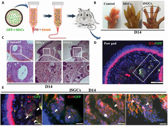
Much effort has been made to generate human skin organ in the laboratory. Yet, the current models are limited due to the lack of many critical biological and structural features of the skin. Importantly, these in vitro models lack appendages and fail to recapitulate the whole human skin construction. Thus, engineering a human skin with the capacity to generate all components, including appendages, is a major challenge. This review intends to provide an update on the recent efforts underway to regenerate appendage-bearing skin organs based on scaffold-free and scaffold-based bioengineering approaches. Although the mouse skin equivalents containing hair follicles, sebaceous glands, and sweat glands have been established in vitro, there has been limited success in humans. A combination of biofabricated matrices and cell aggregates, such as organoids, can pave the way for generating skin substitutes with human-like biological, structural, and physical features. Accordingly, the formation of human skin organoids and reconstruction of vascularized skin equipped with immune cells prompt calls for more scientific research. The generation of appendage-bearing skin substitutes can be applied in practice for wound healing, hair restoration, and scar treatment.

Keywords: 3D culture; bioengineering; regenerative medicine; tissue engineering; wounds.


Conflict of interest statement

The authors declare no conflict of interest.

Figures

Figure 1
Figure 1
Different stages of skin appendage development. Stage 0 starts with the formation of a single layer of basal keratinocytes, which are multipotent cells. In stage 1, areas of thickening epithelium appear— known as hair placode that will end up hair follicles (HFs). The main trigger behind the formation of placodes is frequent interactions between mesenchymal cells and the epithelium. Stage 2 is associated with the extension of the hair placode downward toward the dermis. At the same time, dermal fibroblasts (Fbs) attract and create an aggregation below the placode condense as a result of an epithelial signal from the placode. During stage 3, a dermal message from the dermal aggregation induces the proliferation of the placode cells, which, in turn, surround the dermal aggregation that ultimately develops into the dermal papilla (DP). In stage 4, subsequent growth and differentiation of the epithelial cells lead to the development of the mature hair shaft. Also, the DP becomes more compact and is fully engulfed by the growing HF cells. While the sebaceous gland is evident alongside the HF, the eccrine gland begins as a basal layer bud. In stage 5, the hair shaft tip invades the hair canal. Furthermore, the swear gland lengthens and arranges in a coil.
Figure 2
Figure 2
Schematic representation of A) the human skin layers with their appendages as well as B) the hair follicle structure (Created with Biorender.com)
Figure 3
Figure 3
The timeline of the main basic, translational, and clinical advances for hair follicle (HF) regeneration. Abbreviations: dermal papilla (DP); the United States food and drug administration (FDA); induced pluripotent stem cells (iPSC); pluripotent stem cells (PSCs).
Figure 4
Figure 4
The timing of developmental events during in vitro skin organogenesis using human pluripotent stem cell (hPSCs) derived skin organoid. The human skin organoids are obtained in three main stages. The surface ectoderm and cranial neural crest cells are coinduced in the first stage using hPSCs. As a starting point, these cells form aggregates and then undergo ectodermal induction with critical differentiation factors, including bone morphogenetic protein (BMP) 4, FGF, and a transforming growth factor‐β inhibitor. On day 3, cells erupt outward, leaving the surface ectoderm and undifferentiated hPSC core. When the aggregates are mature (8–12 d), the intermediate layer erupts out, leading to the development of cranial skin. At the end of this stage, skin organoids contain cranial neural crest cells as well as epidermis and dermis precursors. Cystic skin organoids consisting of mesenchymal, neural, and glial progenitor cells surrounded by a sphere of keratinocytes are formed in the second stage. Also, the organoids contain self‐assembled epidermal and dermal layers along with bulb‐like hair follicles. In the third stage, the skin organoids produce a keratinized epidermis with a population of melanocytes, Schwann cells, and sensory neurons. Additionally, hair follicles with sebaceous glands grow outward. Created with Biorender.com.
Figure 5
Figure 5
The capacity of induced sweat gland cells (iSGCs) for directed functional regeneration of sweat glands (SGs). A) A schematic representation of the method, where the 3D bioprinted scaffold containing green fluorescent protein (GFP)‐labeled mesenchymal stem cells (MSCs) was transplanted in burned paws of mice. B) Iodine/starch‐based sweat test of mice treated with different cells; the presence of black dots on footpads was indicative of sweating in iSGCs‐treated mice. C) Hematoxylin and eosin staining of a plantar region with and without the cell treatment; histology results revealed SG regeneration in iSGCs‐treated mice (scale bars, 200 µm). D) Participation of GFP‐labeled iSGCs in directed functional regeneration of SG (K14, red; GFP, green; DAPI, blue; scale bar, 200 µm). E) Detection of K14, K19, K8, and K18 as SG‐specific markers in the regenerated SG tissue (arrows); (K14, K19, K8, and K18, red; GFP, green; scale bars, 50 µm). Reproduced with permission.[ 105 ] Copyright 2021, American Association for the Advancement of Science.

References

    1. a) Chen Y. E., Fischbach M. A., Belkaid Y., Nature 2018, 553, 427; - PMC - PubMed
    2. b) Clevers H., Loh K. M., Nusse R., Science 2014, 346, 1248012. - PubMed
    1. a) Hongbo Z., Maibach H., Dermatotoxicology, CRC Press LCC, Boca Raton, FL: 2004;
    2. b) Nielsen J. B., Benfeldt E., Holmgaard R., Curr. Probl. Dermatol. 2016, 49, 103; - PubMed
    3. c) Tansey E. A., Johnson C. D., Adv. Physiol. Educ. 2015, 39, 139. - PubMed
    1. Hosseini M., Shafiee A., Small 2021, 17, 2101384. - PubMed
    1. a) Gravitz L., Nature 2018, 563, S83; - PubMed
    2. b) Takeo M., Lee W., Ito M., Cold Spring Harbor Perspect. Med. 2015, 5, a023267. - PMC - PubMed
    1. Arda O., Goksugur N., Tuzun Y., Clin. Dermatol. 2014, 32, 3. - PubMed

LinkOut - more resources