. 2022 Oct:56:102449.
doi: 10.1016/j.redox.2022.102449. Epub 2022 Aug 28.

Nrf2 transcriptional upregulation of IDH2 to tune mitochondrial dynamics and rescue angiogenic function of diabetic EPCs

Affiliations

Nrf2 transcriptional upregulation of IDH2 to tune mitochondrial dynamics and rescue angiogenic function of diabetic EPCs

Xiaozhen Dai et al. Redox Biol. 2022 Oct.

Abstract

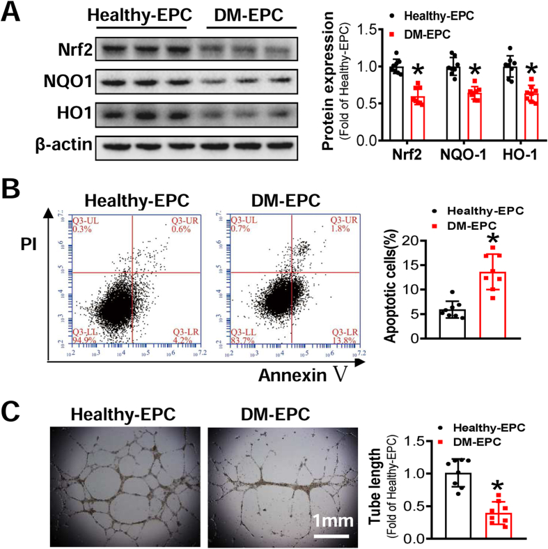
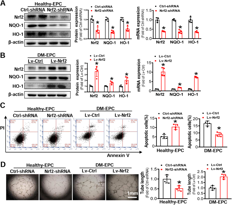
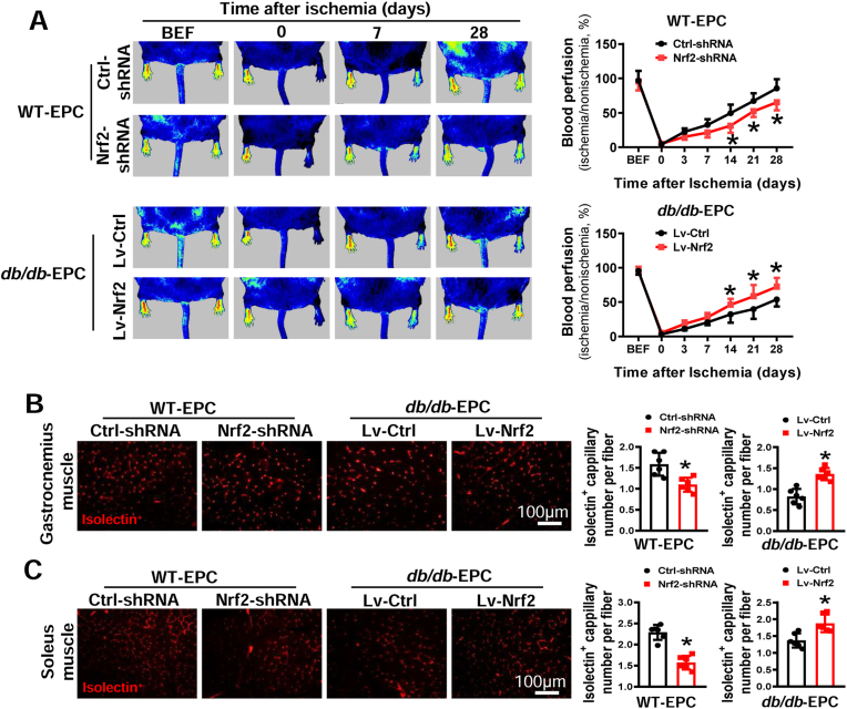
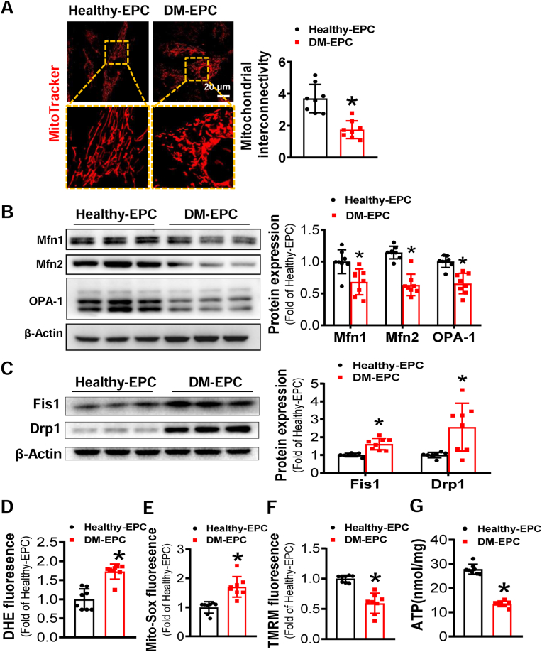
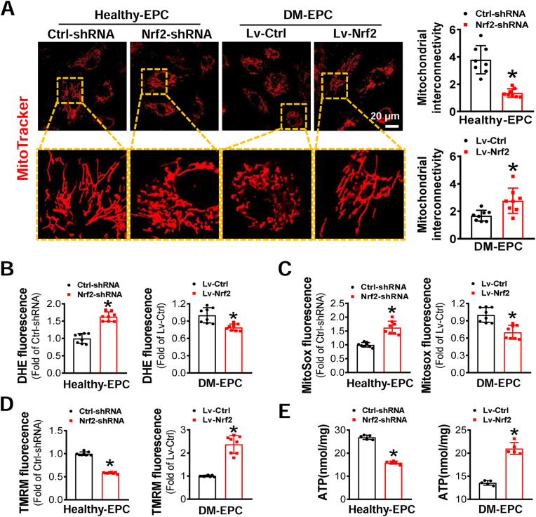
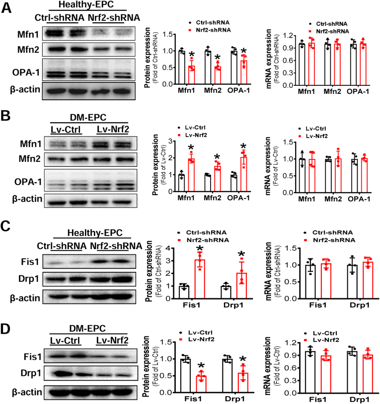
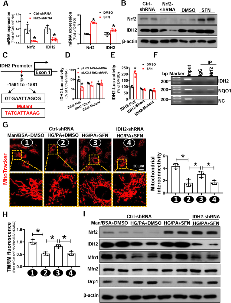
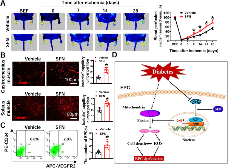
Endothelial progenitor cells (EPCs) are reduced in number and impaired in function in diabetic patients. Whether and how Nrf2 regulates the function of diabetic EPCs remains unclear. In this study, we found that the expression of Nrf2 and its downstream genes were decreased in EPCs from both diabetic patients and db/db mice. Survival ability and angiogenic function of EPCs from diabetic patients and db/db mice also were impaired. Gain- and loss-of-function studies, respectively, showed that knockdown of Nrf2 increased apoptosis and impaired tube formation in EPCs from healthy donors and wild-type mice, while Nrf2 overexpression decreased apoptosis and rescued tube formation in EPCs from diabetic patients and db/db mice. Additionally, proangiogenic function of Nrf2-manipulated mouse EPCs was validated in db/db mice with hind limb ischemia. Mechanistic studies demonstrated that diabetes induced mitochondrial fragmentation and dysfunction of EPCs by dysregulating the abundance of proteins controlling mitochondrial dynamics; upregulating Nrf2 expression attenuated diabetes-induced mitochondrial fragmentation and dysfunction and rectified the abundance of proteins controlling mitochondrial dynamics. Further RNA-sequencing analysis demonstrated that Nrf2 specifically upregulated the transcription of isocitrate dehydrogenase 2 (IDH2), a key enzyme regulating tricarboxylic acid cycle and mitochondrial function. Overexpression of IDH2 rectified Nrf2 knockdown- or diabetes-induced mitochondrial fragmentation and EPC dysfunction. In a therapeutic approach, supplementation of an Nrf2 activator sulforaphane enhanced angiogenesis and blood perfusion recovery in db/db mice with hind limb ischemia. Collectively, these findings indicate that Nrf2 is a potential therapeutic target for improving diabetic EPC function. Thus, elevating Nrf2 expression enhances EPC resistance to diabetes-induced oxidative damage and improves therapeutic efficacy of EPCs in treating diabetic limb ischemia likely via transcriptional upregulating IDH2 expression and improving mitochondrial function of diabetic EPCs.

Keywords: Angiogenesis; Diabetes; Endothelial progenitor cell; Hind limb ischemia; Nuclear factor erythroid 2-related factor 2.


Conflict of interest statement

Declaration of competing interest No potential conflicts of interest relevant to this article were reported.

Figures

Fig. 1
Fig. 1
Diabetes attenuates Nrf2 and impairs the survival and angiogenic function of EPCs. EPCs were isolated from healthy donors (Healthy-EPC) or diabetic patients (DM-EPC). (A) The Expression of Nrf2 and its downstream genes were detected by Western blot, and the quantitative data was normalized by the average of Healthy-EPCs for each protein; β-actin was used as a loading control. (B) The survival abilities of EPCs were determined by Annexin V/PI staining, and the quantitative data was expressed as the percentage of AnnexinV+/PI cells. (C) The angiogenic function of EPCs was evaluated by a Matrigel tube formation assay, and the quantitative data was normalized by the average tube length of Healthy-EPCs. n = 8 per group. Data shown in graphs represents the means ± SD. *p < 0.05, vs Healthy-EPC.
Fig. 2
Fig. 2
Downregulation of Nrf2 impairs the survival and anigogenic function of Healthy-EPCs, while upregulation of Nrf2 rescues the survival and angiogenic function of DM-EPCs. Healthy-EPCs were transfected with lentivirus carrying nonsense shRNA (Ctrl-shRNA) or Nrf2 shRNA (Nrf2-shRNA) and DM-EPCs were transfected with lentivirus carrying Nrf2 (Lv-Nrf2) or control vector (Lv-Ctrl). (A-B) The efficiency of Nrf2 knockdown or upregulation was determined by Western blot and qRT-PCR, and the quantitative data was normalized by the average of the respective control group for each protein; β-actin was used as a loading control. (C) The survival abilities were determined by Annexin V/PI staining, and the quantitative data was expressed as the percentage of AnnexinV+/PI cells. (D) Angiogenic function of EPCs was evaluated by a Matrigel tube formation assay, and the quantitative data was normalized by the average tube length of the respective control group. n = 4 per group. Data shown in graphs represents the means ± SD. *p < 0.05, vs Ctrl-shRNA or Lv-Ctrl.
Fig. 3
Fig. 3
Nrf2 regulates the angiogenic function of mouse EPCs. WT-EPCs were transfected with lentivirus carrying Ctrl-shRNA or Nrf2-shRNA and db/db-EPCs were transfected with lentivirus carrying Lv-Ctrl or Lv-Nrf2. Within 1 h after hind limb ischemia (HLI) surgery, db/db mice (FVB background) were transplanted with different types of EPCs that have a same genetic background. (A) The time-course of blood perfusion before (BEF) and after HLI surgery was monitored by a Pericam Perfusion Speckle Imager, and the blood perfusion was quantified by Image J and expressed as the percentage of the perfusion relative to the collateral nonischemic limb. Transverse sections of ischemic gastrocnemius (B) and soleus muscle (C) were stained with Alexa Fluor® 594 conjugated isolectin to enumerate isolectin-stained cell number as a proxy of capillary density. Capillary density was expressed as isolectin+ capillaries per muscle fiber. n = 6 mice per group. Data shown in graphs represents the means ± SD. *p < 0.05, vs Ctrl-shRNA or Lv-Ctrl.
Fig. 4
Fig. 4
Diabetes induces mitochondrial fragmentation of EPCs and impairs mitochondrial function. (A) Micrographs of mitochondrial morphology of EPCs were visualized by MitoTracker Red CMXRos probe staining, and the morphological alterations were quantified by Image J and expressed as mitochondrial interconnectivity. (B–C) The expression of mitochondrial fusion and fission related proteins was determined by Western blot, the quantitative data was normalized by the average of Healthy-EPC for each protein, and β-actin was used as a loading control. The levels of (D) intracellular ROS, (E) mitochondrial ROS, and (F) mitochondrial membrane potential were detected by DHE, MitoSOX™, and TMRM staining, respectively, and quantified by a flow cytometry and normalized by the average fluorescence density of Healthy-EPCs. (G) ATP concentration in EPCs was measured by an ATP assay Kit. n = 8 per group. Data shown in graphs represents the means ± SD. *p < 0.05, vs Healthy-EPC. (For interpretation of the references to colour in this figure legend, the reader is referred to the Web version of this article.)
Fig. 5
Fig. 5
The effects of Nrf2 expression on mitochondrial dynamics and function of EPCs. Healthy-EPCs were transfected with lentivirus carrying Ctrl-shRNA or Nrf2-shRNA and DM-EPCs were transfected with lentivirus carrying Lv-Ctrl or Lv-Nrf2. (A) Micrographs of mitochondrial morphology were visualized by MitoTracker Red CMXRos probe staining, and the morphological alterations were quantified by Image J and expressed as mitochondrial interconnectivity. The levels of (B) intracellular ROS, (C) mitochondrial ROS, and (D) mitochondrial membrane potential of EPCs were detected by DHE, MitoSOX™, and TMRM staining, respectively, and quantified by a flow cytometry and normalized by the average fluorescence density of the respective control group. (E) ATP concentration in EPCs was measured by an ATP assay Kit. n = 5–8 per group. Data shown in graphs represents the means ± SD. *p < 0.05, vs Ctrl-shRNA or Lv-Ctrl. (For interpretation of the references to colour in this figure legend, the reader is referred to the Web version of this article.)
Fig. 6
Fig. 6
Nrf2 expression affected the protein expression of mitochondrial fusion and fission related proteins but not mRNA expression. Healthy-EPCs were transfected with lentivirus carrying Ctrl-shRNA or Nrf2-shRNA and DM-EPCs were transfected with lentivirus carrying Lv-Ctrl or Lv-Nrf2. The expression of mitochondrial fusion (A-B) and fission (C-D) related molecules at protein and mRNA levels were determined by Western blot and q-RT-PCR, respectively; the quantitative data was normalized by the average of the respective control for each molecule, and β-actin was used as a loading control. n = 4 per group. Data shown in graphs represents the means ± SD. *p < 0.05, vs Ctrl-shRNA or Lv-Ctrl.
Fig. 7
Fig. 7
Nrf2 suppression of diabetes-induced mitochondrial fission of EPCs is dependent on transcriptional upregulation of IDH2. (A-B) Healthy-EPCs were transfected with lentivirus carrying Ctrl-shRNA or Nrf2-shRNA, or treated with DMSO or Nrf2 activator SFN. The (A) mRNA and (B) protein expression of Nrf2 and IDH2 were determined by Western blot and qRT-PCR, respectively; the quantitative data was normalized by the average of the respective control for each group, and β-actin was used as a loading control. (C) The putative binding sites of Nrf2 on IDH2 promoter was predicted by JASPAR. (D) HEK-293T cells were co-transfected with PLG3.0-IDH2-Luc reporter (Full, Short or mutant) and Nrf2-shRNA or Ctrl-shRNA plasmid. Renilla luciferase reporter plasmid (pRL-TK) was used as an internal control of transfection. Firefly luciferase activity and Renilla luciferase activity in media were measured at 48 h post-transfection. (E) HEK-293T cells were transfected PLG3.0-IDH2-Luc reporter (Full, Short or mutant), then treated with or without SFN, a Nrf2 activator. pRL-TK was used as an internal control of transfection. Firefly luciferase activity and Renilla luciferase activity in media were measured at 48h post-transfection. (F) Chromatin immunoprecipitation (ChIP) analyses using an anti-Nrf2 or a normal rabbit IgG were performed in EPC cells. Primers specific for IDH2, NQO1, or NC (Negative control) were used. (GI) Healthy-EPCs were transfected with or without lentivirus carrying Ctrl-shRNA or IDH2-shRNA, respectively, followed by exposure to high glucose (HG, 30 mmol/L) plus palmitate (PA, 100 μmol/L) or Ctrl (mannitol plus bovine serum albumin, Man/BSA) in the presence of SFN or DMSO for 24 h. (G) Micrographs of mitochondrial morphology were visualized by MitoTracker Red CMXRos probe staining, and the morphological alterations were quantified by Image J and expressed as mitochondrial interconnectivity. (H) Mitochondrial membrane potential was detected by TMRM staining, and the data was quantified by a flow cytometry and normalized by the average fluorescence density of the Ctrl-shRNA/Man/BSA+DMSO group. (I) The protein expression of mitochondrial fission and fusion related proteins was determined by Western blot, and β-actin was used as a loading control. At least three independent experiments for each study. Data shown in graphs represents the means ± SD. *p < 0.05. (For interpretation of the references to colour in this figure legend, the reader is referred to the Web version of this article.)
Fig. 8
Fig. 8
SFN enhances blood perfusion and angiogenesis in db/db mice with HLI.db/db mice (FVB background) were pretreated with SFN for 1 week, followed by HLI surgery and continual SFN treatment for additional 4 weeks. (A) The time-course of blood perfusion before (BEF) and after HLI surgery was monitored by a Pericam Perfusion Speckle Imager, and the data was quantified by Image J and expressed as the percentage of the perfusion relative to the collateral nonischemic limb. (B) Transverse sections of ischemic gastrocnemius and soleus muscle were stained with Alexa Fluor® 594 conjugated isolectin to enumerate isolectin-stained cell number as a proxy of capillary density. Capillary density was expressed as isolectin+ capillaries per muscle fiber. (C) At day 7 after HLI surgery, peripheral blood was collected to evaluate the percentage of EPCs (CD34+/VEGFR2+) in circulation by a flow cytometry. n = 6 mice per group. Data shown in graphs represent the means ± SD. *P < 0.05 vs Vehicle group. (D) Schematic illustration of the protective effects of Nrf2 on EPCs under diabetic conditions. Diabetes decreases expression of Nrf2 in EPCs and induces mitochondrial fission and dysfunction, resulting in reactive oxygen species (ROS) overproduction and cell death, ultimately leading to EPC dysfunction. Upregulation of Nrf2 by transgene or supplementation of SFN can rescue diabetes-induced inhibition of Nrf2 transcriptional activity, resulting in upregulation of IDH2 transcription, which inhibits mitochondrial fission, rectifies mitochondrial function, and decreases ROS production and cell death, ultimately rescues EPC dysfunction. ARE, antioxidant responsive element.

References

    1. Jarajapu Y.P., Grant M.B. The promise of cell-based therapies for diabetic complications: challenges and solutions. Circ. Res. 2010;106:854–869. - PMC - PubMed
    1. Fadini G.P., Losordo D., Dimmeler S. Critical reevaluation of endothelial progenitor cell phenotypes for therapeutic and diagnostic use. Circ. Res. 2012;110:624–637. - PMC - PubMed
    1. Dai X., Yan X., Zeng J., Chen J., Wang Y., Chen J., Li Y., Barati M.T., Wintergerst K.A., Pan K., Nystoriak M.A., Conklin D.J., Rokosh G., Epstein P.N., Li X., Tan Y. Elevating cxcr7 improves angiogenic function of epcs via akt/gsk-3beta/fyn-mediated nrf2 activation in diabetic limb ischemia. Circ. Res. 2017;120:e7–e23. - PMC - PubMed
    1. Yan X., Dai X., He L., Ling X., Shao M., Zhang C., Wang Y., Xiao J., Cai L., Li X., Tan Y. A novel cxcr4 antagonist enhances angiogenesis via modifying the ischaemic tissue environment. J. Cell Mol. Med. 2017;21:2298–2307. - PMC - PubMed
    1. Butler J.M., Nolan D.J., Vertes E.L., Varnum-Finney B., Kobayashi H., Hooper A.T., Seandel M., Shido K., White I.A., Kobayashi M., Witte L., May C., Shawber C., Kimura Y., Kitajewski J., Rosenwaks Z., Bernstein I.D., Rafii S. Endothelial cells are essential for the self-renewal and repopulation of notch-dependent hematopoietic stem cells. Cell Stem Cell. 2010;6:251–264. - PMC - PubMed