. 2023 Jan;78(1):28-44.
doi: 10.1016/j.jhep.2022.08.029. Epub 2022 Sep 5.

Neutrophil extracellular traps contribute to liver damage and increase defective low-density neutrophils in alcohol-associated hepatitis

Affiliations

Neutrophil extracellular traps contribute to liver damage and increase defective low-density neutrophils in alcohol-associated hepatitis

Yeonhee Cho et al. J Hepatol. 2023 Jan.

Abstract

Background & aims: In alcohol-associated hepatitis (AH), inflammation and neutrophil counts correlate with poor clinical outcomes. Here, we investigated how neutrophils contribute to liver damage in AH.

Methods: We isolated blood neutrophils from individuals with AH to examine neutrophil extracellular traps (NETs) and performed RNA sequencing to explore their unique characteristics.

Results: We observed a significant increase in NET production in AH. We also observed a unique low-density neutrophil (LDN) population in individuals with AH and alcohol-fed mice that was not present in healthy controls. Transcriptome analysis of peripheral LDNs and high-density neutrophils (HDNs) from individuals with AH revealed that LDNs exhibit a functionally exhausted phenotype, while HDNs are activated. Indeed, AH HDNs exhibited increased resting reactive oxygen species (ROS) production and produced more ROS upon lipopolysaccharide stimulation than control HDNs, whereas AH LDNs failed to respond to lipopolysaccharide. We show that LDNs are generated from HDNs after alcohol-induced NET release in vitro, and this LDN subset has decreased functionality, including reduced phagocytic capacity. Moreover, LDNs showed reduced homing capacity and clearance by macrophage efferocytosis; therefore, dysfunctional neutrophils could remain in the circulation and liver. Depletion of both HDNs and LDNs in vivo prevented alcohol-induced NET production and liver damage in mice. Granulocyte-colony stimulating factor treatment also ameliorated alcohol-induced liver injury in mice.

Conclusion: Neutrophils contribute to liver damage through increased NET formation which increases defective LDNs in AH. Alcohol induces phenotypic changes in neutrophils; HDNs are activated whereas LDNs are defective. Our findings provide mechanistic insights that could guide the development of therapeutic interventions for AH.

Impact and implications: In this study we discovered heterogeneity of neutrophils in alcohol-associated hepatitis, including high-density and low-density neutrophils that show hyper-activated or exhausted transcriptomic profiles, respectively. We found that alcohol induces neutrophil extracellular trap (NET) formation, which contributes to liver damage. NET release by high-density neutrophils resulted in low-density neutrophils that reside in the liver and escape clean-up by macrophages. Our findings help to understand the opposing neutrophil phenotypes observed in individuals with alcohol-associated hepatitis and provide mechanistic insights that could guide therapeutic strategies targeting neutrophils.

Keywords: Low-density neutrophils (LDNs); Neutrophil extracellular traps (NETs); alcoholic hepatitis.


Conflict of interest statement

Conflict of interest

G.S. reports being a paid consult for Durect Corporation, Cyta Therapeutics, Evive, Glympse Bio, Terra Firma, Pandion Therapeutics, Surrozen, Ventyx, Merck, Novartis, Pfizer and Zomagen. I.S.V reports consulting for Mosaic Research Management.

Please refer to the accompanying ICMJE disclosure forms for further details.

Figures

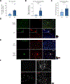
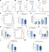
Fig. 1.
Fig. 1.. Neutrophils increase, and contribute to liver damage in AH.
(A, B) Circulating neutrophils (live, CD66+CD11b+) through flow cytometry analysis of whole blood from HC (n = 5) and individuals with AH (n = 6). (C) Liver-infiltrating neutrophils in ALD/individuals with AH (n = 6) and HC (n = 5) by IHC staining with NE. (D) Liver-infiltrating neutrophils through flow cytometry. (E) serum ALT of 4-week chronic alcohol-fed mice and (F) AST in serum of mice receiving 10 days of alcohol diet plus a single binge following neutrophil depletion. Data represent mean with SEM. Statistical significance was determined using t test or one-way ANOVA; *p <0.05, **p <0.01, ***p <0.001. AH, alcohol-associated hepatitis; ALT, alanine aminotransferase; AST, aspartate aminotransferase; HC, healthy controls; IHC, immunohistochemical; PF, pair-fed.
Fig. 2.
Fig. 2.. Neutrophils induce liver damage via increased NET formation in AH.
(A) Citrullinated histone H3 in serum from individuals with AH (n = 8) and HC (n= 6), (B) cell-free circulating DNA extracted from plasma of individuals with AH (n = 18) and HC (n = 7), (C) activity of NE in serum of individuals with AH (n = 8) and HC (n = 6). (D, E) NET production in the liver of (D) ALD/individuals with AH and HC, (E) alcohol-fed mice (4 weeks) and PF mice through co-IF staining. (F, G) NET formation in neutrophils of individuals with AH (n = 6) and HC (n = 6) through (F) microscopy and (G) NET-associated NE activity (NETosis assay). (H) Alcohol-induced NET formation by NETosis assay in human neutrophils. (I, J) NET production through co-IF staining and citrullinated histone H3 in alcohol-fed mice liver following LPS (0.5 mg/kg). (K, L) Monocyte and (M, N) macrophage activation/differentiation following co-culture with NETosis neutrophils through flow cytometry. MFI fold-change of (L) CD86 and (M) CD206. Scale bar, 45 μm. Data represent mean with SEM. Statistical significance was determined using t test or one-way ANOVA; *p <0.05, **p <0.01, ***p <0.001, ****p <0.0001. AH, alcohol-associated hepatitis; HC, healthy controls; LPS, lipopolysaccharides; NET, neutrophil extracellular trap; PF, pair-fed; PMA, phorbol 12-myristate 13-acetate.
Fig. 3.
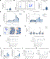
Fig. 3.. An LDN population is significantly increased in individuals with AH and mice.
(A) Neutrophils in LMNCs from PF (n = 10) or alcohol-fed (n = 10) mice. (B,C) Neutrophils in PBMCs of HC (n = 5) and individuals with AH (n = 7). (D) LDN and HDN kinetics in PF (n = 5–6) and EtOH-fed (n = 6) mouse livers. (E) ALT in mouse serum. (F) Giemsa stain of human neutrophils. (G-I) MFI fold-change of surface antigen expression levels between LDNs and HDNs from (G) individuals with AH (n = 9) and HC (n = 6), and (H, I) from PF (n = 5–6) and EtOH-fed (n = 6) mice compared to autologous bone marrow neutrophils. (J, K) Liver-infiltrating HDNs and LDNs following LPS (5 mg/kg) (n = 6) or saline (n = 3–5) injection. (L, M) MFI of Ly6G and CD11b expression on LDNs and HDNs compared with autologous bone marrow neutrophils. Using flow cytometry, human LDNs and HDNs were gated as live, CD11b+CD66b+ cells in PBMC or RBC fractions, respectively, of HC and individuals with AH. Liver-infiltrating LDNs and HDNs were gated as live, CD11b+Ly6G+ cells in LMNC or RBC fractions, respectively, of alcohol-fed or PF mice. Data represent mean with SEM. Statistical significance was determined using t test, one-way or two-way ANOVA; *p <0.05, **p <0.01, ***p <0.001, ****p <0.0001. AH, alcohol-associated hepatitis; ALT, alanine aminotransferase; HC, healthy controls; HDN, high-density neutrophils; LDN, low-density neutrophils; PMBCs, peripheral blood mononuclear cells.
Fig. 4.
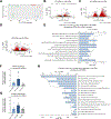
Fig. 4.. Whole transcriptome analysis revealed that HDNs and LDNs exhibit opposite phenotypes in AH.
(A) PCA of the 500 most variable gene from HDNs and LDNs from individuals with AH (n = 4) compared to HC HDNs (n = 3). (B-D) Volcano plot depicting DE analysis results between (B) HC HDNs vs. AH HDNs, (C) AH HDNs vs. AH LDN, (D) HC HDNs vs. AH LDNs. Blue and red mark genes with q value <0.05 and q value <0.05 & |log2(fold-change)| >1, respectively. Pathways enriched in (E) significantly upregulated genes in AH HDNs compared to HC HDNs and (H) significantly downregulated genes in AH LDNs compared to AH HDNs. (F, G) Evaluation of (F) PAD4 and (G) p22phox mRNA expression by RT-qPCR. (I) Gene expression heatmap of genes involved in cellular respiration (GO:0045333) and inflammatory response (MSigDB annotation of GO:0006954). Data represent mean with SEM. Statistical significance was determined by one-way ANOVA. AH, alcohol-associated hepatitis; GO, gene ontology; HC, healthy control; HDN, high-density neutrophils; LDN, low-density neutrophils; PCA, principal component analysis.
Fig. 5.
Fig. 5.. AH LDNs are generated from HDNs after alcohol-induced vital NET formation.
(A-C) Neutrophil viability after NET formation in human neutrophils through (A, B) flow cytometry and (C) LDH assay. (D) Confocal microscopy live imaging of NET release in human neutrophils with 50 mM ethanol. (E-H) Quantification of nuclear and mitochondrial DNA (E,F) in the supernatant of human neutrophils after alcohol exposure (50 mM, 4 h) and (G, H) plasma of individuals with AH. (I, J) Neutrophils in PBMC compartment and (K, L) neutrophil granularity (K) in human neutrophils after alcohol treatment and in (L) AH LDNs compared to AH HDNs and HC HDNs. (M, N) Quantification of remaining DNA in human neutrophils stained with PI. Data represent mean with SEM. Statistical significance was determined using t test or one-way ANOVA; *p <0.05, **p <0.01, ***p <0.001, ****p <0.0001. HDN, high-density neutrophils; LDN, low-density neutrophils; MFI, mean fluorescence intensity; NET, neotrophil extracellular trap; PBMC, peripheral blood mononuclear cell; PMA, phorbol 12-myristate 13-acetate; SSC, side scatter.
Fig. 6.
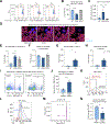
Fig. 6.. AH LDNs show functional defects and reduced homing capacity/clearance.
(A) Baseline ROS and LPS-induced ROS formation in (B) HC HDNs, (C) AH HDNs and (D) AH LDNs. (E, F) Neutrophil phagocytic capacity after alcohol exposure. (G) Flow cytometry analysis of surface antigen expression on human neutrophils upon LPS (100 ng/ml) or alcohol (50 mM). (H) CXCR4 mRNA expression levels in LDNs and HDNs from individuals with AH (n = 4) and HC (n = 4) and (I, J) protein expression change using flow cytometry after alcohol treatment in blood neutrophils. (K) CD47 from transcriptome profile. (L) Annexin V (+) neutrophils to quantify phosphatidylserine on the surface of human neutrophils after alcohol exposure (n = 4). (M) MFI change in THP-1-derived macrophages after neutrophil efferocytosis. Data represent mean with SEM. Statistical significance was determined using t test, one-way or two-way ANOVA; *p <0.05, **p <0.01, ***p <0.001. AH, alcohol-associated hepatitis; HC, healthy controls; HDNs, high-density neutrophils; LDNs, low-density neutrophils; LPS, lipopolysaccharides; MFI, mean fluorescence intensity; PMA, phorbol 12-myristate 13-acetate; ROS, reactive oxygen species.
Fig. 7.
Fig. 7.. PAD4 inhibition or G-CSF treatment ameliorate alcohol-induced liver damage in mice.
(A) Remaining DNA stained with PI and (B) citrullinated histone H3 in human neutrophils with GSK484 (10 mM) (C) ALT activity in serum of alcohol-fed mice with or without in vivo GSK484 treatment (4 mg/kg). (D) ALT activity in serum, (E) liver-infiltrating neutrophils and (F) NET formation in the liver of mice receiving 4-week chronic alcohol with or without G-CSF (200 μg/kg, 7 days). (G) TUNEL assay with mouse liver and (H) its quantification. Scale bar, 45 μm (I) Co-IF staining with TUNEL and Ly6G in mouse liver. Scale bar, 20 μm. (J) MFI of Annexin V in bone marrow neutrophils after ex vivo culture (20 h). (K) Evaluation of G-CSFR in LDNs and HDNs from individuals with AH compared to HC HDNs using RNA-seq data. Data represent mean with SEM. Statistical significance was determined using one-way ANOVA; *p <0.05, **p <0.01, ***p <0.001, ****p <0.0001. AH, alcohol-associated hepatitis; BM, bone marrow; G-CSF, granulocyte-colony stimulating factor; HC, healthy control; NET, neutrophil extracellular traps; PF, pair-fed.

References

    1. Lucey MR, Mathurin P, Morgan TR. Alcoholic hepatitis. N Engl J Med 2009;360(26):2758–2769. - PubMed
    1. Singal AK, Shah VH. Current trials and novel therapeutic targets for alcoholic hepatitis. J Hepatol 2019;70(2):305–313. - PubMed
    1. Forrest EH, Storey N, Sinha R, Atkinson SR, Vergis N, Richardson P, et al. Baseline neutrophil-to-lymphocyte ratio predicts response to corticosteroids and is associated with infection and renal dysfunction in alcoholic hepatitis. Aliment Pharmacol Ther 2019;50(4):442–453. - PubMed
    1. Abu Omar Y, Randhawa T, Attar B, Agrawal R, Wang Y, Pichardo R, et al. Prognostic value of neutrophil-lymphocyte ratio in patients with severe alcoholic hepatitis. Cureus 2019;11(11):e6141. - PMC - PubMed
    1. Kolaczkowska E, Kubes P. Neutrophil recruitment and function in health and inflammation. Nat Rev Immunol 2013;13(3):159–175. - PubMed

Publication types

Substances

LinkOut - more resources