. 2022 Sep 5;22(1):378.
doi: 10.1186/s12903-022-02425-6.

MicroRNA-18a regulates the metastatic properties of oral squamous cell carcinoma cells via HIF-1α expression

Affiliations

MicroRNA-18a regulates the metastatic properties of oral squamous cell carcinoma cells via HIF-1α expression

Shihyun Kim et al. BMC Oral Health. .

Abstract

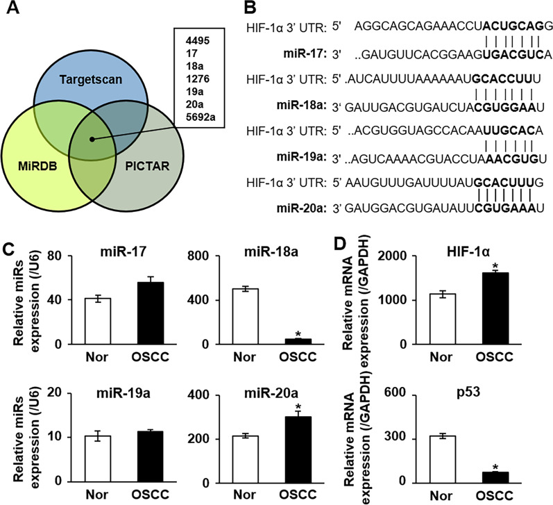
Background: Rapid metastasis of oral squamous cell carcinoma (OSCC) is associated with a poor prognosis and a high mortality rate. However, the molecular mechanisms underlying OSCC metastasis have not been fully elucidated. Although deregulated expression of microRNA (miRNA) has a crucial role in malignant cancer progression, the biological function of miRNA in OSCC progression remains unclear. This study aimed to investigate the function of miRNA-18a in OSCC metastatic regulation via hypoxia-inducible factor 1α (HIF-1α).

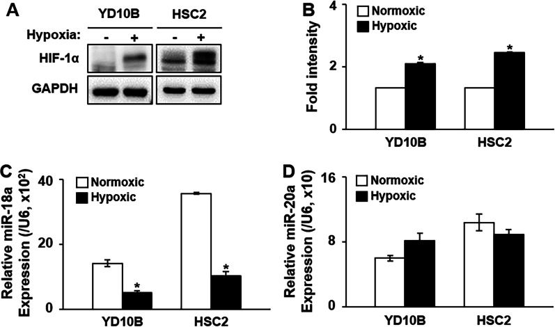
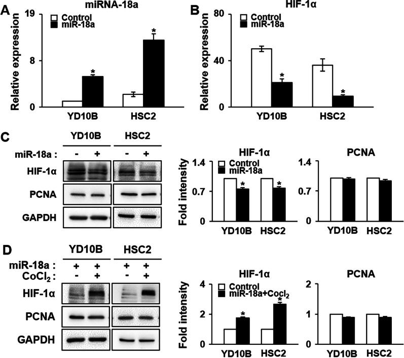
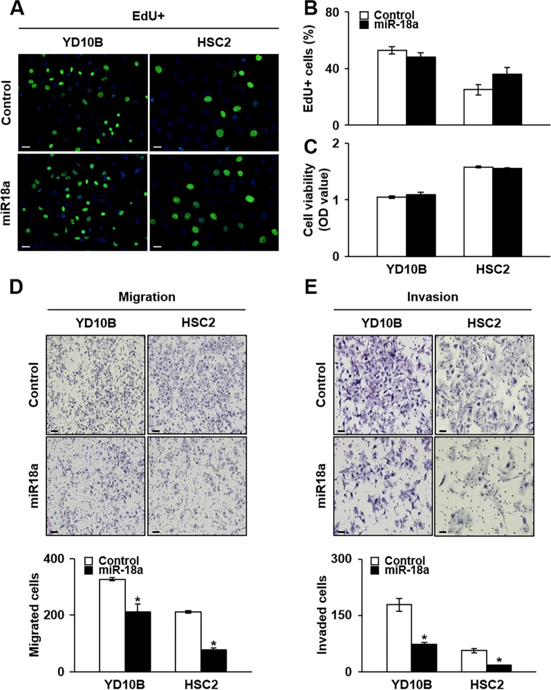
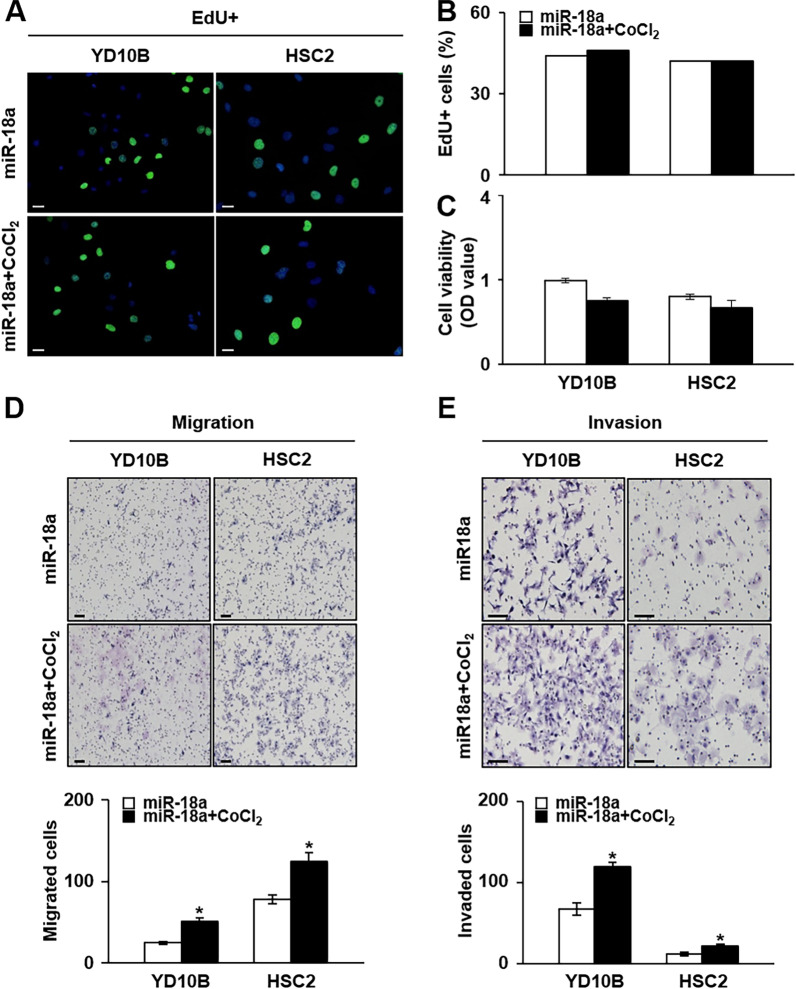
Methods: miRNA-18a-5p (miRNA-18a) expressions in patients with OSCC (n = 39) and in OSCC cell lines (e.g., YD-10B and HSC-2 cells) were analyzed using quantitative real-time polymerase chain reaction. HIF-1α protein expressions in OSCC cells treated with miRNA-18a mimics or combined with cobalt chloride were analyzed using western blotting. The miRNA-18a expression-dependent proliferation and invasion abilities of OSCC cells were analyzed using MTT assay, EdU assay, and a Transwell® insert system.

Results: miRNA-18a expression was significantly lower in OSCC tissue than in the adjacent normal tissue. In OSCC cell lines, HIF-1α expression was significantly decreased by miRNA-18a mimic treatment. Furthermore, the migration and invasion abilities of OSCC cells were significantly decreased by miRNA-18a mimics and significantly increased by the overexpression of HIF-1α under hypoxic conditions relative to those abilities in cells treated only with miRNA-18a mimics.

Conclusions: miRNA-18a negatively affects HIF-1α expression and inhibits the metastasis of OSCC, thereby suggesting its potential as a therapeutic target for antimetastatic strategies in OSCC.

Keywords: HIF-1α; Hypoxia; Invasion; MicroRNA; Migration; Oral squamous cell carcinoma.


Conflict of interest statement

The authors declare that they have no competing interests.

Figures

Fig. 1
Fig. 1
Identification of miRNA-18a associated with HIF-1α in OSCC. A Predictive analysis of HIF-1α and miRNAs according to three miRNA databases (Targetscan, MiRDB, and PICTAR). B Schematic drawing showing the specific binding sites between HIF-1α and the miR-17-92 cluster according to the results of a sequence alignment assay. C mRNA expression of miR-17, -18a, -19a, and -20a in OSCC relative to that in adjacent normal tissues, as assessed using qRT-PCR. U6 was used as the control gene. D mRNA expression of HIF-1α and p53 in OSCC tissues relative to that in adjacent normal tissues according to qRT-PCR analysis. GAPDH was used as the control gene. *Significant difference in expression between the nontumor and tumor conditions (p < 0.05). Tissue samples were pooled. All experiments were performed at least in triplicate. miR-17, microRNA-17; miR-18a, microRNA-18a; miR-20, microRNA-20a; miRs, microRNAs; Nor, normal tissue; OSCC, oral squamous cell carcinoma
Fig. 2
Fig. 2
Expression of miR-18a in OSCC cell lines under hypoxic conditions. A Protein expression of HIF-1α in YD-10B and HSC-2 cells under hypoxic conditions according to western blot analysis. B Intensity graph showing the expression levels of proteins under hypoxic conditions in OSCC cell lines. GAPDH was used as the control gene. C mRNA expression of miR-18a and D miR-20a in OSCC cell lines under hypoxic conditions. U6 was used as the control gene. *Significant difference in expression between the normoxic and hypoxic conditions (p < 0.05). miR-18a, microRNA-18a; miR-20, microRNA-20a; Normoxic, normoxic conditions; Hypoxic, hypoxic conditions. The original picture of western blots of A was shown in Additional file 2: Fig. S1
Fig. 3
Fig. 3
Expression of HIF-1α and PCNA induced by miRNA-18a in OSCC under hypoxic conditions. mRNA expression of A miRNA-18a and B HIF-1α induced by miRNA-18a mimics in OSCC cell lines according to qRT-PCR analysis. U6 was used as the control gene. C Protein expression of HIF-1α and PCNA induced by C miR-18a mimics or D miRNA-18a and CoCl2 in OSCC cell lines according to western blot analysis. Intensity graph showing the expression level of proteins after treatment with C miR-18a mimics or D miRNA-18a and CoCl2 in OSCC cell lines, as determined with western blotting. *Significant difference in expression between control- and miRNA-18a-treated or miRNA-18a and CoCl2-treated OSCC cell lines (p < 0.05). GAPDH was used as the control gene. All experiments were performed at least in triplicate. miR-18a, microRNA-18a; CoCl2, cobalt chloride. The original pictures of western blots of C and D were shown in Additional file 2: Fig. S2, S3 and Fig. S4, S5.
Fig. 4
Fig. 4
Cell viability, migration, and invasion abilities in OSCC cell lines according to miRNA-18a treatment. A Representative images acquired after an EdU assay in YD-10B and HSC-2 cells with and without miRNA-18a treatment (scale bar: 200 μm; original magnification: × 10). B Graphs showing the mean positive cell number for proliferation in five random regions in each well in the two tested OSCC cell lines. C Graphs showing cell viability, as measured using an MTT assay in YD-10B and HSC-2 cells. Representative images of D migration ability (scale bar: 200 μm; original magnification: × 10) and E invasion ability (scale bar: 200 μm; original magnification: × 20) in YD-10B and HSC-2 cells transfected with miRNA-18a. Graphs represent the mean migrated and invaded cells induced by miRNA-18a treatment in YD-10B and HSC-2 cells in five random regions in each well (bottom panels). *Significant difference between the control- and miRNA-18a-treated conditions (p < 0.05). miR-18a, microRNA-18a
Fig. 5
Fig. 5
Cell viability, migration, and invasion abilities of OSCC cell lines under hypoxic conditions with miRNA-18a treatment. A Representative images from the EdU assay in which YD-10B and HSC-2 cells were treated with CoCl2 and miR-18a or miRNA-18a alone (scale bar: 200 μm; original magnification: × 10). B Graphs showing the mean positive cell number for proliferation in five random regions in each well in the two tested OSCC cell lines. C Graphs showing cell viability, as measured by an MTT assay in YD-10B and HSC-2 cells. Representative images of D migration ability (scale bar: 200 μm; original magnification: × 10) and E invasion ability (scale bar: 200 μm; original magnification: × 20) in YD-10B and HSC-2 cells under CoCl2 treatment induced with miRNA-18a treatment. Graphs showing the mean number of migrated and invaded cells induced by miRNA-18a treatment in YD-10B and HSC-2 cells under hypoxic conditions (bottom panels). These values were obtained from five random regions in each well. *Significant difference between cells treated with miRNA-18a alone and those treated with miRNA-18a under hypoxic conditions (p < 0.05). miR-18a, microRNA 18a; CoCl2, cobalt chloride

Similar articles

Cited by

References

    1. Suh JD, Cho JH. Trends in head and neck cancer in South Korea between 1999 and 2012. Clin Exp Otorhinolaryngol. 2016;9(3):263–269. doi: 10.21053/ceo.2015.01123. - DOI - PMC - PubMed
    1. Siegel RL, Miller KD, Jemal A. Cancer statistics, 2019. CA Cancer J Clin. 2019;69(1):7–34. doi: 10.3322/caac.21551. - DOI - PubMed
    1. Jung KW, Won YJ, Kong HJ, Lee ES. Cancer statistics in Korea: incidence, mortality, survival, and prevalence in 2015. Cancer Res Treat. 2018;50(2):303–316. doi: 10.4143/crt.2018.143. - DOI - PMC - PubMed
    1. Bray F, Ferlay J, Soerjomataram I, Siegel RL, Torre LA, Jemal A. Global cancer statistics 2018: GLOBOCAN estimates of incidence and mortality worldwide for 36 cancers in 185 countries. CA Cancer J Clin. 2018;68(6):394–424. doi: 10.3322/caac.21492. - DOI - PubMed
    1. Kuroshima T, Onozato Y, Oikawa Y, Ohsako T, Kugimoto T, Hirai H, Tomioka H, Michi Y, Miura M, Yoshimura R, et al. Prognostic impact of lingual lymph node metastasis in patients with squamous cell carcinoma of the tongue: a retrospective study. Sci Rep. 2021;11(1):20535. doi: 10.1038/s41598-021-99925-2. - DOI - PMC - PubMed

Publication types

MeSH terms

Substances