Case Reports
. 2022 Sep 5;22(1):359.
doi: 10.1186/s12886-022-02566-w.

Central serous chorioretinopathy and angioid streaks: coincidental?

Affiliations
Case Reports

Central serous chorioretinopathy and angioid streaks: coincidental?

Susana Costa Penas et al. BMC Ophthalmol. .

Abstract

Background: To report an unusual case of central serous chorioretinopathy in a patient with angioid streaks.

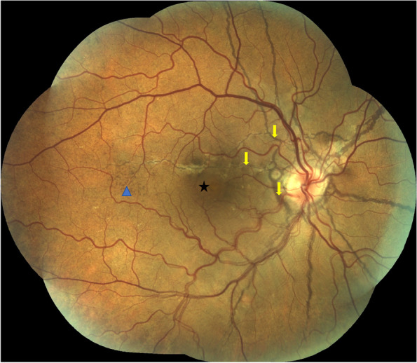
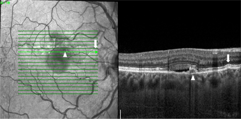
Case presentation: The authors describe a case report of a 26-year old male patient presenting acute scotoma and metamorphopsia in OD. He had been diagnosed with angioid streaks complicated with choroidal neovascularization and referred to us for treatment. The patient presented an ETDRS score of 85 letters (20/20) in OD and in OS. The anterior segment examination was unremarkable. Fundoscopy revealed bilateral angioid streaks (AS) and peau d'orange, as well as a small neurosensory retinal detachment in the macula of OD. A multimodal retinal analysis, including fundus photography, infra-red and fundus autofluorescence imaging, spectral-domain optical coherence tomography, optical coherence tomography angiography, fluorescein and indocyanine green angiography was performed. The diagnosis of central serous chorioretinopathy was made in the absence of any identifiable choroidal neovascularization. He was submitted to half-dose photodynamic therapy with verteporfin. One month later, he reported no visual complaints, his vision was 85 letters (20/20) in OD and a complete resolution of the sub-retinal fluid was registered. No signs of choroidal neovascularization were detected on the optical coherence tomography angiography (OCTA). A complete medical workup evaluation was made to exclude systemic diseases usually associated with AS.

Conclusions: To the authors' knowledge, this is the second reported case of CSC associated with angioid streaks. The focal abnormalities in the Bruch's membrane and the irregular vascular choriocapillary network associated with AS might predispose to CSC.

Keywords: Angioid streaks; CSC; Central serous chorioretinopathy; OCTA.


Conflict of interest statement

Susana Penas: Participation in advisory boards for Alimera, Bayer, Novartis and Roche. Angela Carneiro: Participation in advisory boards for Allergan, Alimera, Bayer, Novartis and Roche. The other authors have no financial disclosures.

Figures

Fig. 1
Fig. 1
Colour fundus photograph montage of the right eye. Multiple brownish angiod streaks are visible radiating from the optic disc (yellow arrows). A small serous detachment is visible in the central macula (black star). Peau d’orange is present in the temporal mid-periphery (blue arrowhead)
Fig. 2
Fig. 2
An horizontal optical coherence tomography scan crossing the fovea shows a small neurosensory retinal detachment (white star), with some shedding of the photorreceptors’ outer segments. An interruption of the retinal pigment epithelium and Bruch’s membrane is visible in correspondance with the angioid streaks (arrowhead). Some hyperreflective material is seen above the RPE (white arrow), probably corresponding to fibrovascular tissue scaring. Although the choroid is not particularly thick (sub-foveal thickness of 308 μm), it presents some dilated Haller vessels with attenuation of the inner choroid (orange octagon), suggesting a pachychoroid feature
Fig. 3
Fig. 3
Optical coherence tomography showing a small retinal pigment epithelium (RPE) detachment underlying the sub-retinal fluid, corresponding to the leaking spot (arrowhead). A small deflection of the Bruch’s membrane and RPE (arrow) results from a small angioid streak, visible in the infra-red image on the left
Fig. 4
Fig. 4
A multimodal angiographic evaluation enhances a small hyperfluorescent spot (white arrowhead) on the early phase fluorescein angiography (FA) (A), not contiguous with the angioid streak (AS), and not visible in the indocyanine green angiography (ICG) (B). Late phase angiograms show mild FA leakage (C), with no hot-spots on the ICG (D). The AS are hyperfluorescent, but more visible in the FA than in the ICG
Fig. 5
Fig. 5
Optical coherence tomography angiography (OCTA) imaging. The infra-red image (A) and the structural enface OCT (B) highlight the angioid streaks (AS) and sub-retinal fluid location. The leaking spot (white arrowhead) is visible both in enface (B) and cross-sectional OCT (C), showing no signs of intralesional vascular flow (C). OCTA images at the avascular (D), choriocapilaris (E) and choroidal (F) layers show no signs of neovessels. A slight choriocapilaris rarefaction is seen in the AS vicinity (black arrowhead)
Fig. 6
Fig. 6
After half-dose photodynamic therapy, the serous retinal detachment resolved completely on the optical coherence tomography foveal-centered scan (A) and there where no signs of sub or intra-retinal fluid around the previous leaking spot (arrowhead) (B)

References

    1. Kaye R, Chandra S, Sheth J, Boon CJF, Sivaprasad S, Lotery A. Central serous chorioretinopathy: an update on risk factors, pathophysiology and imaging modalities. Prog Retin Eye Res. 2020;79:100865. doi: 10.1016/j.preteyeres.2020.100865. - DOI - PubMed
    1. Chatziralli I, Saitakis G, Dimitriou E, Chatzirallis A, Stoungioti S, Theodossiadis G, Theodossiadis P. ANGIOID STREAKS: a comprehensive review from pathophysiology to treatment. Retina. 2019;39(1):1–11. doi: 10.1097/IAE.0000000000002327. - DOI - PubMed
    1. Georgalas I, Papaconstantinou D, Koutsandrea C, Kalantzis G, Karagiannis D, Georgopoulos G, Ladas I. Angioid streaks, clinical course, complications, and current therapeutic management. Ther Clin Risk Manag. 2009;5(1):81–89. - PMC - PubMed
    1. Sarmad A, Alfaqawi F, Chakrabarti M, Elsherbiny S. Central serous chorioretinopathy with angioid streaks: a rare combination. GMS Ophthalmol Cases. 2018;8:Doc01. doi: 10.3205/oc000083. - DOI - PMC - PubMed
    1. Corbelli E, Carnevali A, Marchese A, Cicinelli MV, Querques L, Sacconi R, Bandello F, Querques G. Optical coherence tomography angiography features of ANGIOID streaks. Retina. 2018;38(11):2128–2136. doi: 10.1097/IAE.0000000000001859. - DOI - PubMed

Publication types

MeSH terms