Randomized Controlled Trial
. 2022 Sep 5;13(1):455.
doi: 10.1186/s13287-022-03156-9.

Translational development of ABCB5+ dermal mesenchymal stem cells for therapeutic induction of angiogenesis in non-healing diabetic foot ulcers

Affiliations
Randomized Controlled Trial

Translational development of ABCB5+ dermal mesenchymal stem cells for therapeutic induction of angiogenesis in non-healing diabetic foot ulcers

Andreas Kerstan et al. Stem Cell Res Ther. .

Abstract

Background: While rapid healing of diabetic foot ulcers (DFUs) is highly desirable to avoid infections, amputations and life-threatening complications, DFUs often respond poorly to standard treatment. GMP-manufactured skin-derived ABCB5+ mesenchymal stem cells (MSCs) might provide a new adjunctive DFU treatment, based on their remarkable skin wound homing and engraftment potential, their ability to adaptively respond to inflammatory signals, and their wound healing-promoting efficacy in mouse wound models and human chronic venous ulcers.

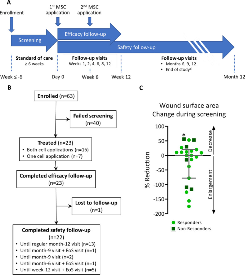
Methods: The angiogenic potential of ABCB5+ MSCs was characterized with respect to angiogenic factor expression at the mRNA and protein level, in vitro endothelial trans-differentiation and tube formation potential, and perfusion-restoring capacity in a mouse hindlimb ischemia model. Finally, the efficacy and safety of ABCB5+ MSCs for topical adjunctive treatment of chronic, standard therapy-refractory, neuropathic plantar DFUs were assessed in an open-label single-arm clinical trial.

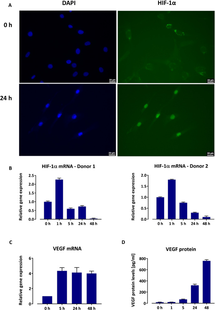
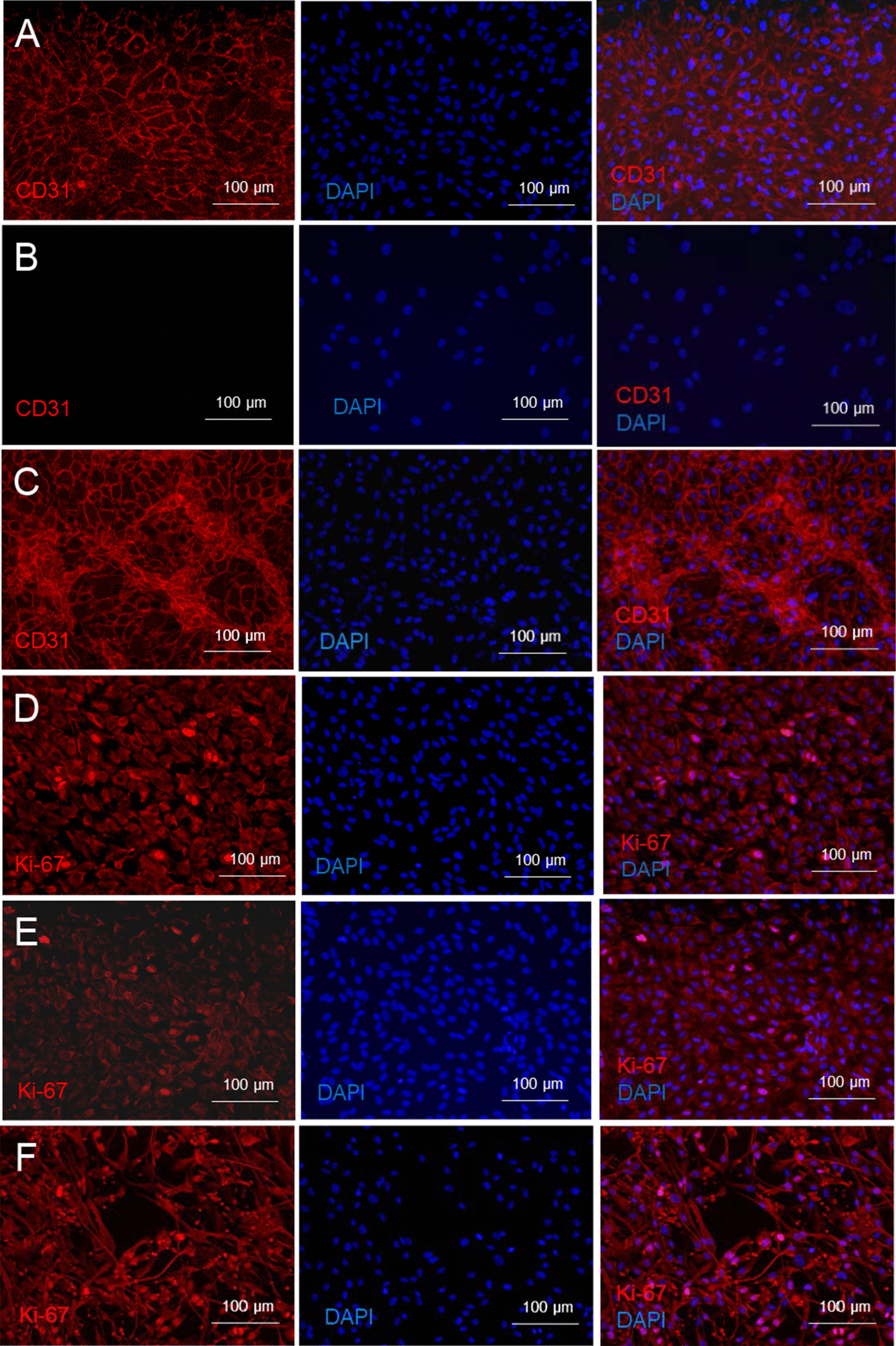
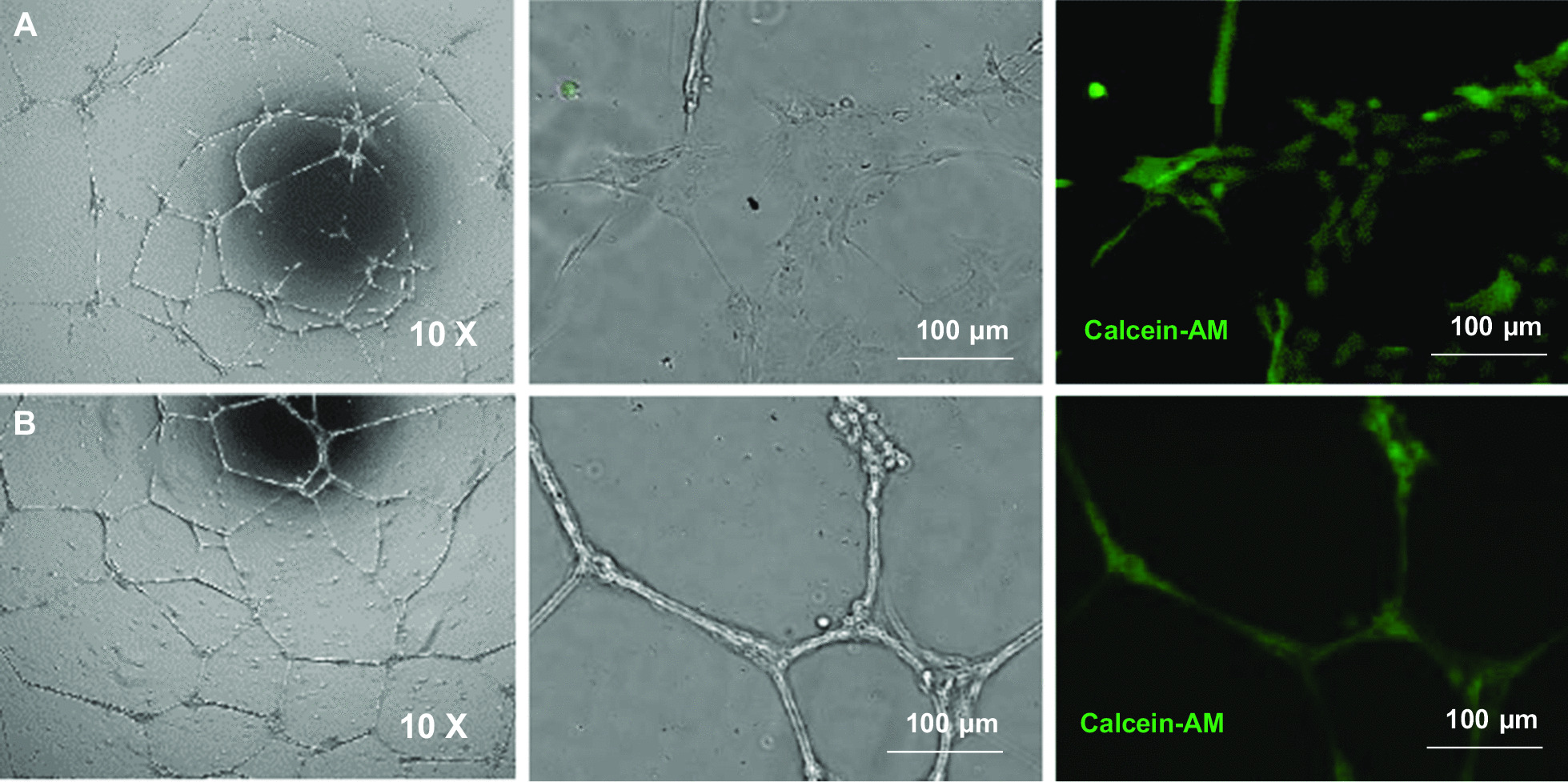
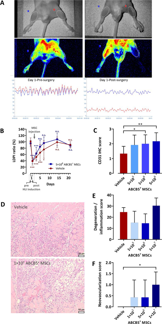
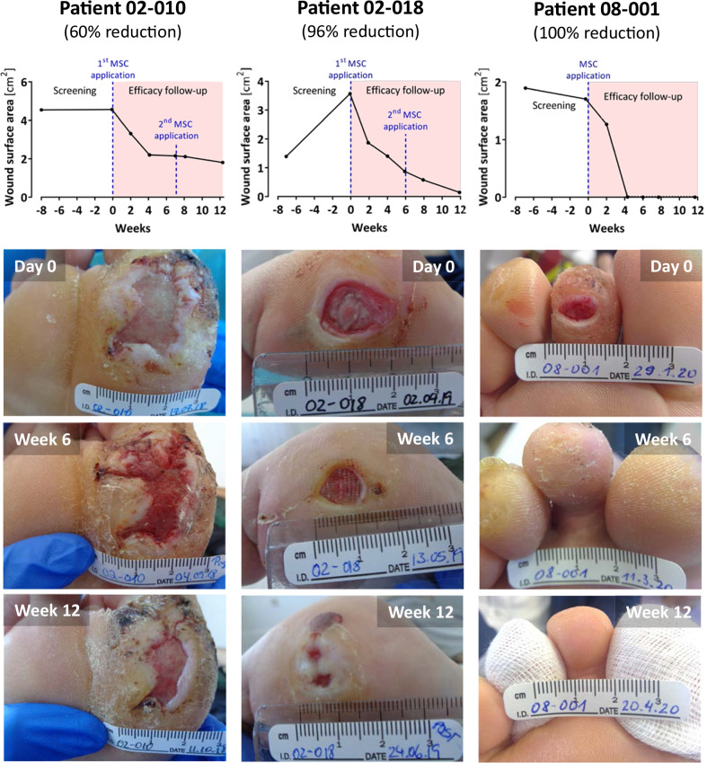
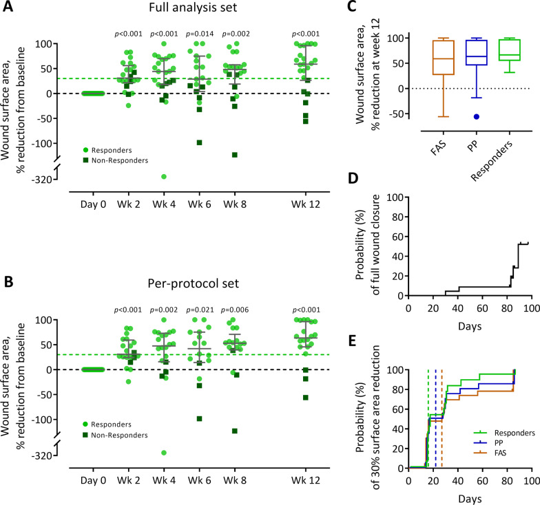
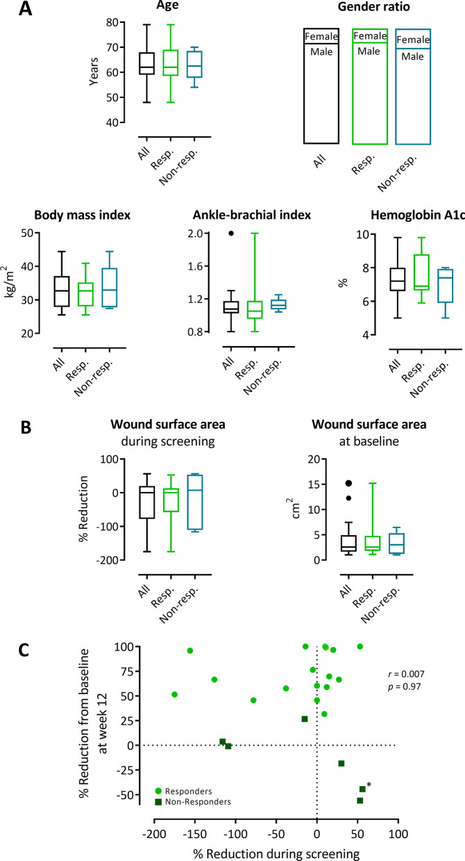
Results: Hypoxic incubation of ABCB5+ MSCs led to posttranslational stabilization of the hypoxia-inducible transcription factor 1α (HIF-1α) and upregulation of HIF-1α mRNA levels. HIF-1α pathway activation was accompanied by upregulation of vascular endothelial growth factor (VEGF) transcription and increase in VEGF protein secretion. Upon culture in growth factor-supplemented medium, ABCB5+ MSCs expressed the endothelial-lineage marker CD31, and after seeding on gel matrix, ABCB5+ MSCs demonstrated formation of capillary-like structures comparable with human umbilical vein endothelial cells. Intramuscularly injected ABCB5+ MSCs to mice with surgically induced hindlimb ischemia accelerated perfusion recovery as measured by laser Doppler blood perfusion imaging and enhanced capillary proliferation and vascularization in the ischemic muscles. Adjunctive topical application of ABCB5+ MSCs onto therapy-refractory DFUs elicited median wound surface area reductions from baseline of 59% (full analysis set, n = 23), 64% (per-protocol set, n = 20) and 67% (subgroup of responders, n = 17) at week 12, while no treatment-related adverse events were observed.

Conclusions: The present observations identify GMP-manufactured ABCB5+ dermal MSCs as a potential, safe candidate for adjunctive therapy of otherwise incurable DFUs and justify the conduct of a larger, randomized controlled trial to validate the clinical efficacy.

Trial registration: ClinicalTrials.gov, NCT03267784, Registered 30 August 2017, https://clinicaltrials.gov/ct2/show/NCT03267784.

Keywords: ABCB5; Advanced-therapy medicinal product; Angiogenesis; Chronic wound; Diabetic foot ulcer; Mesenchymal stem cells; Wound healing.


Conflict of interest statement

MHF and NYF are inventors or co-inventors of US and international patents assigned to Brigham and Women’s Hospital and/or Boston Children’s Hospital (Boston, MA, USA), licensed to TICEBA GmbH (Heidelberg, Germany) and RHEACELL GmbH & Co. KG (Heidelberg, Germany). MHF and KSK serve as scientific advisors to TICEBA and RHEACELL and participate in corporate-sponsored research collaborations with RHEACELL. ENR, SK, JE, SS and SB are employees of TICEBA. KD, KK and HMS are employees of RHEACELL. CG is CEO, and MAK is CSO of TICEBA and RHEACELL. The remaining authors declare no competing interests.

Figures

Fig. 1
Fig. 1
HIF-1α and VEGF expression by ABCB5+ MSCs during hypoxic culture. A Representative immunofluorescence staining of ABCB5+ MSCs revealing nuclear translocation of HIF-1α at 24 h. Nuclei were counterstained with DAPI. Scale bars: 20 µm. B HIF-1α mRNA expression by ABCB5+ MSCs from two donors, shown as fold expression from baseline (normoxic conditions, 0 h). Data are means + SD of three replicates. C VEGF mRNA expression by ABCB5+ MSCs, shown as fold expression from baseline (normoxic conditions, 0 h). Data are means + SD of three donors. D VEGF protein secretion by ABCB5+ MSCS, measured as VEGF protein concentration in culture supernatant. Data are means + SD of three replicates from a representative donor
Fig. 2
Fig. 2
Endothelial trans-differentiation of ABCB5+ MSCs. A Co-stimulation for 96 h with 200 ng/ml VEGF, 1000 ng/ml FGF-2 and 1000 ng/ml PDGF-BB elicited angiogenic trans-differentiation of ABCB5+ MSCs as revealed by CD31-positive (red) staining. B ABCB5+ MSCs cultured without growth factor supplementation served as negative control. C HUVECs served as positive control. D–F Proliferative activity of ABCB5+ MSCs stimulated to undergo endothelial trans-differentiation. D ABCB5+ MSCs were stimulated for 96 h with 200 ng/ml VEGF, 1000 ng/ml FGF-2 and 1000 ng/ml PDGF-BB. Proliferative activity was assessed by Ki67 staining (red). E ABCB5+ MSCs cultured without growth factor supplementation served as negative control. F HUVECs served as positive control. Nuclei were counterstained with DAPI (blue). Representative images of three independent experiments
Fig. 3
Fig. 3
Tube formation assays. Human ABCB5+ MSCs and B HUVECs were cultured for 18–20 h on Geltrex™ matrix. Calcein staining (green) demonstrates viability (i.e., metabolic activity) of tubular structure-forming cells
Fig. 4
Fig. 4
Blood flow recovery and neovascularization following surgically induced HLI in OF1 mice. A Representative LDPI acquisition before and immediately after HLI induction, illustrating the experimental setup. Scanned areas are marked by ellipses; the warmest color (intense red) represents 200 perfusion units. Graphs show mean perfusion unit during 1 min in the non-ischemic (blue) and ischemic (red) thigh. B LPDI ratio between the ischemic and the non-ischemic limb in mice treated with 5 × 106 ABCB5+ MSCs or vehicle. Means with SD of n = 10 (day 1), n = 9 (day 3), n = 8 (day 5; days 7–21 MSCs) and n = 7 (days 7–21 vehicle) animals. C–F Immunohistochemical and histopathological evaluation of the ischemic hindlimb muscles at 6 days after HLI induction in mice treated with ABCB5+ MSCs or vehicle injected into the ischemic limb 24 h after surgery. C CD31 expression in the thigh muscles, presented as mean (SD) IHC score, with 0 = none, 1 = minimal, 2 = slight, and 3 = moderate, of n = 12 animals. D Representative H&E sections of the gastrocnemius muscle from a vehicle- and an MSC-treated mouse, showing inflammatory and degenerative lesions in both mice and increased neovascularization in the MSC-treated mouse. Scale bars: 50 µm. E Degenerative and inflammatory processes in the gastrocnemius muscle, presented as mean (SD) summary score according to ISO 10993–6:2007 of n = 6 (vehicle) and n = 7 (MSCs) animals. F Neovascularization in the gastrocnemius muscle, presented as mean (SD) score, with 0 = none, 1 = 1–3 focal buds, 2 = groups of 4–7 capillaries with supporting fibroblastic structures, 3 = broad band and 4 = extensive band of capillaries with supporting fibroblastic structures, of n = 6 (vehicle) and n = 7 (MSCs) animals. *p < 0.05, **p < 0.01, ***p < 0.001 versus baseline (B) or vehicle (C, E, F); one-way ANOVA with Dunnett’s post hoc test
Fig. 5
Fig. 5
Trial design, study patients and wound surface area during screening. A Schematic representation of the trial design. aOnly patients who did not reach month-12 visit before 30 June 2020 and were not scheduled for a planned safety follow-up visit in June 2020 were subjected to an end-of-study visit. B Study patient flow chart. EoS visit, end-of-study visit [see (a)]. C Percent reduction of wound surface area during a ≥ 6-week screening period (median 49 days, range 42–68 days; except for one outlier, whose screening period lasted 118 days, denoted by an asterisk). Error bar represents median and interquartile range
Fig. 6
Fig. 6
Wound healing progress during the treatment and efficacy follow-up period. Shown are three representative patients in the subgroup of responders. All patients had consented to publication of the photographs
Fig. 7
Fig. 7
Wound surface area reduction in DFU patients treated with ABCB5+ MSCs. A–B Percent wound surface area reduction from baseline during the treatment and efficacy follow-up period in the full analysis set (A) and per-protocol set (B). Patients who presented with wound surface area reductions of at least 30% from baseline (indicated by light green dashed lines) at week 12 were considered responders. Error bars indicate median and interquartile range; p values (two-sided Wilcoxon signed rank test) indicate statistical significance of changes from baseline. C Tukey’s boxplots of the primary efficacy outcome parameter, % wound surface area reduction from baseline at week 12, in the full analysis set (FAS, N = 23), per-protocol set (PP, N = 20) and responders (i.e., patients who presented with at least 30% wound surface area at week 12; N = 17). D-E Kaplan–Meier plots for the time to full wound closure (D) and first 30% surface area reduction (E) in the FAS, PP and responders. Patients without event were censored at the date of the last available wound surface area assessment (indicated by small vertical ticks). Vertical dashed lines indicate median time to event (not reached for full wound closure)
Fig. 8
Fig. 8
Assessment of potential influences of baseline patient characteristic, baseline wound size and wound surface area reduction during the screening period on response to treatment. Baseline patient characteristics and baseline wound surface area in all treated patients, responders and non-responders. A–B Comparisons of baseline patient characteristics (A) and of wound surface area reduction during screening and of wound surface area at baseline (B) between all treated patients, responders and non-responders. Depicted are Tukey’s boxplots (except for gender ratio); n = 23 (all patients; ankle-brachial index: n = 22), n = 17 (responders; ankle-brachial index: n = 16), n = 6 (non-responders). Kruskal–Wallis tests followed by Dunn’s multiple comparisons revealed no statistically significant differences between groups (p > 0.999 for all comparisons except for ankle-brachial index responders vs. non-responders: p = 0.697). C Spearman’s rank correlation analysis between wound surface area reduction during screening and wound surface area reduction from baseline at week 12. *Asterisk denotes a patient whose screening period lasted 118 days, as compared to 42–68 days (median 49 days) for the other patients

References

    1. Armstrong DG, Boulton AJM, Bus SA. Diabetic foot ulcers and their recurrence. N Engl J Med. 2017;376:2367–2375. doi: 10.1056/NEJMra1615439. - DOI - PubMed
    1. Jupiter DC, Thorud JC, Buckley CJ, Shibuya N. The impact of foot ulceration and amputation on mortality in diabetic patients. I: From ulceration to death, a systematic review. Int Wound J. 2016;13:892–903. doi: 10.1111/iwj.12404. - DOI - PMC - PubMed
    1. Xiang J, Wang S, He Y, Xu L, Zhang S, Tang Z. Reasonable glycemic control would help wound healing during the treatment of diabetic foot ulcers. Diabetes Ther. 2019;10:95–105. doi: 10.1007/s13300-018-0536-8. - DOI - PMC - PubMed
    1. Walsh JW, Hoffstad OJ, Sullivan MO, Margolis DJ. Association of diabetic foot ulcer and death in a population-based cohort from the United Kingdom. Diabet Med. 2016;33:1493–1498. doi: 10.1111/dme.13054. - DOI - PubMed
    1. Young MJ, McCardle JE, Randall LE, Barclay JI. Improved survival of diabetic foot ulcer patients 1995–2008: possible impact of aggressive cardiovascular risk management. Diabetes Care. 2008;31:2143–2147. doi: 10.2337/dc08-1242. - DOI - PMC - PubMed

Publication types

MeSH terms

Substances

Associated data