. 2022 Sep 5;13(1):449.
doi: 10.1186/s13287-022-03142-1.

Systemic proteomics and miRNA profile analysis of exosomes derived from human pluripotent stem cells

Affiliations

Systemic proteomics and miRNA profile analysis of exosomes derived from human pluripotent stem cells

Youkun Bi et al. Stem Cell Res Ther. .

Abstract

Background: Increasing studies have reported the therapeutic effect of mesenchymal stem cell (MSC)-derived exosomes by which protein and miRNA are clearly characterized. However, the proteomics and miRNA profiles of exosomes derived from human embryonic stem cells (hESCs) and human-induced pluripotent stem cells (hiPSCs) remain unclear.

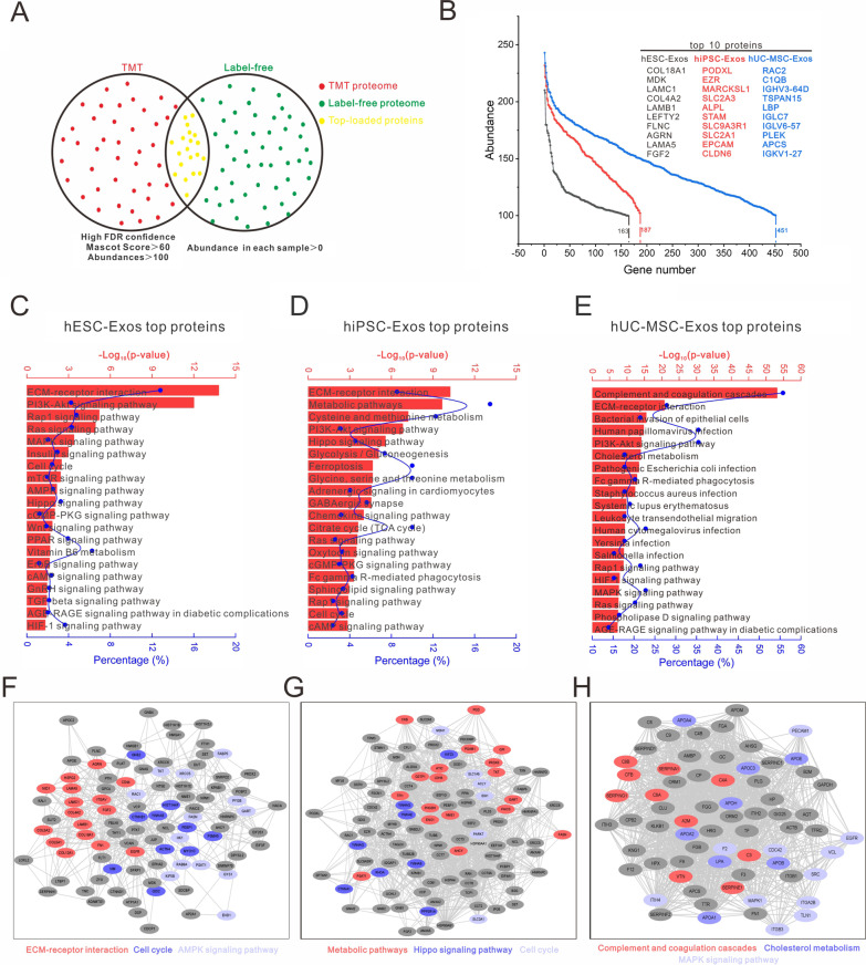
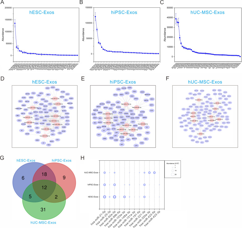
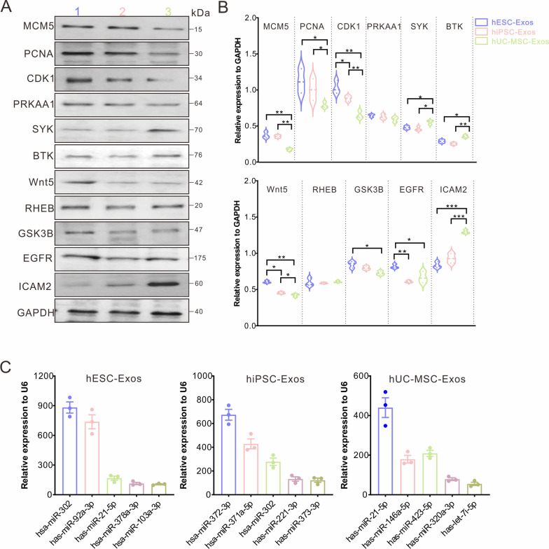
Methods: In this study, we isolated exosomes from hESCs, hiPSCs, and human umbilical cord mesenchymal stem cells (hUC-MSCs) via classic ultracentrifugation and a 0.22-μm filter, followed by the conservative identification. Tandem mass tag labeling and label-free relative peptide quantification together defined their proteomics. High-throughput sequencing was performed to determine miRNA profiles. Then, we conducted a bioinformatics analysis to identify the dominant biological processes and pathways modulated by exosome cargos. Finally, the western blot and RT-qPCR were performed to detect the actual loads of proteins and miRNAs in three types of exosomes.

Results: Based on our study, the cargos from three types of exosomes contribute to sophisticated biological processes. In comparison, hESC exosomes (hESC-Exos) were superior in regulating development, metabolism, and anti-aging, and hiPSC exosomes (hiPSC-Exos) had similar biological functions as hESC-Exos, whereas hUC-MSCs exosomes (hUC-MSC-Exos) contributed more to immune regulation.

Conclusions: The data presented in our study help define the protein and miRNA landscapes of three exosomes, predict their biological functions via systematic and comprehensive network analysis at the system level, and reveal their respective potential applications in different fields so as to optimize exosome selection in preclinical and clinical trials.

Keywords: Exosomes; Human embryonic stem cells; Human umbilical cord mesenchymal stem cells; Human-induced pluripotent stem cells; Proteomics; miRNA.


Conflict of interest statement

The authors declare no potential conflict of interest.

The authors declare no potential conflict of interest.

Figures

Fig. 1
Fig. 1
Quality analysis of isolated exosomes. A Representative TEM micrograph of exosomes derived from hESCs, hiPSCs, and hUC-MSCs. Scale bar = 100 nm. B DLS system describing the diameter of isolated exosomes. C Western blot assay determining the expression of Calnexin, TSG101, CD63, and HSP70 in cells and exosomes. GAPDH was set as the internal reference. D Evaluation of mean exosomes yield per cell. All statistical data are presented as means ± standard deviation of two-tailed unpaired Student’s t-tests. *P < 0.05
Fig. 2
Fig. 2
The top-loaded proteins in exosomes were involved in sophisticated network regulation. A Screening of the top-expressed candidate proteins from TMT and label-free protein pools under the conditions of high FDR confidence, mascot score > 60, and abundance > 100 (red dots) and abundance in each sample > 0 (green dots). Yellow dots represent the top-loaded proteins of exosomes. B Expression abundance curve of the three exosome types and the top ten loaded proteins in exosomes. CE KEGG analysis of the top-expressed proteins in hESC-Exos, hiPSC-Exos, and hUC-MSC-Exos, respectively, ranked from high to low -Log10(P-value). The red bar represents the -Log10 (P-value) and the blue dot the proportion of candidate genes in the total pathway-related gene pool. FH PPI analysis of top-expressed proteins in hESC-Exos, hiPSC-Exos, and hUC-MSC-Exos, respectively, according to the KOBAS algorithm. The color of the histogram corresponds to the color of the gene clusters in the PPI network
Fig. 3
Fig. 3
The shared proteins had different signaling regulation abilities when performing pairwise bioinformatics analysis. A, D, and G KEGG analyses of overlapping proteins in hESC-Exos and hiPSC-Exos A, hESC-Exos and hUC-MSC-Exos B, and hiPSC-Exos and hUC-MSC-Exos G. The red bar and black dot represent the -Log10 (P-value) and the proportion of candidate genes in the total pathway-related gene pool, respectively. B, E, and H Volcano diagrams of differentially expressed proteins in hESC-Exos vs hiPSC-Exos B, hESC-Exos vs hUC-MSC-Exos E, and hiPSC-Exos vs hUC-MSC-Exos E at P < 0.05 and |log2FC|≥ 1. Histogram of the GO and KEGG analyses of upregulated protein clusters ranked from high to low significance. C, F, and I Heatmap of the levels of shared proteins between hESC-Exos and hiPSC-Exos C, hESC-Exos and hUC-MSC-Exos F, and hiPSC-Exos and hUC-MSC-Exos I. The significantly enriched pathways are shown on the right
Fig. 4
Fig. 4
The overlapping proteins of three exosome samples were involved in complex biological regulation. A Venn diagram of the shared proteins in hESC-Exos, hiPSC-Exos, and hUC-MSC-Exos. B and C GO and KEGG analyses of the shared proteins among the three exosome types. The red bar represents the -Log10 (P-value) and the blue dot the proportion of candidate genes in the total pathway-related gene pool. D Crosstalk between the shared proteins in hESC-Exos, hiPSC-Exos, and hUC-MSC-Exos and regulated the signaling pathways they regulate. E Heatmap of the levels of shared proteins among the three exosome types. The significantly enriched pathways are shown on the right
Fig. 5
Fig. 5
The top miRNAs derived from three exosome samples finely regulate complex signaling network. AC Abundance of the top 20 miRNAs in hESC-Exos (A), hiPSC-Exos (B), and hUC-MSC-Exos (C). DF Pathways regulated by the top miRNAs (read > 1000) in hESC-Exos (D), hiPSC-Exos (E), and hUC-MSC-Exos (F). GI Biological processes regulated by the top miRNAs (read > 1000) in hESC-Exos (G), hiPSC-Exos (H), and hUC-MSC-Exos (I). The red bar represents the -Log10 (P-value)
Fig. 6
Fig. 6
The signal regulation characteristics of specific miRNAs derived from three exosome samples. A Venn diagram of the miRNAs in hESC-Exos, hiPSC-Exos, and hUC-MSC-Exos. BD Pathways regulated by the unique miRNAs in hESC-Exos (B), hiPSC-Exos (C), and hUC-MSC-Exos (D). The red bar represents -Log10 (P-value). E Heatmap of the shared miRNAs among the three exosome types. F Significantly enriched pathways regulated by the different miRNA clusters in (E). The bubble size represents -Log10 (P-value)
Fig. 7
Fig. 7
Three exosome samples had different miRNA profiles related to pluripotency regulation. AC Abundance of the miRNAs related to pluripotency regulation. DF Regulatory network of the top ten miRNAs and their target genes in pluripotency regulation. G Venn diagram of the miRNAs involved in pluripotency regulation and shared among the three exosome types. (F) Abundance of the miRNAs in (G). The bubble size represents abundance
Fig. 8
Fig. 8
Detection of specific proteins and miRNAs in the three exosome types. A Representative western blot graphs regarding the detection of representative proteins in hESC-Exos (1), hiPSC-Exos (2), and hUC-MSC-Exos (3). GAPDH was set as the internal reference. Three independent replicates were performed. B Quantification of protein levels in (A). All statistical data are presented as means ± standard deviation of two-tailed unpaired Student’s t-tests. *P < 0.05, **P < 0.01, and ***P < 0.001. C RT-qPCR detection of the expression of the top five miRNAs in the three exosome types. Three independent replicates were performed

References

    1. Abbaszadeh H, Ghorbani F, Derakhshani M, Movassaghpour A, Yousefi M. Human umbilical cord mesenchymal stem cell-derived extracellular vesicles: a novel therapeutic paradigm. J Cell Physiol. 2020;235(2):706–717. doi: 10.1002/jcp.29004. - DOI - PubMed
    1. Abdelgawad M, Bakry NS, Farghali AA, Abdel-Latif A, Lotfy A. Mesenchymal stem cell-based therapy and exosomes in COVID-19: current trends and prospects. Stem Cell Res Ther. 2021;12(1):469. doi: 10.1186/s13287-021-02542-z. - DOI - PMC - PubMed
    1. Abels ER, Breakefield XO. Introduction to extracellular vesicles: biogenesis, rna cargo selection, content, release, and uptake. Cell Mol Neurobiol. 2016;36(3):301–312. doi: 10.1007/s10571-016-0366-z. - DOI - PMC - PubMed
    1. Baumann K. EVs promote stemness. Nat Rev Mol Cell Biol. 2021;22(2):72–73. doi: 10.1038/s41580-020-00327-5. - DOI - PubMed
    1. Bei Y, Das S, Rodosthenous RS, Holvoet P, Vanhaverbeke M, Monteiro MC, Monteiro VVS, Radosinska J, Bartekova M, Jansen F, Li Q, Rajasingh J, Xiao J. Extracellular vesicles in cardiovascular theranostics. Theranostics. 2017;7(17):4168–4182. doi: 10.7150/thno.21274. - DOI - PMC - PubMed

Publication types

LinkOut - more resources