

Factors secreted by monosodium urate crystal-stimulated macrophages promote a proinflammatory state in osteoblasts: a potential indirect mechanism of bone erosion in gout
- PMID: 36064735
- PMCID: PMC9442999
- DOI: 10.1186/s13075-022-02900-z
Factors secreted by monosodium urate crystal-stimulated macrophages promote a proinflammatory state in osteoblasts: a potential indirect mechanism of bone erosion in gout
Abstract
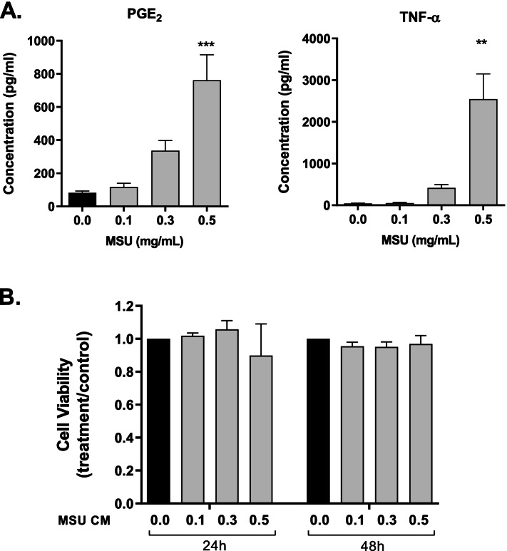
Background: Tophi are lesions commonly present at sites of bone erosion in gout-affected joints. The tophus comprises a core of monosodium urate (MSU) crystals surrounded by soft tissue that contains macrophages and other immune cells. Previous studies found that MSU crystals directly reduce osteoblast viability and function. The aim of the current study was to determine the indirect, macrophage-mediated effects of MSU crystals on osteoblasts.
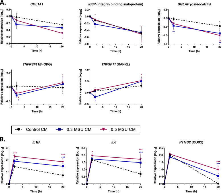
Methods: Conditioned medium from the RAW264.7 mouse macrophage cell line cultured with MSU crystals was added to the MC3T3-E1 mouse osteoblastic cell line. Conditioned medium from the THP-1 human monocytic cell line cultured with MSU crystals was added to primary human osteoblasts (HOBs). Matrix mineralization was assessed by von Kossa staining. Gene expression was determined by real-time PCR, and concentrations of secreted factors were determined by enzyme-linked immunosorbent assay.
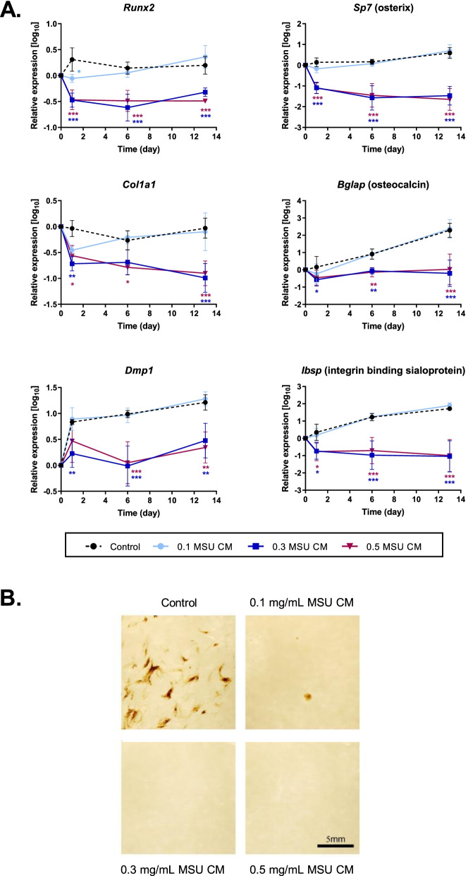
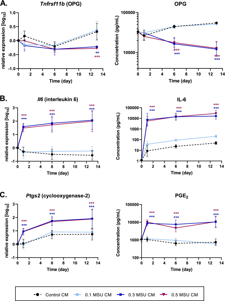
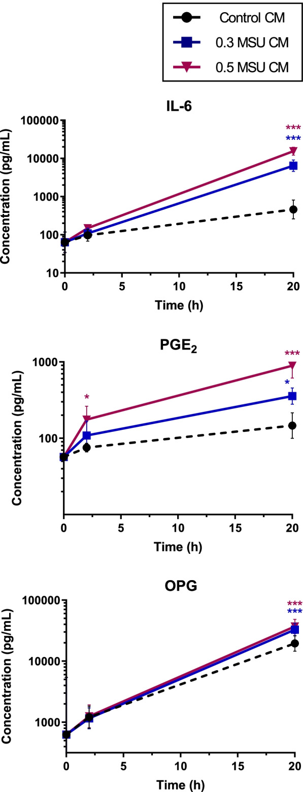
Results: In MC3T3-E1 cells cultured for 13 days in an osteogenic medium, the expression of the osteoblast marker genes Col1a1, Runx2, Sp7, Bglap, Ibsp, and Dmp1 was inhibited by a conditioned medium from MSU crystal-stimulated RAW264.7 macrophages. Mineral staining of MC3T3-E1 cultures on day 21 confirmed the inhibition of osteoblast differentiation. In HOB cultures, the effect of 20 h incubation with a conditioned medium from MSU crystal-stimulated THP-1 monocytes on osteoblast gene expression was less consistent. Expression of the genes encoding cyclooxygenase-2 and IL-6 and secretion of the proinflammatory mediators PGE2 and IL-6 were induced in MC3T3-E1 and HOBs incubated with conditioned medium from MSU crystal-stimulated macrophages/monocytes. However, inhibition of cyclooxygenase-2 activity and PGE2 secretion from HOBs indicated that this pathway does not play a major role in mediating the indirect effects of MSU crystals in HOBs.
Conclusions: Factors secreted from macrophages stimulated by MSU crystals attenuate osteoblast differentiation and induce the expression and secretion of proinflammatory mediators from osteoblasts. We suggest that bone erosion in joints affected by gout results from a combination of direct and indirect effects of MSU crystals.
Keywords: Bone; Gout; Osteoblast; Tophus; Urate.
© 2022. The Author(s).
Conflict of interest statement
Nicola Dalbeth has received consulting fees, speaker fees or grants from AstraZeneca, Dyve Biosciences, Horizon, Amgen, Selecta, Arthrosi, JW Pharmaceutical Corporation, PK Med, PTC Therapeutics, Protalix, Cello Health, Abbvie, and Janssen, outside the submitted work. The other authors declare that they have no competing interests.
Figures


References
-
- Chhana A, Callon KE, Pool B, Naot D, Watson M, Gamble GD, McQueen FM, Cornish J, Dalbeth N. Monosodium urate monohydrate crystals inhibit osteoblast viability and function: implications for development of bone erosion in gout. Ann Rheum Dis. 2011;70(9):1684–1691. doi: 10.1136/ard.2010.144774. - DOI - PubMed
-
- Chhana A, Pool B, Callon KE, Tay ML, Musson D, Naot D, McCarthy G, McGlashan S, Cornish J, Dalbeth N. Monosodium urate crystals reduce osteocyte viability and indirectly promote a shift in osteocyte function towards a proinflammatory and proresorptive state. Arthritis Res Ther. 2018;20(1):208. doi: 10.1186/s13075-018-1704-y. - DOI - PMC - PubMed
Publication types
MeSH terms
Substances
Grants and funding
LinkOut - more resources
Full Text Sources
Medical
Research Materials
Miscellaneous
