. 2022 Sep 5;13(1):5222.
doi: 10.1038/s41467-022-32760-9.

Allosteric inhibition of HTRA1 activity by a conformational lock mechanism to treat age-related macular degeneration

Affiliations

Allosteric inhibition of HTRA1 activity by a conformational lock mechanism to treat age-related macular degeneration

Stefan Gerhardy et al. Nat Commun. .

Abstract

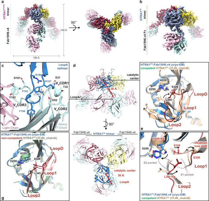
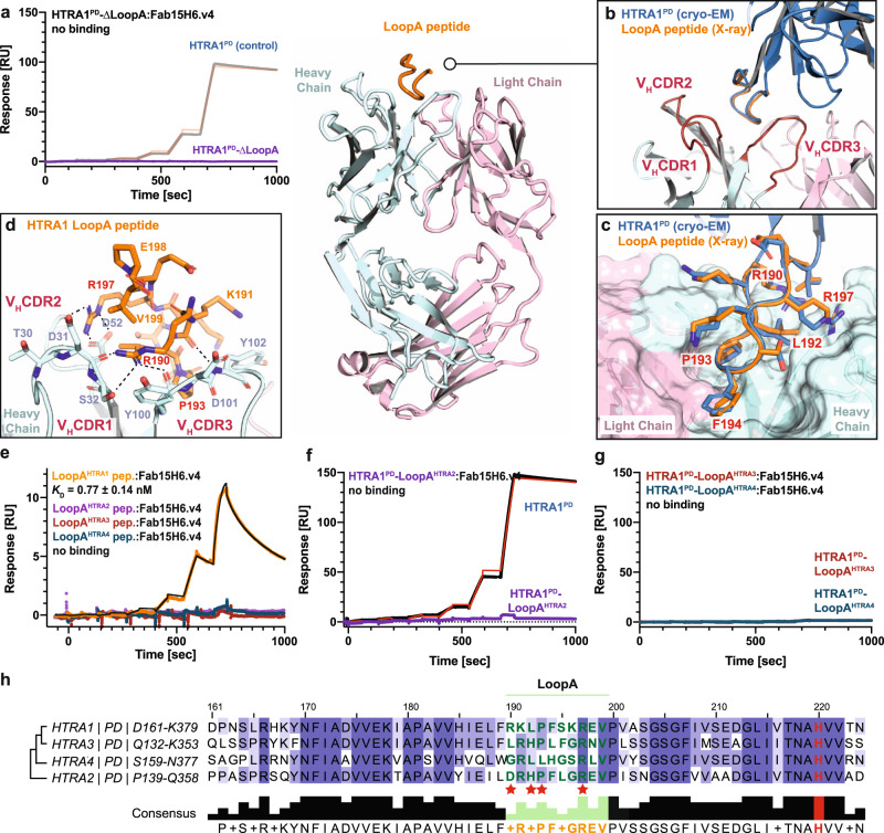
The trimeric serine protease HTRA1 is a genetic risk factor associated with geographic atrophy (GA), a currently untreatable form of age-related macular degeneration. Here, we describe the allosteric inhibition mechanism of HTRA1 by a clinical Fab fragment, currently being evaluated for GA treatment. Using cryo-EM, X-ray crystallography and biochemical assays we identify the exposed LoopA of HTRA1 as the sole Fab epitope, which is approximately 30 Å away from the active site. The cryo-EM structure of the HTRA1:Fab complex in combination with molecular dynamics simulations revealed that Fab binding to LoopA locks HTRA1 in a non-competent conformational state, incapable of supporting catalysis. Moreover, grafting the HTRA1-LoopA epitope onto HTRA2 and HTRA3 transferred the allosteric inhibition mechanism. This suggests a conserved conformational lock mechanism across the HTRA family and a critical role of LoopA for catalysis, which was supported by the reduced activity of HTRA1-3 upon LoopA deletion or perturbation. This study reveals the long-range inhibition mechanism of the clinical Fab and identifies an essential function of the exposed LoopA for activity of HTRA family proteases.


Conflict of interest statement

All authors were employees of Genentech Inc., a for-profit institution, at the time when the studies were performed.

Figures

Fig. 1
Fig. 1. Fab15H6.v4 inhibits the enzymatic activity of HTRA1FL and HTRA1PD through an allosteric mechanism.
a HTRA1FL domain architecture. Enzyme activity of HTRA1FL, HTRA1PD, and catalytically inactive HTRA1PD/SA in the presence of Fab15H6.v4. Control Fab33 (anti-PCSK9) does not affect enzymatic activity. b Cleavage of macromolecular substrates Dickkopf-related protein 3 (DKK3), BIGLYCAN, and DECORIN by HTRA1FL (51 kDa) and HTRA1PD (24 kDa) in the presence of Fab15H6.v4. Right panel: cleavage assay control with individual proteins. Asterisks (*) indicate the contaminant of the control Fab33 preparation. c Labeling of HTRA1FL, HTRA1PD, and HTRA1PD/SA active site using a small fluorescent activity-based probe (TAMRA-ABP) in the presence of Fab15H6.v4. HTRA1FL undergoes self-cleavage and therefore appears as two bands in the TAMRA-ABP assay. d Size-exclusion profiles of HTRA1PD/SA:Fab15H6.v4 (red, calc. mass = 228 kDa) and full length HTRA1FL/SA:Fab15H6.v4 (blue, calc. mass = 311 kDa) complexes. Protein standards for estimated size comparison are in grey. e Kinetics of Fab15H6.v4 binding to HTRA1PD and to HTRA1PD pre-incubated with 7-mer ABP determined by Surface Plasmon Resonance (SPR). Bar graphs in (a) and kinetic data in (e) are presented as the mean ± S.D. of three independent experiments. Images in (b, c), as well as chromatogram in (d) are representative of two independent experiments. Source data are provided as a Source Data file.
Fig. 2
Fig. 2. Fab15H6.v4 binds to the distant LoopA of the HTRA1 trimer and stabilizes a non-competent conformation.
a Cryo-EM map of HTRA1PD/SA trimer with three Fabs (Fab15H6.v4) bound to the exposed LoopA at a resolution of 3.3 Å. The monomers of the HTRA1PD/SA trimer are in blue, yellow and red and Fab light and heavy chains are in pink and cyan, respectively. b Cryo-EM map of wildtype HTRA1PD:Fab15H6.v4 complex at 3.3 Å resolution, in which the flexible constant regions (CH1/CL) of the Fab15H6.v4 were masked out during refinement. c Detailed view on the LoopA epitope and Fab15H6.v4 interaction. Critical residues are represented as sticks. d Ribbon representation of wildtype HTRA1PD bound by Fab15H6.v4 in side view (top) and top view (bottom). The LoopA epitope is more than 30 Å away from the catalytic center. e Comparison of the Fab-bound wildtype HTRA1PD (blue) with the competent apo-HTRA1 in the active conformation (wheat, pdb 3TJN_chainB) showing severe loop distortions in the catalytic center (loops in red). f Detailed view of the distorted active center loops (red) and catalytic residues of Fab-bound wildtype HTRA1PD (blue) in comparison to the competent apo-HTRA1 conformation (wheat, pdb 3TJN_chainB); the catalytic serine S328 and H220 are out of position, the oxyanion hole (indicated by residue G326) is not formed and the L345 occludes the S1 pocket. g Comparison of Fab-bound HTRA1PD (blue) and apo-HTRA1PD (green, pdb 3TJN_chainA) with LoopD, Loop1, and Loop2 in similar, non-competent conformations (red, salmon).
Fig. 3
Fig. 3. Molecular dynamics (MD) simulations show that binding of Fab15H6.v4 to LoopA locks HTRA1 in a non-competent state.
a Detailed conformational differences within the catalytic center of competent (brown, pdb 3TJN_chainB) and Fab-bound HTRA1PD (blue). The distance (dashed line) between L345 and S328 decreases from competent (green) to the non-competent conformation (red) where it occludes the S1 pocket. The distance (dashed line) between H220 and S328 increases when transitioning from the active (green) to the inactive (red) conformation, impeding the hydrogen transfer during catalysis. b, c Measured distance between L345Cγ1 and S328 (b) and between H220Nε2 and S328 (c) during the MD simulations of competent HTRA1PD (wheat) with the calculated distances of competent and non-competent conformation indicated by green and red dotted lines, respectively. d, e Measured distance between L345Cγ1 and S328 (d) and between H220Nε2 and S328 (e) during the MD simulations of Fab-bound HTRA1PD (blue). Expected distances for competent and non-competent conformations are indicated by green and red dotted lines, respectively. f Principal component analysis of MD simulations of 3TJN_chainB and the Fab-bound HTRA1PD, showing cross plots of the first two individual principal components.
Fig. 4
Fig. 4. Fab15H6.v4 binds to HTRA1 LoopA epitope with high specificity.
a SPR binding kinetics show no interaction between Fab15H6.v4 and HTRA1-∆LoopA mutant. Overview of the co-crystal structure of Fab15H6.v4 with LoopA peptide (center). b Detail of co-crystal structure showing an excellent alignment of the LoopA peptide (orange) with the LoopA of the cryo-EM structural model (blue). In the co-crystal structure the LoopA is exclusively contacted by the Fab heavy chain (CDR1-3, red). c Side chain comparison between LoopA peptide of the co-crystal structure (orange) and LoopA of the cryo-EM structural model (blue) with key residues labeled in red. d Key residues between LoopA and Fab15H6.v4 based on the co-crystal structure include R190, L192, P193 and R197 (labeled red). e SPR binding kinetics of immobilized Fab15H6.v4 to LoopA peptides derived from HTRA family members. f, g SPR binding kinetics experiments show no interaction between Fab15H6.v4 and the chimeric proteins HTRA1-LoopAHTRA2, HTRA1-LoopAHTRA3 and HTRA1-LoopAHTRA4. h Alignment of partial protease domain sequences of HTRA family members highlighting the poor conservation of LoopA. Red stars indicate residues important for Fab interaction (see Supplementary Tables 4 and 5). Phylogram on the left based on sequence conservation of the HTRA1-4 protease domains. Boundaries of the protease domains are indicated on the left. SPR kinetic data in (a, eg) are presented as the mean ± S.D. of three independent experiments.
Fig. 5
Fig. 5. Deletion or perturbation of LoopA diminishes catalytic activity within the HTRA family.
a Enzymatic activity of wildtype HTRA1PD, HTRA2PD/PDZ, and HTRA3PD/PDZ compared to their ΔLoopA mutants; Fab15H6.v4 and the control Fab33 did not modify the mutant activities. b Size-exclusion chromatography profiles of HTRA1PD (blue), HTRA1PD-∆LoopA (purple), HTRA2PD/PDZ (light purple) and HTRA2PD/PDZ-∆LoopA (brown) indicate formation of trimers (protein standards in grey). c Enzymatic activity of LoopA chimeras of HTRA1PD in the presence or absence of Fab15H6.v4. d In-vitro DKK3 cleavage assay using purified HTRA1PD or HTRA1PD-ΔLoop mutant in the presence or absence of Fab15H6.v4. e In-vitro DKK3 cleavage assay using wildtype HTRA1PD and the LoopA swap chimera HTRA1PD-LoopAHTRA2. f Design and enzymatic activity of single amino acid substitutions within LoopA of HTRA1PD and of GSG/GSGSG linkers. g Control gels showing individual proteins incubated under identical conditions as used for cleavage assays in d, e Asterisk (*) in (d, e, g) indicates contaminant in the Fab33 preparations that overlaps with the cleaved DKK3 band. Bar graphs in a, c, f are presented as the mean ± S.D. of three independent experiments. Images in d, e, g as well as chromatogram in b are representative of two independent experiments. For experiments in a, b the HTRA2 and HTRA3 constructs comprised the protease and PDZ domains (HTRA2/3PD/PDZ). Source data are provided as a Source Data file.
Fig. 6
Fig. 6. Transfer of the Fab15H6.v4 allosteric inhibition mechanism to HTRA2 and HTRA3.
a Enzymatic activity of wildtype HTRA2PD/PDZ (pink) and HTRA3PD/PDZ (blue) compared to the chimeric HTRA2PD/PDZ-LoopAHTRA1 (green) and HTRA3PD/PDZ-LoopAHTRA1 (purple) proteins in the presence of Fab15H6.v4 or control Fab33. b SPR binding kinetics of Fab15H6.v4 interaction with wildtype HTRA2PD/PDZ and HTRA3PD/PDZ and their LoopA chimeras. c, d Labeling of HTRA2PD/PDZ and HTRA3PD/PDZ wildtype proteins compared to the LoopA chimeras using fluorescent activity-based probe (TAMRA-ABP) in the absence or presence of Fab15H6.v4 or control Fab33. e In-vitro cleavage of DKK3 substrate using wildtype HTRA2PD/PDZ or the chimeric HTRA2PD/PDZ-LoopAHTRA1 in the presence of Fab15H6.v4. No cleavage was detected using HTRA3PD/PDZ or HTRA3PD/PDZ-LoopAHTRA1 (not shown) f Control gel showing individual proteins incubated under identical conditions as used for cleavage assay in e Asterisks (*) in (e, f) indicate contaminant in the Fab33 preparation that overlaps with the cleaved DKK3 band. Bar graphs in a and kinetic data in b are presented as the mean ± S.D. of three independent experiments. Images in cf are representative of two independent experiments. For all experiments in Fig. 6 the HTRA2 and HTRA3 constructs comprised the protease and PDZ domains (HTRA2/3PD/PDZ). Source data are provided as a Source Data file.

References

    1. Fleckenstein M, et al. Ophthalmology. 2018;125:369–390. doi: 10.1016/j.ophtha.2017.08.038. - DOI - PubMed
    1. Fritsche LG, et al. A large genome-wide association study of age-related macular degeneration highlights contributions of rare and common variants. Nat. Genet. 2016;48:134–43.. doi: 10.1038/ng.3448. - DOI - PMC - PubMed
    1. Yan Q, et al. Genome-wide analysis of disease progression in age-related macular degeneration. Hum. Mol. Genet. 2018;27:929–940. doi: 10.1093/hmg/ddy002. - DOI - PMC - PubMed
    1. Edwards AO, et al. Complement factor H polymorphism and age-related macular degeneration. Science. 2005;308:421–424. doi: 10.1126/science.1110189. - DOI - PubMed
    1. Haines JL, et al. Complement factor H variant increases the risk of age-related macular degeneration. Science. 2005;308:419–421. doi: 10.1126/science.1110359. - DOI - PubMed

Publication types

MeSH terms