. 2022 Sep 5;12(1):363.
doi: 10.1038/s41398-022-02128-0.

Layer III pyramidal cells in the prefrontal cortex reveal morphological changes in subjects with depression, schizophrenia, and suicide

Affiliations

Layer III pyramidal cells in the prefrontal cortex reveal morphological changes in subjects with depression, schizophrenia, and suicide

Nick Y Larsen et al. Transl Psychiatry. .

Abstract

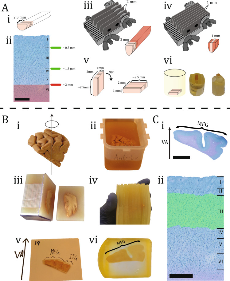
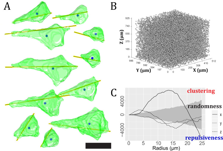
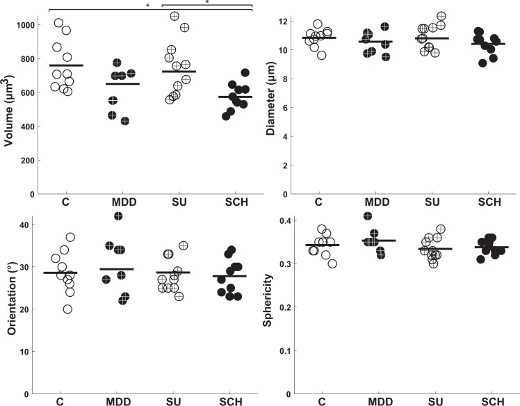
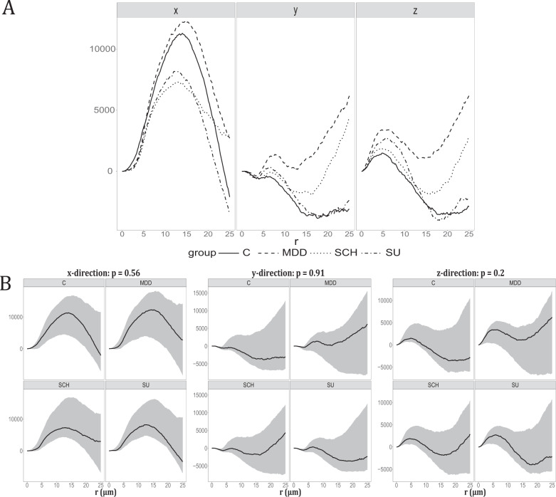
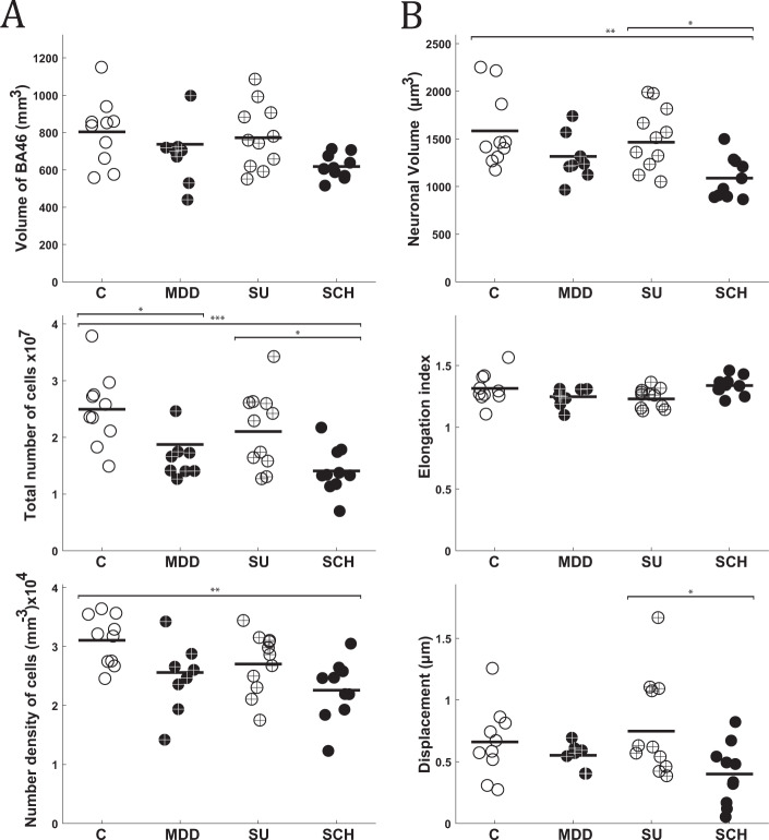
Brodmann Area 46 (BA46) has long been regarded as a hotspot of disease pathology in individuals with schizophrenia (SCH) and major depressive disorder (MDD). Pyramidal neurons in layer III of the Brodmann Area 46 (BA46) project to other cortical regions and play a fundamental role in corticocortical and thalamocortical circuits. The AutoCUTS-LM pipeline was used to study the 3-dimensional structural morphology and spatial organization of pyramidal cells. Using quantitative light microscopy, we used stereology to calculate the entire volume of layer III in BA46 and the total number and density of pyramidal cells. Volume tensors estimated by the planar rotator quantified the volume, shape, and nucleus displacement of pyramidal cells. All of these assessments were carried out in four groups of subjects: controls (C, n = 10), SCH (n = 10), MDD (n = 8), and suicide subjects with a history of depression (SU, n = 11). SCH subjects had a significantly lower somal volume, total number, and density of pyramidal neurons when compared to C and tended to show a volume reduction in layer III of BA46. When comparing MDD subjects with C, the measured parameters were inclined to follow SCH, although there was only a significant reduction in pyramidal total cell number. While no morphometric differences were observed between SU and MDD, SU had a significantly higher total number of pyramidal cells and nucleus displacement than SCH. Finally, no differences in the spatial organization of pyramidal cells were found among groups. These results suggest that despite significant morphological alterations in layer III of BA46, which may impair prefrontal connections in people with SCH and MDD, the spatial organization of pyramidal cells remains the same across the four groups and suggests no defects in neuronal migration. The increased understanding of pyramidal cell biology may provide the cellular basis for symptoms and neuroimaging observations in SCH and MDD patients.


Conflict of interest statement

The authors declare no competing interests.

Figures

Fig. 1
Fig. 1. Sample preparation.
A Biopsy preparation of the laminar site of the biopsy from BA46 is depicted in this illustration, with an average height of 2.5 mm (i). The six layers of BA46 can be seen in a cross-section of a biopsy after it has been stained with toluidine blue. Scale bar = 500 µm. Layer III is located within 2 mm of the pial surface (ii). A razor knife with a 2 mm gap between each blade was flipped on top of the biopsy. The red area shows the removed tissue (iii). A razor knife with a 1 mm gap between each blade was flipped on top of the biopsy. The red area shows the removed tissue (iv). The dimension of the tissue can be seen after the excess tissue has been removed. Before inserting the tissue within the embedding mold, it was flipped 90 to make sure the layers were parallel to the cutting direction (v). The illustration shows the tissue in the embedding mold. The resin block was photographed after it was trimmed with a glass knife, so it only contained layers I–IV (vi). B Tissue block preparation of DLPFC containing BA46 was rotated uniformly around a vertical axis (VA), perpendicular to the block’s central pial surface (i). The tissue block was embedded in a box filled with 7% agarose (ii). Next, the tissue block was cut systematically into uniformly random into 2.5 mm slabs orthogonal to the anterior-posterior direction (iii). The tissue block was then reassembled and every second the slab was systematically sampled (iv). After that, each slab was documented and various gyri positions were specified, e.g., medial frontal gyrus (MFG), inferior frontal gyrus (IFG) (v). Finally, the slabs were embedded in glycolmethacrylate (vi). C The section were stained with toluidine blue and VA was positioned based on documentation. Scale bar = 6 mm (i). The stereological analysis was then performed in layer III of BA46 based on the cytoarchitecture. Scale bar = 500 µm (ii).
Fig. 2
Fig. 2. Quantitative measurements of 3D-reconstruction.
A Pyramidal cell reconstruction in 3D, with yellow lines denoting orientation and solid circles denotes the centroid of pyramidal cells. Scale bar = 20 µm. B Point pattern consisting of centroids of pyramidal cells in layer III of BA46 acquired from a biopsy. C The results of a spatial point pattern analysis of pyramidal cell centroids using the cylindrical K-function. The empirical cylindrical K-function in the directions of the three axes is shown as three curves: x-axis (solid line), y-axis (dotted line), or z-axis (dashed line). The observed point pattern is completely random only if the curves fall within the envelope (gray area). When a curve falls above the envelope, it indicates evidence of clustering; when a curve falls below the envelope, it indicates evidence of repulsiveness. If the curve in one direction is particularly high, it indicates cylinder-shaped clusters in this direction.
Fig. 3
Fig. 3. Morphological results of the 3D-analysis of BA46 in layer III.
The estimated volume, diameter, orientation, and sphericity of pyramidal cells in layer III of BA46 between the four groups. Control (C), Major depressive disorder (MDD), Suicide (SU), and Schizophrenia (SCH). P < 0.05, ∗∗P < 0.01, ∗∗∗P < 0.001.
Fig. 4
Fig. 4. Spatial point pattern analysis.
A Plot of the weighted mean of each group. The weighted mean of the empirical cylindrical K-functions minus the theoretical value under CSR in the direction shown at the top. The line type corresponds to the groups: Control (C), Major depressive disorder (MDD), Suicide (SU), and Schizophrenia (SCH) as stated in the legend. B Test the difference between groups in the x, y, and z-axes.95% Global envelope test for testing the hypothesis of no differences in the cylindrical K-functions of the four groups Control (C), Major depressive disorder (MDD), Suicide (SU), and Schizophrenia (SCH). There is a test for the cylindrical K-function in each of the directions of the x-, y-, and z-axes. The 95% global envelopes (gray area) were based on 8000 random permutations. The solid curves are the estimates obtained from data after the theoretical value under CSR was subtracted. The direction is stated at the top of each plot, as well as the p-value of the test.
Fig. 5
Fig. 5. Morphological results of the stereological analysis of BA46 in layer III.
A Stereological measurements of the total volume, total neuronal number of pyramidal cells, and number density of pyramidal cells in layer III of BA46. B Volume tensor estimates of pyramidal cell somal size and shape in layer III of BA46, including volume, elongation index, and displacement. Control (C), Major depressive disorder (MDD), Suicide (SU), and Schizophrenia (SCH). P < 0.05, ∗∗P < 0.01, ∗∗∗P < 0.001.

References

    1. Samsom JN, Wong AHC. Schizophrenia and depression comorbidity: what we have learned from animal models. Front Psychiatry 2015;6. 10.3389/fpsyt.2015.00013. - PMC - PubMed
    1. Witt SH, Streit F, Jungkunz M, Frank J, Awasthi S, Reinbold CS, et al. Genome-wide association study of borderline personality disorder reveals genetic overlap with bipolar disorder, major depression and schizophrenia. Transl Psychiatry. 2017;7:e1155–e1155. doi: 10.1038/tp.2017.115. - DOI - PMC - PubMed
    1. Miller EK, Cohen. JD. An integrative theory of prefrontal cortex function. Annu Rev Neurosci. 2001;24:167–202. doi: 10.1146/annurev.neuro.24.1.167. - DOI - PubMed
    1. Lewis DA, Curley AA, Glausier JR, Volk DW. Cortical parvalbumin interneurons and cognitive dysfunction in schizophrenia. Trends Neurosci. 2012;35:57–67. doi: 10.1016/j.tins.2011.10.004. - DOI - PMC - PubMed
    1. Arnsten AFT, Jin LE. Molecular influences on working memory circuits in dorsolateral prefrontal cortex. Prog Mol Biol Transl Sci. 2014:211–31. 10.1016/b978-0-12-420170-5.00008-8. - PubMed

Publication types