Review
. 2022 Nov;24(11):2000-2018.
doi: 10.1002/ejhf.2669. Epub 2022 Oct 6.

Myocarditis following COVID-19 vaccine: incidence, presentation, diagnosis, pathophysiology, therapy, and outcomes put into perspective. A clinical consensus document supported by the Heart Failure Association of the European Society of Cardiology (ESC) and the ESC Working Group on Myocardial and Pericardial Diseases

Affiliations
Review

Myocarditis following COVID-19 vaccine: incidence, presentation, diagnosis, pathophysiology, therapy, and outcomes put into perspective. A clinical consensus document supported by the Heart Failure Association of the European Society of Cardiology (ESC) and the ESC Working Group on Myocardial and Pericardial Diseases

Bettina Heidecker et al. Eur J Heart Fail. 2022 Nov.

Erratum in

Abstract

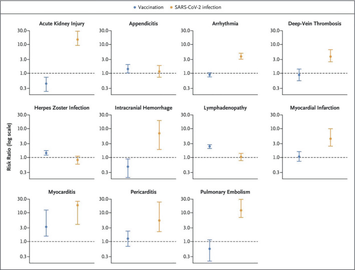
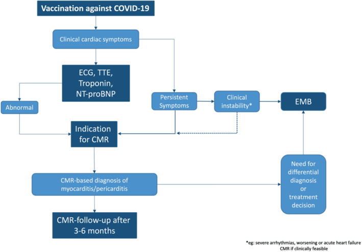
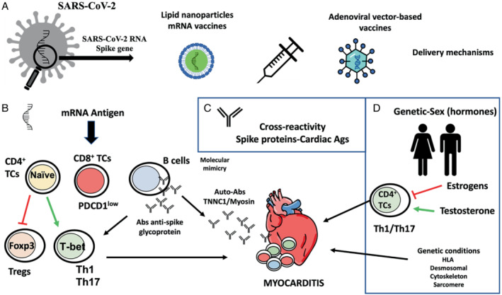
Over 10 million doses of COVID-19 vaccines based on RNA technology, viral vectors, recombinant protein, and inactivated virus have been administered worldwide. Although generally very safe, post-vaccine myocarditis can result from adaptive humoral and cellular, cardiac-specific inflammation within days and weeks of vaccination. Rates of vaccine-associated myocarditis vary by age and sex with the highest rates in males between 12 and 39 years. The clinical course is generally mild with rare cases of left ventricular dysfunction, heart failure and arrhythmias. Mild cases are likely underdiagnosed as cardiac magnetic resonance imaging (CMR) is not commonly performed even in suspected cases and not at all in asymptomatic and mildly symptomatic patients. Hospitalization of symptomatic patients with electrocardiographic changes and increased plasma troponin levels is considered necessary in the acute phase to monitor for arrhythmias and potential decline in left ventricular function. In addition to evaluation for symptoms, electrocardiographic changes and elevated troponin levels, CMR is the best non-invasive diagnostic tool with endomyocardial biopsy being restricted to severe cases with heart failure and/or arrhythmias. The management beyond guideline-directed treatment of heart failure and arrhythmias includes non-specific measures to control pain. Anti-inflammatory drugs such as non-steroidal anti-inflammatory drugs, and corticosteroids have been used in more severe cases, with only anecdotal evidence for their effectiveness. In all age groups studied, the overall risks of SARS-CoV-2 infection-related hospitalization and death are hugely greater than the risks from post-vaccine myocarditis. This consensus statement serves as a practical resource for physicians in their clinical practice, to understand, diagnose, and manage affected patients. Furthermore, it is intended to stimulate research in this area.

Keywords: COVID-19; Inflammation; Myocarditis; Outcomes; Pathology; Vaccination.


Figures

Figure 1
Figure 1
Risk of complications after COVID‐19 vaccine versus with COVID‐19 infection: data were obtained from a national study in Israel. Each cohort consisted of more than 800 000 individuals. Relative risk for developing myocarditis after vaccine was 3.2, while it is 18.3 after getting COVID‐19. From Barda et al.
Figure 2
Figure 2
Twelve‐lead electrocardiogram of a 24‐year‐old man who developed severe, stabbing chest pain radiating to both shoulders and markedly elevated high‐sensitivity troponin T levels (peak 635 ng/L) after receiving the BNT162b2 mRNA vaccine as a booster. There are conduction abnormalities (aVL, III, aVF) and non‐specific ST‐segment changes in the precordial leads.
Figure 3
Figure 3
Cardiac magnetic resonance (CMR) images of a patient with signs of a myopericarditis after mRNA SARS‐CoV‐2 vaccination with Spikevax (Moderna). Full description of this case can be found in Jahnke et al. One day after vaccination the patient complained about chest pain and discomfort, shortness of breath, limited physical capacity and malaise. High‐sensitivity troponin T was elevated up to 526 ng/L (normal <14 ng/L). C‐reactive protein, N‐terminal pro‐B‐type natriuretic peptide, electrocardiogram, echocardiography, coronary angiogram and computed tomography pulmonary angiography were normal. CMR was normal for function (including strain) (A), but demonstrated slight pericardial effusion (red arrow in A). T2 weighted images indicated a regional oedema anterolateral/inferolateral (basal) with corresponding elevated quantitative myocardial T2‐mapping parameters up to 70 ms (normal up to 51 ms at 3 Tesla) (C, E – red arrows). Patchy subepicardial late gadolinium enhancement (LGE) indicating inflammatory myocardial necrosis (G). Pericardial enhancement in the T2‐weighted and LGE images in corresponding locations indicated a pericardial involvement as well (C, E). The findings resolved at 4‐month CMR follow‐up (green arrow in B, D, F, H).
Figure 4
Figure 4
(A) Patient with signs of a myocarditis after mRNA SARS‐CoV‐2 vaccination with Comirnaty (Pfizer‐BioNTech). Three days after the second dose of the vaccine, the patient presented to the emergency room of a referring hospital with chest pain and discomfort, shortness of breath, and decreased exercise capacity. High‐sensitivity troponin T level was elevated at 380 ng/L; N‐terminal pro‐B‐type natriuretic peptide 250 ng/L (<88). Cardiac magnetic resonance (CMR) demonstrated normal left and right ventricular ejection fraction (A, B), with reduced global longitudinal strain. T2‐weighted images indicated a regional oedema inferior/inferolateral (basal) (in D, E) with corresponding elevated quantitative myocardial T2‐mapping parameters (C) and corresponding subepicardial focal late gadolinium enhancement (F, G). (B) The findings at baseline (indicated by red arrow) resolved almost completely (small epicardial fibrosis inferolateral basal) at 4‐month CMR follow‐up (second figure – green arrow). Full description of this case can be found in Jahnke et al.
Figure 5
Figure 5
Potential workflow for the use of advanced cardiac magnetic resonance (CMR) in patients post‐vaccination and suspected myo‐/pericarditis. ECG, electrocardiogram; EMB, endomyocardial biopsy; NT‐proBNP, N‐terminal pro‐B‐type natriuretic peptide; TTE, transthoracic echocardiography. Modified from Doeblin and Kelle61
Figure 6
Figure 6
(A) In acute enteroviral myocarditis, myocyte necrosis in the presence of numerous CD3+ T cells and CD68+ macrophages (32‐year‐old male). (B) Some patients with COVID‐19 develop acute/subacute myocarditis (17‐year‐old female). (C) The majority of patients develop low levels of T‐cell infiltrates, but numerous macrophages (38‐year‐old male). Similar findings are observed in mRNA vaccinated patients. Rare cases present with acute/subacute myocarditis (D, 37‐year‐old male) in endomyocardial biopsy, but most patients reveal mild inflammation and suffer from pre‐existing diseases such as hypertensive heart disease (E, 56‐year‐old male). All images magnification ×200.
Figure 7
Figure 7
Potential molecular mechanisms for the development of myocarditis following vaccines against COVID‐19. (A) COVID‐19 vaccines are developed from the modified SARS‐CoV‐2 Spike gene. The mRNA vaccines are introduced via lipid nanoparticles, while the adenoviral vector‐based vaccines deliver the Spike sequence as a codon‐optimized DNA. (B) The mRNA can act as an antigen, so it can be recognized by the immune system and activate specific responses of the adaptive immune system. Some of these responses are capable of activating cardiotropic clones of T and B cells triggering cardiac inflammation. (C) Molecular mimicry between Spike glycoprotein and myosin heavy chain or troponin C1, may trigger cross‐reactivity between IgM antibodies against SARS‐CoV‐2 Spike glycoprotein and cardiac autoantigens and potentially induce myocardial inflammation. (D) Development of SARS‐CoV‐2 vaccine myocarditis is associated with young men, suggesting a role for sex hormones. Testosterone activates specific T helper cell responses, whereas oestrogen inhibits pro‐inflammatory T‐cell responses. In addition, viral myocarditis is associated with genetic variants of genes encoding for different HLA factors and structural proteins of the heart.

References

    1. Johns Hopkins Corona Virus Resource Center . COVID‐19 Dashboard. https://coronavirus.jhu.edu/map.html (last accessed 8 September 2022).
    1. Shay DK, Shimabukuro TT, DeStefano F. Myocarditis occurring after immunization with mRNA‐based COVID‐19 vaccines. JAMA Cardiol. 6:1115–7. - PubMed
    1. Kim HW, Jenista ER, Wendell DC, Azevedo CF, Campbell MJ, Darty SN, et al. Patients with acute myocarditis following mRNA COVID‐19 vaccination. JAMA Cardiol. 2021;6:1196–201. - PMC - PubMed
    1. Shiyovich A, Witberg G, Aviv Y, Eisen A, Orvin K, Wiessman M, et al. Myocarditis following COVID‐19 vaccination: magnetic resonance imaging study. Eur Heart J Cardiovasc Imaging. 2021;23:1075–82. - PubMed
    1. Gargano JW, Wallace M, Hadler SC, Langley G, Su JR, Oster ME, et al. Use of mRNA COVID‐19 vaccine after reports of myocarditis among vaccine recipients: update from the Advisory Committee on Immunization Practices ‐ United States, June 2021. MMWR Morb Mortal Wkly Rep. 2021;70:977–82. - PMC - PubMed

Substances