. 2022 Nov 1;222(3):iyac136.
doi: 10.1093/genetics/iyac136.

A balancing act: interactions within NuA4/TIP60 regulate picNuA4 function in Saccharomyces cerevisiae and humans

Affiliations

A balancing act: interactions within NuA4/TIP60 regulate picNuA4 function in Saccharomyces cerevisiae and humans

Phoebe Y T Lu et al. Genetics. .

Abstract

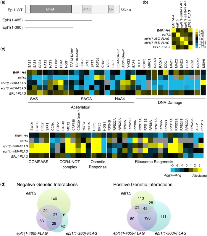
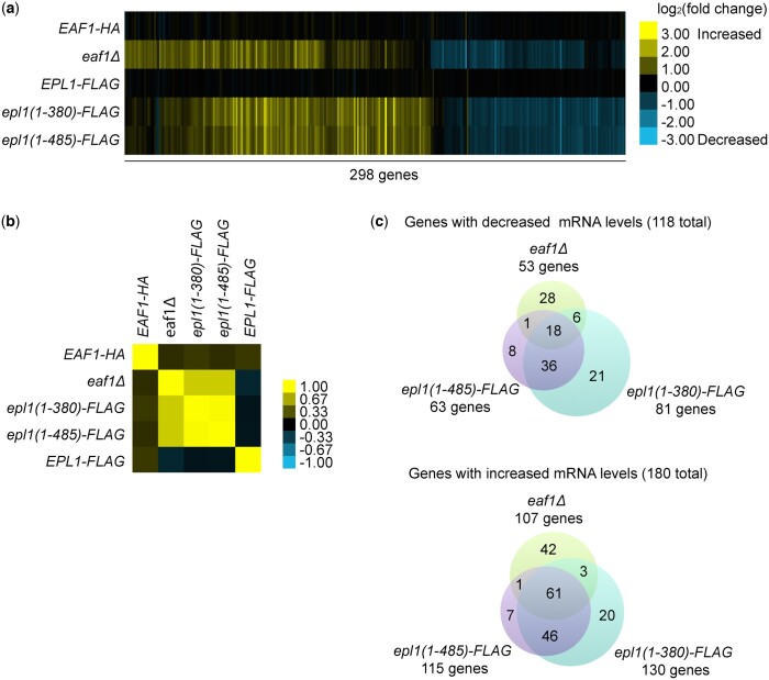
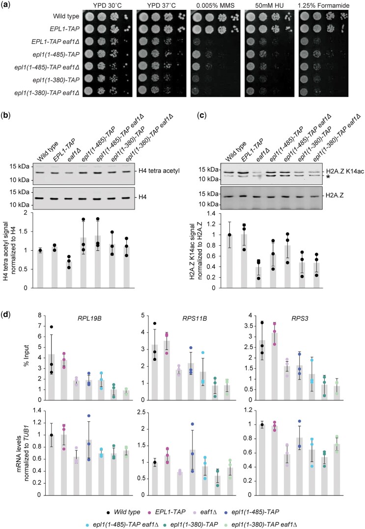
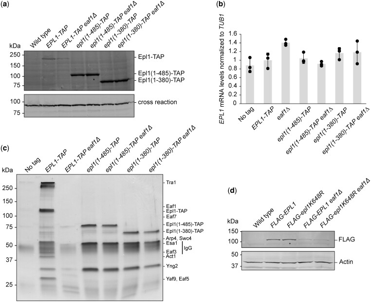
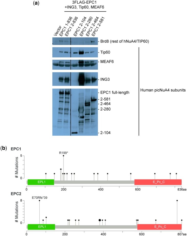
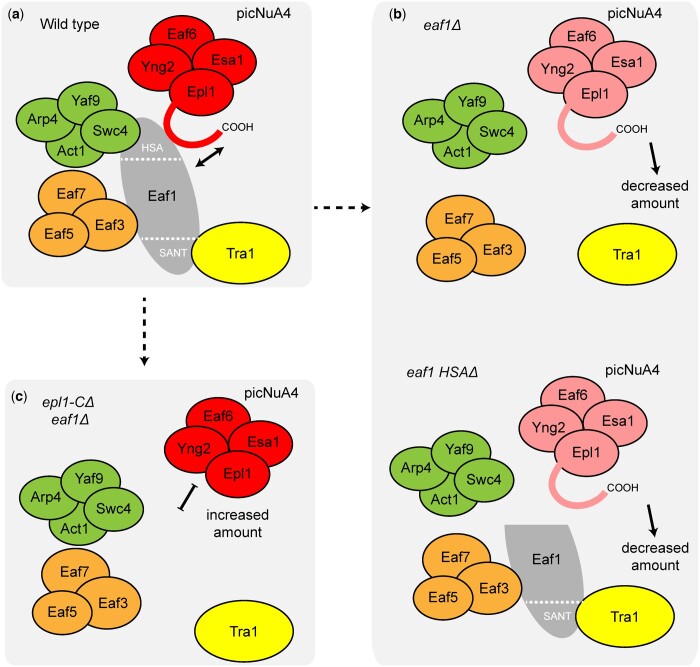
The NuA4 lysine acetyltransferase complex acetylates histone and nonhistone proteins and functions in transcription regulation, cell cycle progression, and DNA repair. NuA4 harbors an interesting duality in that its catalytic module can function independently and distinctly as picNuA4. At the molecular level, picNuA4 anchors to its bigger brother via physical interactions between the C-terminus of Epl1 and the HSA domain of Eaf1, the NuA4 central scaffolding subunit. This is reflected at the regulatory level, as picNuA4 can be liberated genetically from NuA4 by disrupting the Epl1-Eaf1 interaction. As such, removal of either Eaf1 or the Epl1 C-terminus offers a unique opportunity to elucidate the contributions of Eaf1 and Epl1 to NuA4 biology and in turn their roles in balancing picNuA4 and NuA4 activities. Using high-throughput genetic and gene expression profiling, and targeted functional assays to compare eaf1Δ and epl1-CΔ mutants, we found that EAF1 and EPL1 had both overlapping and distinct roles. Strikingly, loss of EAF1 or its HSA domain led to a significant decrease in the amount of picNuA4, while loss of the Epl1 C-terminus increased picNuA4 levels, suggesting starkly opposing effects on picNuA4 regulation. The eaf1Δ epl1-CΔ double mutants resembled the epl1-CΔ single mutants, indicating that Eaf1's role in picNuA4 regulation depended on the Epl1 C-terminus. Key aspects of this regulation were evolutionarily conserved, as truncating an Epl1 homolog in human cells increased the levels of other picNuA4 subunits. Our findings suggested a model in which distinct aspects of the Epl1-Eaf1 interaction regulated picNuA4 amount and activity.

Keywords: chromatin; gene regulation; histone acetylase; histone acetylation; yeast genetics.


Figures

Fig. 1.
Fig. 1.
Genetic interaction analysis revealed similarities and distinctions between eaf1 and epl1 mutants. a) Schematic representation of the epl1-CΔ mutations. The conserved EpcA domain is shown, along with the alanine and glutamine rich regions. b) Spearman’s rho correlation of genetic interaction profiles of wild type and the indicated mutant strains. Yellow and blue indicate positive and negative correlations respectively. c) Representative genetic interactions for the eaf1Δ and epl1-CΔ mutants with genes encoding subunits of the indicated complexes. Blue indicates aggravating interactions, yellow represents alleviating interactions, and gray denotes missing data. d) Venn diagrams showing significant negative and positive genetic interactions between eaf1Δ, epl1(1-485), and epl1(1-380).
Fig. 2.
Fig. 2.
Gene expression profiles supported shared and unique roles for EAF1 and EPL1. a) Heat map showing the log2 fold changes for 298 genes with significant changes in expression in at least one of the eaf1Δ or epl1-CΔ mutants. Absolute fold changes greater than 1.7 (corresponding to values greater than 0.77 or less than −0.77 on the heatmap) were considered significant. Yellow indicates upregulated genes, and blue represents downregulated genes. b) Spearman’s rho correlation of the gene expression profile of wild type and mutant strains. c) Venn diagrams showing significantly upregulated and downregulated genes in eaf1Δ, epl1(1-485), and epl1(1-380).
Fig. 3.
Fig. 3.
Epl1 C-terminus truncation masked some of the effects of deleting EAF1. a) Logarithmic serial dilutions of the indicated strains were plated on YPD media, with or without genotoxic agents, and incubated for 3 days at the indicated temperature. The eaf1Δ and epl1-CΔ mutants showed decreased growth in the presence of the genotoxic agents. The eaf1Δ epl1(1-485) mutant showed milder growth defects under MMS compared to the eaf1Δ single mutant. Bulk histone acetylation was examined from whole cell extracts of the indicated strains, analyzed by protein blotting with (b) anti-tetra-acetylated H4 and (c) anti-H2A.Z K14ac antibodies. Antibodies against H4 and H2A.Z were used as loading controls. * denotes a background band in the H2A.Z K14ac immunoblot; we note that it fluctuates for unknown reasons in the epl1-CΔ mutants. The tetra-acetylated H4 and H2A.Z K14ac signals were quantified relative to the H4 and H2A.Z signals, respectively, and normalized to the wild-type strain. Averages were taken across 3 replicates, with error bars indicating the standard deviation. d) Disruption of NuA4 function in the eaf1Δ and epl1-CΔ single and double mutants resulted in both decreased mRNA levels and H4 promoter acetylation at RP candidate genes: RPL19B, RPS11B, and RPS3. Enrichment of tetra-acetylated H4 was normalized to % input. mRNA levels were measured by RT-qPCR and normalized to TUB1 mRNA levels. Error bars represent standard deviation of the means for 3 independent experiments.
Fig. 4.
Fig. 4.
The Epl1 C-terminus was required for its interaction with chromatin, independent of EAF1 status. a) Epl1’s association with chromatin was assessed by western blot analysis of cellular fractions generated by centrifugation of whole cell extract over a sucrose gradient. W, whole cell extract; S, soluble nonchromatin fraction; C, chromatin pellet. In each case, the fractionation efficiency was judged by the levels of H4 and Pgk1, found in the chromatin and the supernatant fraction, respectively. b) Epl1 enrichment at the promoters of RP genes was lost in the epl1-CΔ mutants, based on ChIP-qPCR analysis of the representative genes RPL21A, RPL39, RPL17B, RPL19B, RPS11B, and RPS3. Results are depicted as box plots overlaid with the individual measurements from each replicate.
Fig. 5.
Fig. 5.
Truncating Epl1 stabilized picNuA4 and increased Epl1 protein levels independent of Eaf1 status. a) Epl1 protein levels were strongly decreased in the eaf1Δ mutant but increased in the epl1-CΔ mutants. A cross-reactive band was used as a loading control. b) EPL1 expression was similar across all strains. The mRNA levels of EPL1 was measured by RT-qPCR and normalized to levels of TUB1 mRNA. Error bars represent the standard error of 3 independent biological replicates. c) Immunopurification of NuA4 using Epl1-TAP demonstrated a decrease in the amount of Epl1-associated NuA4 subunits in the eaf1Δ mutant, while only subunits found in picNuA4 were observed in the epl1-CΔ mutants. Purified fractions from indicated strains were loaded onto a 4–20% gradient SDS-PAGE gel and visualized by silver staining. Bands corresponding to NuA4 subunits are indicated on the left. Untagged Epl1 was used as a negative control. d) The Epl1 K648R point mutation did not restore Epl1 protein levels in the eaf1Δ mutant, as measured in whole cell extracts from strains expressing the indicated plasmid-derived FLAG-Epl1 constructs. PGK1 was used as a loading control.
Fig. 6.
Fig. 6.
The effect of truncating Epl1 on picNuA4 levels was conserved from yeast to humans. a) The EPC1(2-280) mutant that is homologous to Epl1(1-380) led to increased picNuA4 levels. A series of C-terminal truncation mutants of 3FLAG-EPC1 were immunopurified after cotransfection with HA-Tip60/Kat5, HA-ING3, and HA-MEAF6 and analyzed by SDS-PAGE followed by immunoblotting using antibodies against FLAG and the indicated hNuA4/TIP60 subunits. b) The human homologs of EPL1, EPC1, and EPC2 were truncated in cancer patients. Lollipop plots of the EPC1 and EPC2 proteins show the location of previously identified truncating mutations, along with annotated protein domains. Analysis and images were generated using the cBio Cancer Genomics Portal (Cerami et al. 2012; Gao et al. 2013).
Fig. 7.
Fig. 7.
The Eaf1 HSA domain was required for NuA4 stability and function. a) Schematic representation of Eaf1 internal deletion alleles: HSAΔ, SANTΔ, and HSAΔ/SANTΔ. b) The loss of the HSA domain led to a similar defect in bulk histone acetylation as the complete loss of EAF1. Whole cell extracts of the indicated strains were analyzed by protein blotting with anti-tetra-acetylated H4 or anti-H2A.Z K14ac antibodies. Antibodies against H4 and H2A.Z were used as a loading controls. * indicates a background band. c) Logarithmic serial dilutions of the indicated strains were plated onto SC-URA media containing the indicated genotoxic agents and incubated for 3 days at the indicated temperature. Strains lacking the Eaf1 HSA domain recapitulated the eaf1Δ mutant phenotype. d) Purification of Epl1 revealed decreased association of NuA4 subunits in the eaf1 HSAΔ strain. Purified fractions from the indicated strains were loaded onto a 4–20% gradient SDS–PAGE gel and visualized by silver staining. Bands corresponding to NuA4 subunits are indicated on the left. Untagged Epl1 was used as a negative control. e) Epl1 purifications of the indicated strains were visualized by immunoblotting to verify Epl1-immunoprecipitation and Esa1/Kat5 association. IgG and anti-Esa1/Kat5 antibodies were used to detect Epl1-TAP and Esa1/Kat5, respectively.
Fig. 8.
Fig. 8.
Model for the regulatory mechanisms of Eaf1 and the Epl1 C-terminus in picNuA4 stability. a) The catalytic submodule (picNuA4) associates with NuA4 through the Epl1 C-terminus and the Eaf1 HSA domain. b) Deletion of Eaf1 or its HSA domain leads to reduced picNuA4 levels, perhaps indicative of decreased protein stability. c) Truncation of the Epl1 C-terminus in an eaf1Δ background increases picNuA4 levels and restores H4 acetylation.

Similar articles

Cited by

References

    1. Allard S, Utley RT, Savard J, Clarke A, Grant P, Brandl CJ, Pillus L, Workman JL, Côté J.. NuA4, an essential transcription adaptor/histone H4 acetyltransferase complex containing Esa1p and the ATM-related cofactor Tra1p. EMBO J. 1999;18(18):5108–5119. - PMC - PubMed
    1. Auger A, Galarneau L, Altaf M, Nourani A, Doyon Y, Utley RT, Cronier D, Allard S, Côté J.. Eaf1 is the platform for NuA4 molecular assembly that evolutionarily links chromatin acetylation to ATP-dependent exchange of histone H2A variants. Mol Cell Biol. 2008;28(7):2257–2270. - PMC - PubMed
    1. Ausubel M, Brent R, Kingston RE, Moore DD, Seidman JG, Smith JA, Struhl K. Current Protocols in Molecular Biology. PA: John Wiley & Sons, Inc., Media; 1987.
    1. Avvakumov N, Lalonde M-E, Saksouk N, Paquet E, Glass KC, Landry A-J, Doyon Y, Cayrou C, Robitaille GA, Richard DE, et al.Conserved molecular interactions within the HBO1 acetyltransferase complexes regulate cell proliferation. Mol Cell Biol. 2012;32(3):689–703. - PMC - PubMed
    1. Babiarz JE, Halley JE, Rine J.. Telomeric heterochromatin boundaries require NuA4-dependent acetylation of histone variant H2A.Z in Saccharomyces cerevisiae. Genes Dev. 2006;20(6):700–710. - PMC - PubMed

Publication types

MeSH terms