. 2022 Dec;530(18):3179-3192.
doi: 10.1002/cne.25401. Epub 2022 Sep 6.

The parabigeminal nucleus is a source of "retinogeniculate replacement terminals" in mice that lack retinofugal input

Affiliations

The parabigeminal nucleus is a source of "retinogeniculate replacement terminals" in mice that lack retinofugal input

Kyle L Whyland et al. J Comp Neurol. 2022 Dec.

Abstract

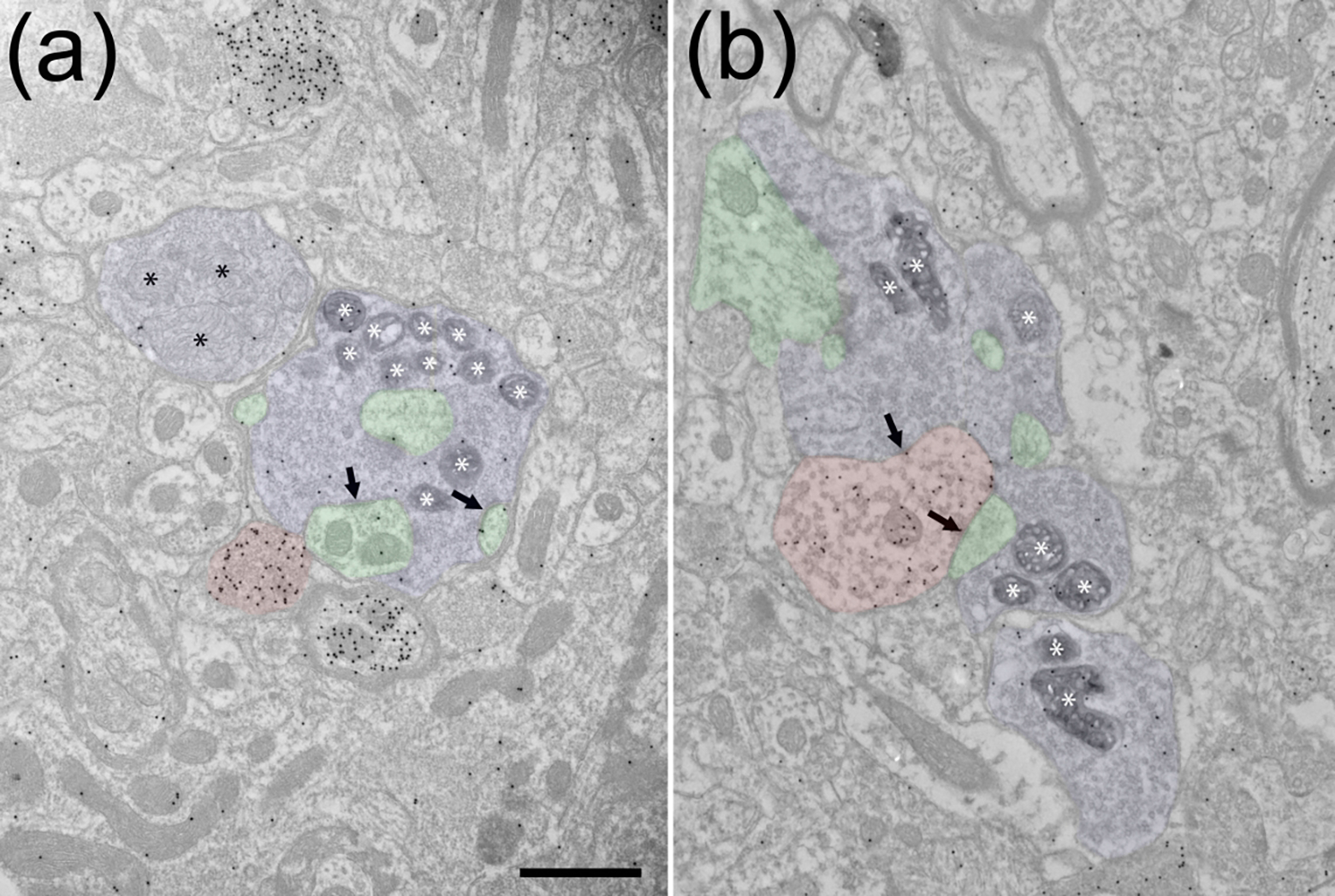
In the dorsal lateral geniculate nucleus (LGN) of mice that lack retinal input, a population of large terminals supplants the synaptic arrangements normally made by the missing retinogeniculate terminals. To identify potential sources of these "retinogeniculate replacement terminals," we used mutant mice (math5-/- ) which lack retinofugal projections due to the failure of retinal ganglion cells to develop. In this line, we labeled LGN terminals that originate from the primary visual cortex (V1) or the parabigeminal nucleus (PBG), and compared their ultrastructure to retinogeniculate, V1 or PBG terminals in the dLGN of C57Blk6 (WT) mice (schematically depicted above graph). Corticogeniculate terminals labeled in WT and math5-/- mice were similar in size and both groups were significantly smaller than WT retinogeniculate terminals. In contrast, the PBG projection in math5-/- mice was extensive and there was considerable overlap in the sizes of retinogeniculate terminals in WT mice and PBG terminals in math5-/- mice (summarized in histogram). The data indicate that V1 is not a source of "retinogeniculate replacement terminals" and suggests that large PBG terminals expand their innervation territory to replace retinogeniculate terminals in their absence.

Keywords: RpB4-cre; corticogeniculate; dorsal lateral geniculate nucleus; math5-/-; pedunculopontine tegmentum; synapse; tectogeniculate; ultrastructure.


Conflict of interest statement

Conflict of interest: The authors declare no competing financial interests.

Figures

Figure 1:
Figure 1:. Retinogeniculate terminals in WT mice.
Electron micrographs illustrate ipsilateral (a) and contralateral (b) retinogeniculate terminals (blue overlay) labeled via monocular virus injections in C57Blk6 mice that induced the expression of peroxidase in mitochondria (white asterisks). The tissue was stained to detect GABA with gold particles. Unlabeled retinogeniculate terminals contain pale mitochondria (black asterisks, a). Retinogeniculate terminals both contain and synapse (black arrows) on the dendrites of nonGABAergic thalamocortical cells (a, green overlay, identified by a low density of gold particles) that often protrude into the terminals. Retinogeniculate terminals also contact the dendritic terminals of GABAergic interneurons (b, F2 profiles, red overlay, GABA content identified by a high density of overlying gold particles). The illustrated F2 profile synapses (black arrow) on a thalamocortical cell dendrite (green overlay, identified by a low density of overlying gold particles). Scale bar = 1 μm and applies to both panels.
Figure 2:
Figure 2:. Corticogeniculate terminals in WT and math5−/− mice.
Electron micrographs illustrate corticogeniculate terminals labeled via V1 BDA injections in C57Blk6 (a) and math5−/− (b-d) mice. The tissue was stained to detect GABA with gold particles. In both WT and math5−/− mice, V1 terminals are small profiles that contain round synaptic vesicles (RS profiles) that synapse (white arrows) on thalamocortical cell dendrites (green overlay, low density of gold particles). In the electron micrograph illustrating the WT corticogeniculate terminal, an adjacent unlabeled retinal terminal (blue overaly, identified by its pale mitochondria, black asterisks) also contacts (black arrow) a thalamocortical cell dendrite (green overlay). Scale bar = 800nm and applies to all panels.
Figure 3:
Figure 3:
Retinogeniculate terminals in C57Blk6 (WT) mice are significantly larger than corticogeniculate terminals in WT mice and math5−/− mice (**** p < 0.0001). There is no significant difference in the size of ipsilateral and contralateral retinogeniculate terminals in WT mice and no significant difference in the sizes of corticogeniculate terminals in WT and math5−/− mice (ns).
Figure 4:
Figure 4:. Cre-dependent V1 and SC projections to the dLGN of Rpb4-cre and Rpb4-cre/math5−/− mice.
Cre-dependent (cell-filling) virus injections in V1 in Rpb4-cre (a,b) and Rpb4-cre/ math5−/− mice (e,f) labeled a dense population of cells in layer V that extended dendrites into layers I-IV, a smaller population of cells in layer VI, dense population of terminals in the ipsilateral pulvinar (PUL) and ventral lateral geniculate nucleus (vLGN), and axons that course through the ipsilateral dorsal lateral geniculate nucleus (dLGN). In Rpb4-cre mice, most labeled axons avoid the optic tract (b, arrow). In Rbb4-cre/ math5−/− mice, the position of many of the labeled axons shifted dorsally to occupy the position of the missing optic tract (f, arrow). Cre-dependent (cell-filling) virus injections in the SC of Rpb4-cre (c,d) and Rbb4-cre/math5−/− mice (g,h) labeled subpopulation of cells in the stratum griseum superficiale (SGS), stratum griseum intermediale (SGI) and stratum griseum profundum (SGP) and a sparse population of terminals in the ipsilateral dorsolateral shell of the dLGN (d,h arrows). Scale in a = 500 μm and also applies to e. Scale in h = 100 μm and also applies to b-d and f-g.
Figure 5:
Figure 5:
Electron micrographs illustrate the ultrastructure of the dLGN in Rpb4-cre mice (a,c,e) and Rpb4-cre/Math5−/− mice (b,d,f) that received cre-dependent (cell-filling) virus injections in the ipsilateral V1. In Rpb4-cre mice, the optic tract at the dorsal surface of the dLGN is a thick layer of unlabeled myelinated axons (a). In Rpb4/math5−/− mice, the dorsal surface of the dLGN contains labeled axons in the location of the missing optic tract (b). Labeled axons course through the body of the dLGN in both Rpb4-cre (c) and Rpb4/math5−/− (d) mice. In both Rpb4-cre and Rpb4-cre/math5−/− mice, sparse labeled terminals that displayed RS morphology (e, white arrow indicates synapse, RPb4-cre) and unmyelinated fibers that contained vesicles (f, white arrow Rpb4-cre/math5−/−) were occasionally observed. Scale bars: a = 8 μm, b= 2 μm, c = 4 μm, d = 2 μm, e = 800 nm, f = 1 μm.
Figure 6:
Figure 6:. SC and PPT projections to the dLGN of WT and math5−\− mice.
Tectogeniculate projections labeled via virus injections in the ipsilateral SC innervate the dorsolateral shell of the dLGN in WT (a; from Bickford et al. 2015) and math5−\− mice (b). An antibody against the vesicular acetylcholine transporter labels small diffusely distributed terminals in both WT (c; from Sokhadze et al. 2022) and math5−\− mice (d). Scale in a = 100 μm and also applies to b. Scale in c = 50 μm and also applies to d.
Figure 7:
Figure 7:. PBG terminals in the dLGN of WT and math5−/− mice.
Electron micrographs illustrate the ulstrastructure of dLGN terminals labeled via unilateral PBG (cell-filling) virus injections in math5−/− (a, ipsilateral dLGN c, contralateral dLGN) and C57Blk6 mice (c, contralateral dLGN). PBG terminals in the dLGN are large profiles that often contain and synapse (white arrows) on dendrites (green overlay) that protrude into the labeled terminals. Scale bar = 800nm and applies to all panels.
Figure 8:
Figure 8:
Retinogeniculate terminals in C57Blk6 (WT) mice are significantly larger than PBG-dLGN terminals in math5−\− mice (**** p < 0.0001). There is no significant difference in the size of ipsilateral and contralateral retinogeniculate terminals in WT mice (ns). Ipsilateral PBG-dLGN terminals are significantly larger than contralateral PBG-dLGN terminals in math5−\− mice (** p = 0.0083).
Figure 9:
Figure 9:
There is little overlap in the sizes of retinogeniculate terminals in C75Blk6 mice (blue, ipsilateral and contralateral measurements combined) and corticogeniculate terminals in math5−\− mice (green). However, there is considerable overlap in the sizes of retinogeniculate terminals in C57Blk6 mice and PBG-dLGN terminals in math5−\− mice (red, ipsilateral and contralateral measurements combined).
Figure 10:
Figure 10:
Ipsilateral PBG-dLGN terminals in math5−\− mice are significantly larger than contralateral PBG-dLGN terminals in both C57Blk6 (WT) and math5−\− mice (** p = 0.0038; **** p < 0.0001). There is no significant difference between contralateral PBG-dLGN terminals in WT mice and contralateral PBG-dLGN terminals in math5−\− mice (ns).

References

    1. Abbott CW, Kozanian OO, & Huffman KJ (2015). The effects of lifelong blindness on murine neuroanatomy and gene expression. Frontiers in Aging Neuroscience, 7(JUL). 10.3389/fnagi.2015.00144 - DOI - PMC - PubMed
    1. Adesnik H (2018). Layer-specific excitation/inhibition balances during neuronal synchronization in the visual cortex. Journal of Physiology, 596(9), 1639–1657. 10.1113/JP274986 - DOI - PMC - PubMed
    1. Augustinaite S, & Kuhn B (2020). Complementary Ca2+ Activity of Sensory Activated and Suppressed Layer 6 Corticothalamic Neurons Reflects Behavioral State. Current Biology, 30(20), 3945–3960.e5. 10.1016/j.cub.2020.07.069 - DOI - PubMed
    1. Basso MA, Bickford ME, & Cang J (2021). Unraveling circuits of visual perception and cognition through the superior colliculus. In Neuron (Vol. 109, Issue 6, pp. 918–937). Cell Press. 10.1016/j.neuron.2021.01.013 - DOI - PMC - PubMed
    1. Bhandari A, Ward TW, Smith J, & van Hook MJ (2022). Structural and Functional Plasticity in the Dorsolateral Geniculate Nucleus of Mice following Bilateral Enucleation. Neuroscience, 488, 44–59. 10.1016/j.neuroscience.2022.01.029 - DOI - PMC - PubMed

Publication types