Review
. 2022 Sep 6;79(10):511.
doi: 10.1007/s00018-022-04536-3.

The heterogeneity of microglial activation and its epigenetic and non-coding RNA regulations in the immunopathogenesis of neurodegenerative diseases

Affiliations
Review

The heterogeneity of microglial activation and its epigenetic and non-coding RNA regulations in the immunopathogenesis of neurodegenerative diseases

Chaoyi Li et al. Cell Mol Life Sci. .

Abstract

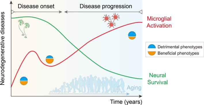
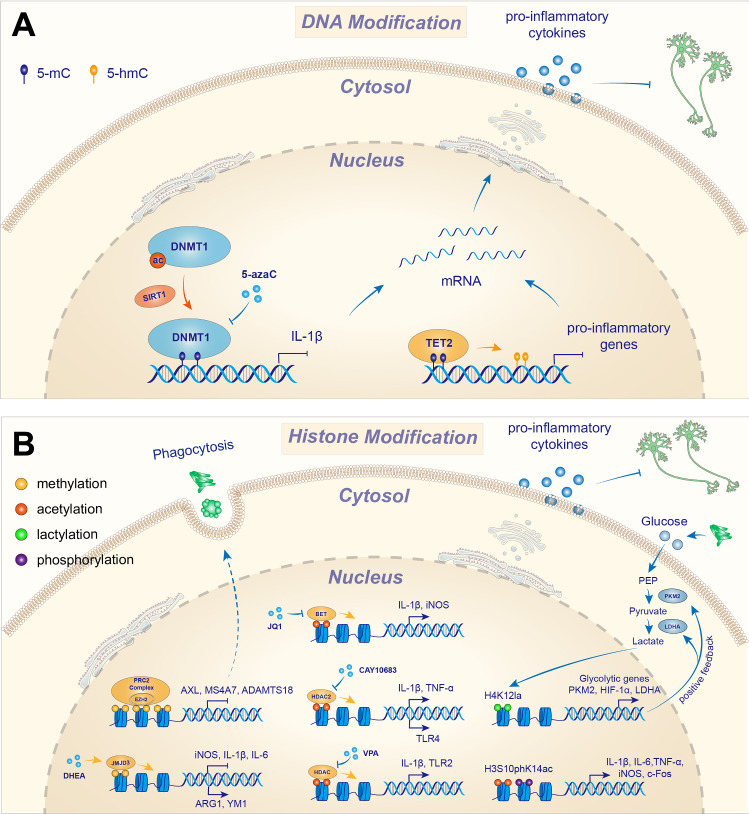
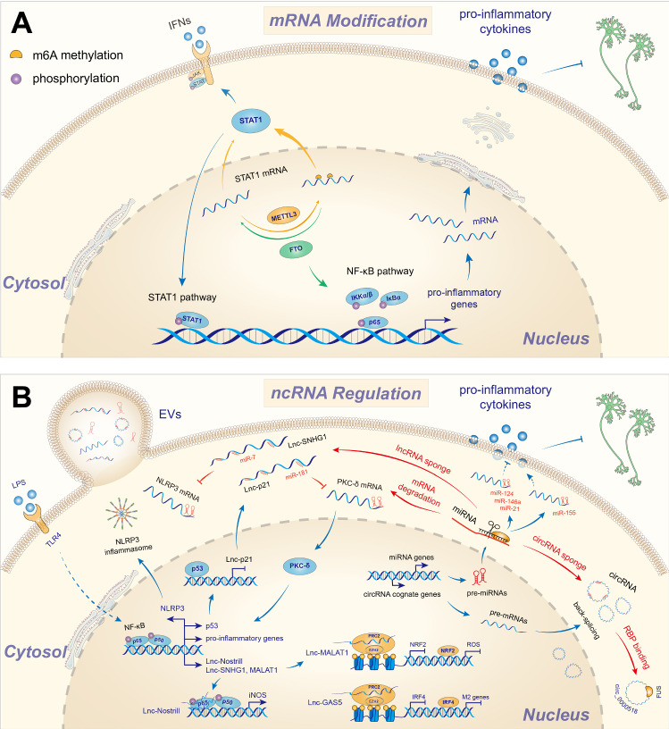
Microglia are resident immune cells in the brain and play a central role in the development and surveillance of the nervous system. Extensive gliosis is a common pathological feature of several neurodegenerative diseases, such as Alzheimer's disease (AD), the most common cause of dementia. Microglia can respond to multiple inflammatory insults and later transform into different phenotypes, such as pro- and anti-inflammatory phenotypes, thereby exerting different functions. In recent years, an increasing number of studies based on both traditional bulk sequencing and novel single-cell/nuclear sequencing and multi-omics analysis, have shown that microglial phenotypes are highly heterogeneous and dynamic, depending on the severity and stage of the disease as well as the particular inflammatory milieu. Thus, redirecting microglial activation to beneficial and neuroprotective phenotypes promises to halt the progression of neurodegenerative diseases. To this end, an increasing number of studies have focused on unraveling heterogeneous microglial phenotypes and their underlying molecular mechanisms, including those due to epigenetic and non-coding RNA modulations. In this review, we summarize the epigenetic mechanisms in the form of DNA and histone modifications, as well as the general non-coding RNA regulations that modulate microglial activation during immunopathogenesis of neurodegenerative diseases and discuss promising research approaches in the microglial era.

Keywords: DNA modification; Epigenetic regulation; Histone modification; Microglial activation; Microglial phenotypes; Neurodegenerative disease; Non-coding RNA; mRNA modification.


Conflict of interest statement

The authors have declared that no competing interest exists.

Figures

Fig. 1
Fig. 1
The proposed model of dynamic microglial phenotypes during the development of neurodegenerative diseases. The initial effects of microglial activation could be beneficial in clearing misfolded protein aggregates and cellular debris. However, persistent and chronic overactivation of microglia triggered by these aggregates, sustained immune stimuli, or the aging process contributes critically to the development of neurodegenerative processes. During immunopathogenesis, microglial activation gradually switches from a beneficial to a detrimental phenotype, leading to extensive neuronal death. Interfering with the microglial phenotype promises to halt chronic immunopathogenesis and create a more balanced immune milieu in the CNS
Fig. 2
Fig. 2
Heterogeneity of microglia in neurodegenerative diseases. Harmful microglial phenotypes usually release various pro-inflammatory factors, and cause extensive neuronal death, eventually leading to cognitive deficits, memory impairment and movement disorders. MGnD is essentially induced by apoptotic neurons and plays a pro-inflammatory role in neuronal injury. LDAM is characterized by the accumulation of lipid droplets specifically in aging microglia and releases pro-inflammatory cytokines that exacerbate neuronal death. In contrast, beneficial microglial phenotypes generally induce multiple anti-inflammatory factors and enhance phagocytosis of cellular debris or protein aggregates, to promote neuronal survival and improve memory and cognitive function. PAM may be a neuroprotective phenotype that can physically prevent Aβ deposition. WAM appears to play an anti-inflammatory role that may enhance the phagocytosis of microglia. DAM was discovered by single-cell RNA sequencing specifically in microglia adjacent to amyloid deposits in AD. It is thought to be neuroprotective by enhancing phagocytosis, but may also be neurotoxic by upregulating AD risk genes and enhancing the APOE-TREM2 signaling in late-stage AD, which has been shown to be detrimental in neurodegenerative diseases
Fig. 3
Fig. 3
DNA modification and histone modification during neurodegeneration. A DNA modification in microglia. SIRT1 and 5-azacytide (5-aza) inhibit DNMT1, increasing the expression of IL-1β, thereby exacerbating pro-inflammation in microglia. TET2 oxidizes 5-mC to 5-hmC, increasing the release of various pro-inflammatory cytokines. B Histone modification in microglia. PRC2 is a histone methylase complex that inhibits the expression of clearance genes such as AXL, MS4A7, and ADAMTS18, thereby impairing phagocytosis of microglia. JMJD3 as a histone demethylase could induce the expression of ARG1 and in turn suppress the production of pro-inflammatory iNOS, IL-1β, and IL-6, to attenuate microglia-induced neuroinflammation. Moreover, VPA and CAY10683 inhibit HDACs to decrease the expression of IL-1β and TNF-α to attenuate neuroinflammation. Histone phosphorylation is also an important epigenetic modulation in microglia. The elevated histone H3S10phK14ac level can increase the expression of pro-inflammatory genes such as IL-1β, IL-6, TNF-α, iNOS, and c-Fos in activated microglia. Histone lactylation, as an emerging epigenetic modification, was increased in the brain of both 5xFAD mice and patients with AD. The elevated level of H4K12la was specifically detected in Aβ plaque-adjacent microglia, which composed the glycolysis/H4K12la/glycolytic genes positive feedback loop that aggravates microglial dysfunction in AD. Particularly, H4K12la modification is enriched at the promoters of glycolytic genes, such as PKM2, HIF-1α and LDHA, and further activates their transcription and increases glycolytic activity. PEP: phosphoenolpyruvate
Fig. 4
Fig. 4
RNA modification and non-coding RNA regulation in macrophage/microglia. A m6A modification. METTL3 methylates STAT1 mRNA to increase its stability, thereby increasing STAT1 expression and exacerbating pro- inflammation. In contrast, FTO deficiency could decrease the phosphorylation levels of IKKα/β, IκBα, and p65, which underlie NF-κB signaling, thereby increasing the production of pro-inflammatory cytokines. B Non-coding RNA regulations. LncRNAs function by binding to RBPs or serving as miRNA sponges to regulate gene expression. Lnc-p21 is induced by LPS and could sequester miR-181/PKC-δ to promote NF-κB signaling and increase the release of pro-inflammatory cytokines. The lnc-MALAT1 interacts with EZH2 to suppress the expression of NRF2, which, in turn, inhibits the release of ROS. The lnc-GAS5 binds to PRC2 to suppress the expression of IRF4, thereby reducing the activation of M2 microglia. Lnc-Nostrill is physically associated with NF-kB subunit p65 for the induction of iNOS expression and NO production. Lnc-SNHG1 acted as a competing endogenous RNA for miR-7 to promote NLRP3 expression and activated NLRP3 inflammasome. circRNAs have similar properties to lncRNAs in terms of their mechanisms of action as miRNA sponges or RBP-binding docks. circ_0000518 serves as the scaffold for binding with FUS protein. miRNAs have been extensively studied as pro-inflammatory or anti-inflammatory in microglia. They act by binding to the 3’UTR of target mRNAs to destabilize and knockdown transcripts, and by binding to lncRNAs or circRNAs. MiR-124, miR-146a and miR-21 can bind with downstream genes to eventually suppress the release of pro-inflammatory cytokines, while miR-155 basically promotes the expression of pro-inflammatory genes. Note that non-coding RNAs could be packaged into EVs that communicate between cells and could also be used as important therapeutic vehicles
Fig. 5
Fig. 5
Perspectives in studying microglial biology. Emerging studies have shown that several key factors such as genetic variations, gut microbiota, and aging may modulate microglial activation with different mechanisms likely controlled by epigenetic regulations. (i) The gut microbiota is required for the maintenance of proper microglial properties and is likely involved in neuroinflammation-induced diseases. (ii) Using high-throughput sequencing techniques such as GWAS and multi-omics analysis numerous risk genes associated with neurodegenerative diseases have been detected selectively or preferentially in microglia rather than neurons in large populations of diseased and healthy patients. It is suggested that risk variations in microglia may alter the microglial phenotype and immune profile per se and eventually lead to chronic microglia overactivation and the development of neurodegenerative diseases. (iii) To circumvent the differences between mouse and human microglia, novel methods have been developed to differentiate wild-type hPSCs, as well as isogenic hPSCs edited with CRISPR/Cas9 into microglia that can provide abundant mature microglia from humans, especially patients, for disease modeling and studying the mechanisms of microglial activation

Similar articles

Cited by

References

    1. Lane CA, Hardy J, Schott JM (2018) Alzheimer’s disease. Eur J Neurol 25(1):59–70. 10.1111/ene.13439 - PubMed
    1. Poewe W et al (2017) Parkinson disease. Nat Rev Dis Primers 3:17013. 10.1038/nrdp.2017.13 - PubMed
    1. Ascherio A, Schwarzschild MA (2016) The epidemiology of Parkinson’s disease: risk factors and prevention. Lancet Neurol 15(12):1257–1272. 10.1016/S1474-4422(16)30230-7 - PubMed
    1. Selkoe DJ (2012) Preventing Alzheimer’s disease. Science 337(6101):1488–1492. 10.1126/science.1228541 - PubMed
    1. Zaletel I et al (2018) Early impairments of hippocampal neurogenesis in 5xFAD mouse model of Alzheimer’s disease are associated with altered expression of SOXB transcription factors. J Alzheimers Dis 65(3):963–976. 10.3233/JAD-180277 - PubMed

Substances

LinkOut - more resources