. 2022 Oct 24;7(20):e161078.
doi: 10.1172/jci.insight.161078.

Resident macrophage subpopulations occupy distinct microenvironments in the kidney

Affiliations

Resident macrophage subpopulations occupy distinct microenvironments in the kidney

Matthew D Cheung et al. JCI Insight. .

Abstract

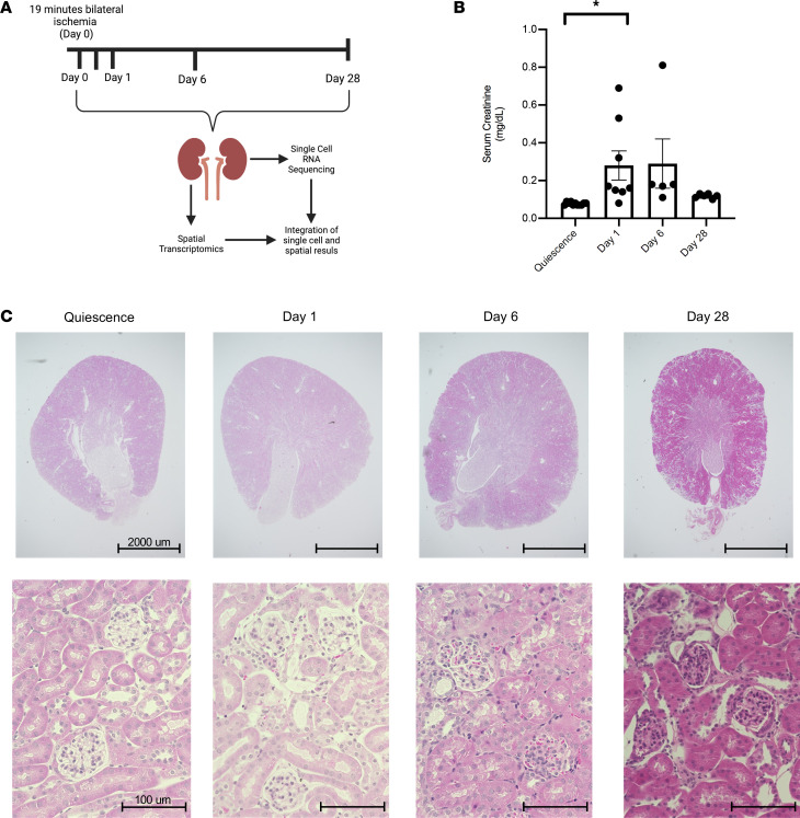
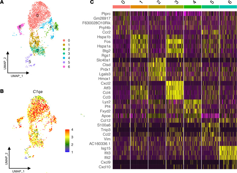
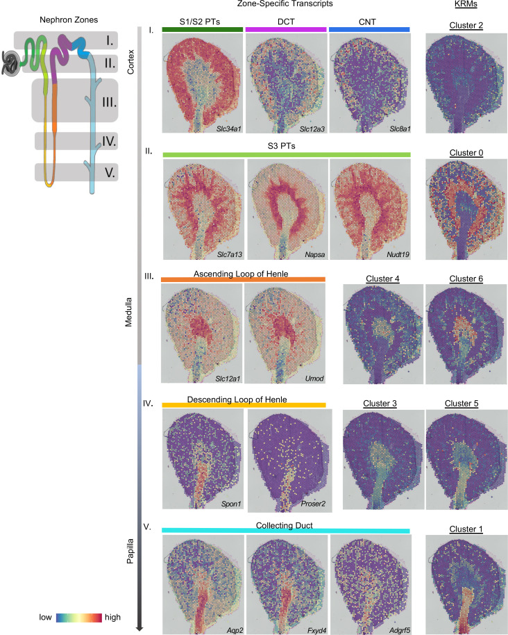
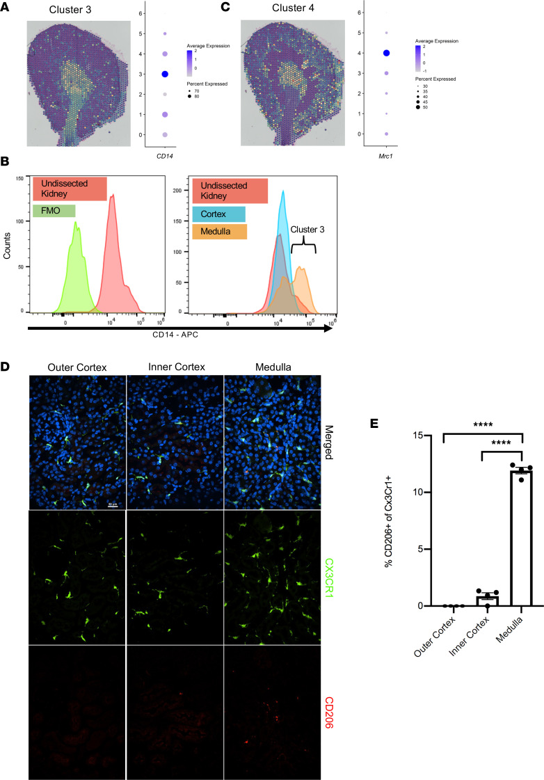
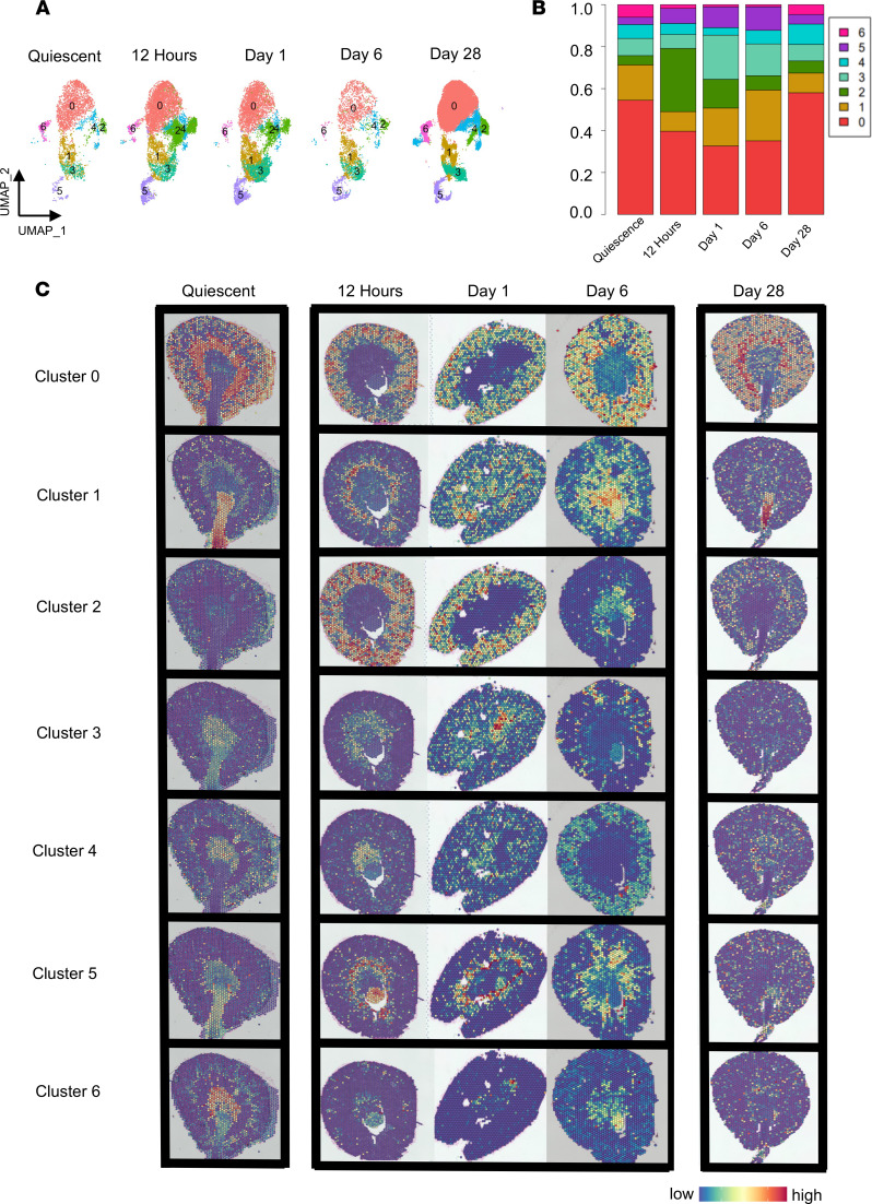
The kidney contains a population of resident macrophages from birth that expands as it grows and forms a contiguous network throughout the tissue. Kidney-resident macrophages (KRMs) are important in homeostasis and the response to acute kidney injury. While the kidney contains many microenvironments, it is unknown whether KRMs are a heterogeneous population differentiated by function and location. We combined single-cell RNA-Seq (scRNA-Seq), spatial transcriptomics, flow cytometry, and immunofluorescence imaging to localize, characterize, and validate KRM populations during quiescence and following 19 minutes of bilateral ischemic kidney injury. scRNA-Seq and spatial transcriptomics revealed 7 distinct KRM subpopulations, which are organized into zones corresponding to regions of the nephron. Each subpopulation was identifiable by a unique transcriptomic signature, suggesting distinct functions. Specific protein markers were identified for 2 clusters, allowing analysis by flow cytometry or immunofluorescence imaging. Following injury, the original localization of each subpopulation was lost, either from changing locations or transcriptomic signatures. The original spatial distribution of KRMs was not fully restored for at least 28 days after injury. The change in KRM localization confirmed a long-hypothesized dysregulation of the local immune system following acute injury and may explain the increased risk for chronic kidney disease.

Keywords: Bioinformatics; Immunology; Innate immunity; Macrophages; Nephrology.


Figures

Figure 1
Figure 1. Model of acute kidney injury.
(A) Schematic depicting work flow for scRNA-Seq and spatial transcriptomics. Mice were subjected to bilateral ischemia/reperfusion injury for 19 minutes. Kidneys were harvested at day 0, 12 hours, and days 1, 6, and 28 after injury. Kidneys were either utilized for spatial transcriptomics or digested and flow sorted for KRMs and subjected to scRNA-Seq. There were 3 biological replicates per time point. (B) Serum creatinine levels (mg/dL) at quiescence (day 0) and days 1, 6, and 28 after injury from at least 2 independent experiments. Data are reported as mean ± SEM. A 1-way ANOVA was used to determine the statistical significance between groups. *P < 0.05. (C) H&E-stained kidney sections at quiescence and days 1, 6, and 28 after injury. Scale bar: 2000 μm (top row); 100 μm (bottom row).
Figure 2
Figure 2. Single-cell RNA-Seq and spatial transcriptomics reveal distinct subpopulations of kidney-resident macrophages.
(A) Uniform manifold approximation and projection (UMAP) plot of sequenced kidney-resident macrophages (KRMs) demonstrating 13 clusters. Contaminating kidney cells and clusters representing less than 1% were removed to leave 7 unique clusters in quiescence. (B) C1qa expression in all KRM clusters during quiescence. (C) Heatmap of top 5 differentially expressed genes among each subpopulation in quiescence ordered by adjusted P value.
Figure 3
Figure 3. Kidney-resident macrophages are found in distinct regions.
An integrated analysis of the single-cell RNA-Seq (scRNA-Seq) and spatial transcriptomics data was performed to localize the kidney-resident macrophage (KRM) clusters on a kidney section. A diagram of a nephron is color coded to delineate different nephron segments (see nephron zones; left). Gray shading and numbering (I–V) describes nephron zones that would be enriched in areas of a kidney cross-section. The spatial location of the nephron segments is shown by mapping segment-specific transcripts onto the histological image (see zone-specific transcripts; middle). Transcript markers are listed in the bottom right-hand corner of each section. Specific nephron segments are listed above each image. Colored bars correspond to the location of the segments from the nephron shown on the left. Row number (I–V) indicates the nephron zone. The integration of the KRM scRNA-Seq data onto the spatial section plots the location of KRM subpopulations within the quiescent kidney (right). The clusters are aligned with the kidney nephron segments that are found in the same zone to highlight the colocalization between KRMs and kidney cells. CNT, connecting tubule; DCT, distal convoluted tubule; PT, proximal tubule.
Figure 4
Figure 4. Spatial validation of protein markers.
(A) Spatial location of cluster 3 overlaid on the histological image and a dot plot of CD14 transcript expression for each cluster. (B) Kidneys were harvested and dissected to separate the cortex from the medulla to confirm the location of cluster 3 CD14++ cells in the medulla. Flow cytometry analysis of CD14 expression in KRMs of the whole kidney (left) and dissected cortex compared with medulla (right) along with the fluorescence minus one (FMO) control. (C) Spatial location of cluster 4 overlaid on histological image and a dot plot of Mrc1 transcript expression show that cluster 4 is localized in the outer cortex and inner medulla but not the inner cortex. (D) Representative images from immunofluorescence of kidney sections of Cx3Cr1 GFP+/– mice stained with CD206 and the nuclear stain DAPI to validate cluster 4 KRMs by confocal microscopy (original magnification, ×40). Results were averaged from 4 separate fields within each area with 4 mice in total over 2 independent experiments. Scale bar: 20 μm. (E) Quantitation from a blinded observer of CD206+ KRMs in the outer cortex, inner cortex, and medulla, expressed as a proportion of Cx3Cr1+ cells. ****P < 0.0001 by 1-way ANOVA followed by Tukey’s test. Data are shown as mean ± SEM.
Figure 5
Figure 5. Spatial and proportional changes to KRM subpopulations following injury.
(A) Uniform manifold approximation and projection (UMAP) plot of KRM clusters at quiescence and 12 hours, day 1, day 6, and day 28 after injury to assess changes after injury. (B) Changes in proportions of each cluster over time. (C) scRNA-Seq data from each time point integrated with their respective spatial transcriptomic kidney sections to resolve cluster locations. Each row represents a KRM cluster, whereas each column depicts a time point from quiescence to day 28 (left to right). Images were taken with ×4 objective then stitched together so they appear as ×2.

References

    1. Soos TJ, et al. CX3CR1+ interstitial dendritic cells form a contiguous network throughout the entire kidney. Kidney Int. 2006;70(3):591–596. doi: 10.1038/sj.ki.5001567. - DOI - PubMed
    1. Liu F, et al. Distinct fate, dynamics and niches of renal macrophages of bone marrow or embryonic origins. Nat Commun. 2020;11(1):2280. doi: 10.1038/s41467-020-16158-z. - DOI - PMC - PubMed
    1. Lever JM, et al. Resident macrophages reprogram toward a developmental state after acute kidney injury. JCI Insight. doi: 10.1172/jci.insight.125503. doi: 10.1172/jci.insight.125503. [published online January 24, 2019]. - DOI - DOI - PMC - PubMed
    1. Lever JM, et al. Parabiosis reveals leukocyte dynamics in the kidney. Lab Invest. 2018;98(3):391–402. doi: 10.1038/labinvest.2017.130. - DOI - PMC - PubMed
    1. Hume D, Gordon S. Mononuclear phagocyte system of the mouse defined by immunohistochemical localization of antigen F4/80. Identification of resident macrophages in renal medullary and cortical interstitium and the juxtaglomerular complex. J Exp Med. 1983;157(5):1704–1709. doi: 10.1084/jem.157.5.1704. - DOI - PMC - PubMed

Publication types