. 2022 Sep 6;17(9):e0274212.
doi: 10.1371/journal.pone.0274212. eCollection 2022.

A joint ventricle and WMH segmentation from MRI for evaluation of healthy and pathological changes in the aging brain

Affiliations

A joint ventricle and WMH segmentation from MRI for evaluation of healthy and pathological changes in the aging brain

Hans E Atlason et al. PLoS One. .

Abstract

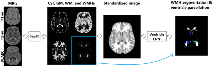
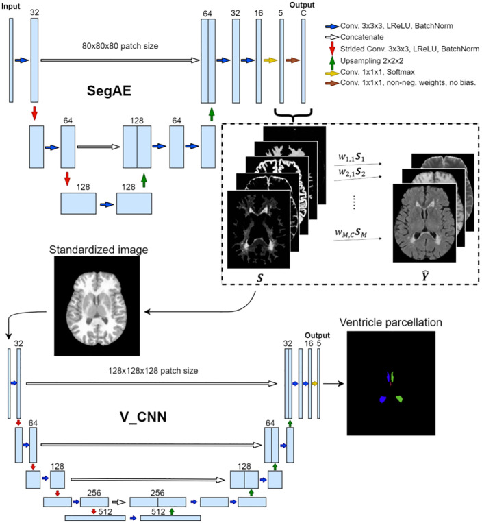
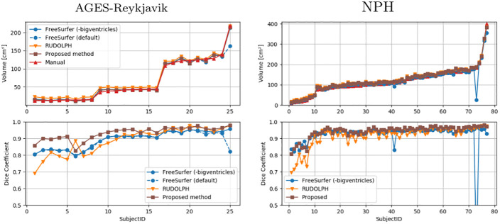
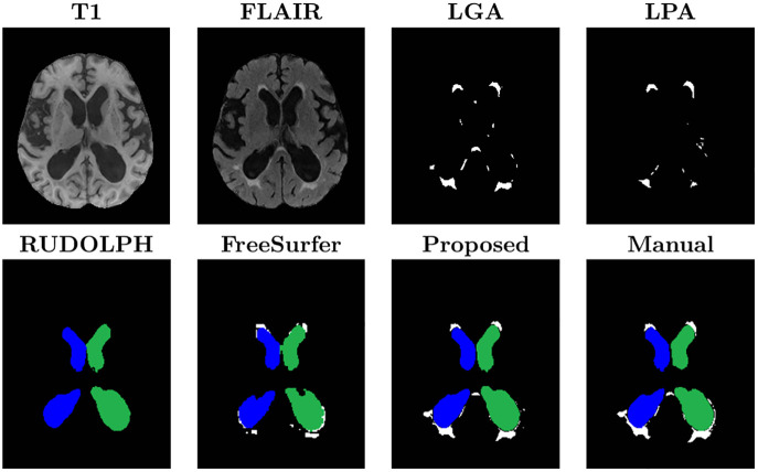
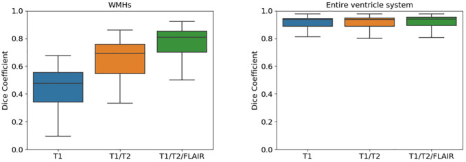
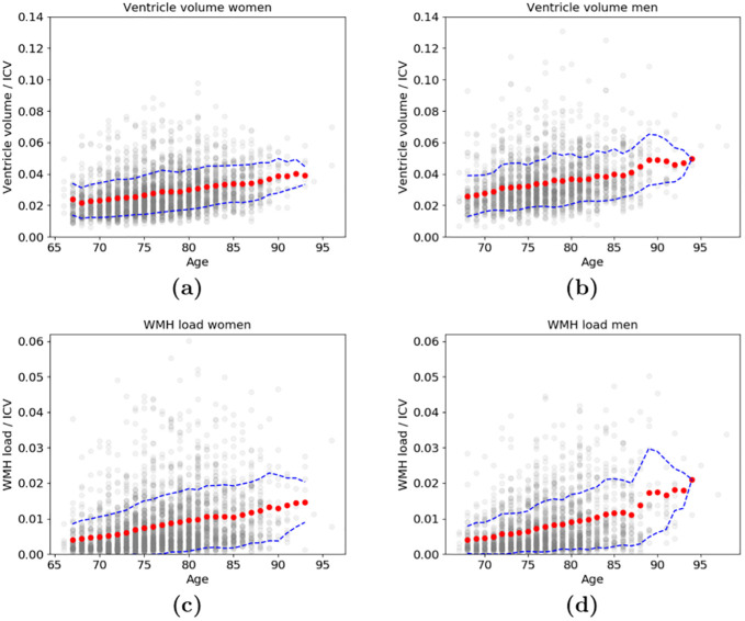
Age-related changes in brain structure include atrophy of the brain parenchyma and white matter changes of presumed vascular origin. Enlargement of the ventricles may occur due to atrophy or impaired cerebrospinal fluid (CSF) circulation. The co-occurrence of these changes in neurodegenerative diseases and in aging brains often requires investigators to take both into account when studying the brain, however, automated segmentation of enlarged ventricles and white matter hyperintensities (WMHs) can be a challenging task. Here, we present a hybrid multi-atlas segmentation and convolutional autoencoder approach for joint ventricle parcellation and WMH segmentation from magnetic resonance images (MRIs). Our fully automated approach uses a convolutional autoencoder to generate a standardized image of grey matter, white matter, CSF, and WMHs, which, in conjunction with labels generated by a multi-atlas segmentation approach, is then fed into a convolutional neural network to parcellate the ventricular system. Hence, our approach does not depend on manually delineated training data for new data sets. The segmentation pipeline was validated on both healthy elderly subjects and subjects with normal pressure hydrocephalus using ground truth manual labels and compared with state-of-the-art segmentation methods. We then applied the method to a cohort of 2401 elderly brains to investigate associations of ventricle volume and WMH load with various demographics and clinical biomarkers, using a multiple regression model. Our results indicate that the ventricle volume and WMH load are both highly variable in a cohort of elderly subjects and there is an independent association between the two, which highlights the importance of taking both the possibility of enlarged ventricles and WMHs into account when studying the aging brain.


Conflict of interest statement

The authors have declared that no competing interests exist.

Figures

Fig 1
Fig 1. The proposed pipeline for joint ventricle and WMH segmentation.
SegAE is used to decompose T1-w, T2-w and FLAIR images into four images, where the proportion of CSF, GM, WM, and WMHs is represented in each voxel. These are in turn used to create a standardized image from which the Ventricle CNN parcellates the ventricular system into the left and right lateral ventricles, and the 3rd and 4th ventricles.
Fig 2
Fig 2. Identification and removal of pulsation artifact.
Image (a) shows a FLAIR image with a pulsation artifact in the third ventricle (yellow arrow). Images (b) and (c) show the corresponding CSF and WMH output, respectively, from SegAE before thresholding. Image (d) shows a pulsation artifact segmentation obtained with element-wise multiplication of the CSF and WMH outputs (non-binarized). Image (e) shows the CSF segmentation that has been corrected for pulsation artifacts by adding the pulsation artifact segmentation shown in (d).
Fig 3
Fig 3. Preparation of training labels.
Image (a) shows a slice of a FLAIR image showing choroid plexus in the right lateral ventricle (yellow arrow). Image (b) shows the corresponding ventricle segmentation from RUDOLPH, and (c) shows the corresponding CSF segmentation from SegAE. Image (d) shows a ventricle segmentation obtained by element-wise multiplication of each label from RUDOLPH with the CSF segmentation in (c) and a morphological closing of the lateral and third ventricles. (e) shows a corresponding manual delineation.
Fig 4
Fig 4. The MRI sequences vs. the standardized image.
Images (a) and (b) show T1-w and a FLAIR images of a subject, respectively, and image (c) shows a standardized image made of SegAE segmentations, which is free of inhomogeneity artifacts and WMHs.
Fig 5
Fig 5. The proposed CNN architecture.
The input into the SegAE network comprises large 3D patches of MRI images that are in turn reconstructed in an unsupervised manner by SegAE (the reconstructed output is denoted with Y^). The estimated components of the reconstruction (denoted with S) provide the segmentation of the input into WMHs, WM, GM, CSF, and meninges (meninges are discarded in subsequent steps), which in turn are used to create a standardized image that is used as the input into the V_CNN. Kernels of size 3 × 3 × 3 are used in all convolutional layers except size 1 × 1 × 1 is used in the final two layers of both SegAE and the V_CNN. The V_CNN output is a segmentation of the four ventricle compartments, which in conjunction with the SegAE output provides a consistent ventricle and WMH segmentation.
Fig 6
Fig 6. Quantitative evaluation of the ventricle segmentation.
The top graphs show the overall ventricle volume for the manual masks (red) and masks generated by FreeSurfer (blue), RUDOLPH (orange), and the proposed method (brown), ordered by the volume of the manual masks. The bottom graphs show the DSC for the same methods compared with the manual masks. Results on the AGES-Reykjavik data are shown on the left and the NPH data on the right.
Fig 7
Fig 7. Visual comparison of the proposed method and the five methods used for comparison.
The images show the left (green) and right (blue) lateral ventricles (the 3rd and 4th ventricles are not visible in these slices), and WMHs (white). LPA and LGA provide WMH labels but not ventricle labels. RUDOLPH and FreeSurfer provide a whole brain segmentation with ventricle labels, however RUDOLPH does not provide WMH labels and the WMH labels from FreeSurfer are not accurate. The proposed method provides accurate ventricle and WMH labels.
Fig 8
Fig 8. Boxplots comparing the DSC when using different number of input sequences in the proposed method.
The left plot shows WMHs and the right plot shows the ventricular system when generating segmentations from: 1) Only T1-w (blue), 2) only T1-w and T2-w (orange), and 3) T1-w, T2-w, and FLAIR images (green).
Fig 9
Fig 9. The 3 year moving average (red dots) and standard deviation (dashed blue line) of ventricle volumes and WMH load.
The association between age and the total volume of all the ventricles divided by ICV (top) for (a) women and (b) men, as well as the association between age and WMH load divided by ICV (bottom) for (c) women and (d) men. The ventricle volume and WMH load of individual subjects, at their corresponding age, are shown in grey.
Fig 10
Fig 10. Resampling may cause erroneous WMH segmentation in the cerebellum.
Images (a), (b), and (c) show an axial slice of the cerebellum in T2-w, FLAIR, and T1-w images, respectively, of a subject in the NPH data set. The T2-w images have a higher in-plane resolution, which shows the thin lines of CSF in the cerebellum. Meanwhile, the upsampling of the lower resolution FLAIR and T1-w images gives them a blurry appearance, leading to brighter voxels instead of fine dark lines corresponding to the CSF in the T2-w image.

References

    1. Hou Y, Dan X, Babbar M, Wei Y, Hasselbalch SG, Croteau DL, et al. Ageing as a risk factor for neurodegenerative disease. Nature Reviews Neurology. 2019;15(10):565–581. doi: 10.1038/s41582-019-0244-7 - DOI - PubMed
    1. Schneider JA, Arvanitakis Z, Bang W, Bennett DA. Mixed brain pathologies account for most dementia cases in community-dwelling older persons. Neurology. 2007;69(24):2197–2204. doi: 10.1212/01.wnl.0000271090.28148.24 - DOI - PubMed
    1. Harvey R, Skelton-Robinson M, Rossor M. The prevalence and causes of dementia in people under the age of 65 years. Journal of Neurology, Neurosurgery & Psychiatry. 2003;74(9):1206–1209. doi: 10.1136/jnnp.74.9.1206 - DOI - PMC - PubMed
    1. Lee S, Viqar F, Zimmerman ME, Narkhede A, Tosto G, Benzinger TL, et al. White matter hyperintensities are a core feature of Alzheimer’s disease: evidence from the dominantly inherited Alzheimer network. Annals of neurology. 2016;79(6):929–939. doi: 10.1002/ana.24647 - DOI - PMC - PubMed
    1. Coutu JP, Goldblatt A, Rosas HD, Salat DH. White matter changes are associated with ventricular expansion in aging, mild cognitive impairment, and Alzheimer’s disease. Journal of Alzheimer’s Disease. 2016;49(2):329–342. doi: 10.3233/JAD-150306 - DOI - PMC - PubMed

Publication types