. 2022 Sep 6;20(9):e3001727.
doi: 10.1371/journal.pbio.3001727. eCollection 2022 Sep.

M-TUBE enables large-volume bacterial gene delivery using a high-throughput microfluidic electroporation platform

Affiliations

M-TUBE enables large-volume bacterial gene delivery using a high-throughput microfluidic electroporation platform

Po-Hsun Huang et al. PLoS Biol. .

Abstract

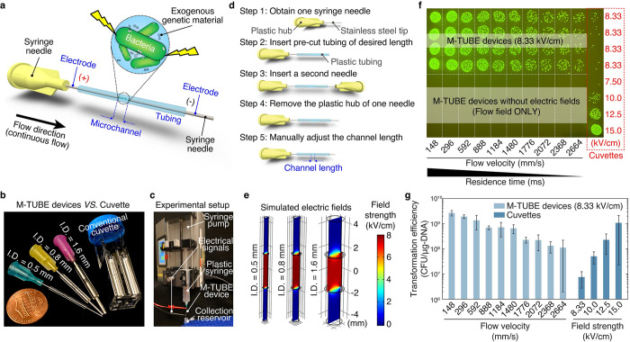
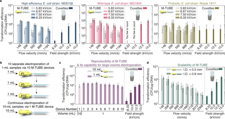
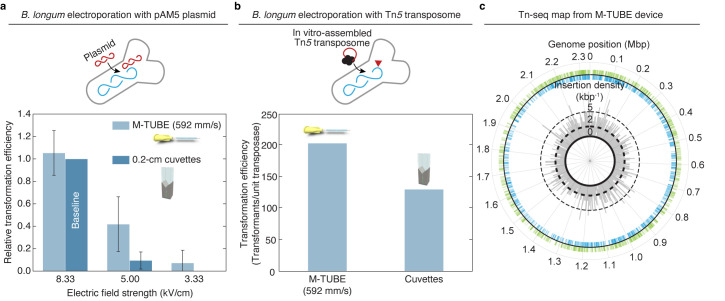
Conventional cuvette-based and microfluidics-based electroporation approaches for bacterial gene delivery have distinct advantages, but they are typically limited to relatively small sample volumes, reducing their utility for applications requiring high throughput such as the generation of mutant libraries. Here, we present a scalable, large-scale bacterial gene delivery approach enabled by a disposable, user-friendly microfluidic electroporation device requiring minimal device fabrication and straightforward operation. We demonstrate that the proposed device can outperform conventional cuvettes in a range of situations, including across Escherichia coli strains with a range of electroporation efficiencies, and we use its large-volume bacterial electroporation capability to generate a library of transposon mutants in the anaerobic gut commensal Bifidobacterium longum.


Conflict of interest statement

The authors have declared that no competing interests exist.

Figures

Fig 1
Fig 1. M-TUBE is a fabrication-free, microfluidics tubing-based bacterial electroporation device that is simple to assemble and exhibits higher electroporation efficiency than cuvettes.
(a) Schematic of the M-TUBE device. The device is composed of 2 syringe needles and 1 piece of plastic tubing of predefined length. The 2 syringe needles and plastic tubing serve as the 2 electrodes and microchannel, respectively. When the 2 electrodes are connected to an external power supply (or electrical signal generator), an electric field is established within the microchannel, where bacterial electroporation can take place. (b) M-TUBE devices with 3 ID are all similar in size to a conventional cuvette. (c) Photograph of the experiment setup when using the M-TUBE device. Since the M-TUBE device is made from standard, commercially available syringe needles and plastic tubing, it can be readily attached to syringe pumps for automated sample delivery, removing the need for manually pipetting samples. (d) Detailed breakdown of the protocol for M-TUBE assembly. One device can be completely assembled in 90–120 s. The total cost of parts is currently less than $0.22 and this price could be lowered if parts are bought in bulk. (e) Simulations of the electric field established in M-TUBE devices using COMSOL Multiphysics predict similar field strengths irrespective of ID. (f) Spot-dilution assay to quantify viability on selective plates when E. coli NEB10β cells were flowed through the device with a plasmid encoding ampicillin resistance and GFP (S4 Table) in the presence or absence of an electric field. Transformation was dependent on the electric field. For M-TUBE devices, a voltage of ±2.50 kV (AC field) was applied, which results in an electric field of 8.33 kV/cm. The same batch of cells was used to conduct cuvette-based electroporation as a comparison. (g) Comparison of transformation efficiency (CFUs per μg of DNA) corresponding to the plates in (f). The electroporation efficiency of M-TUBE decreased as the fluid velocity was increased, as expected due to the shorter duration of exposure to the electric field. Regardless of the fluid velocity, the efficiency of M-TUBE was at least 1 order of magnitude higher than that of cuvettes with the same field strength (8.33 kV/cm). Data represent the average (n ≥ 3) and error bars represent 1 standard deviation. The data underlying Fig 1E and 1G can be found in S1 and S2 Data files, respectively. AC, alternating current; CFU, colony-forming unit; GFP, green fluorescent protein; ID, inner diameter; M-TUBE, microfluidic tubing-based bacterial electroporation.
Fig 2
Fig 2. The M-TUBE device exhibits higher efficiency than cuvettes across E. coli strains, is reproducible, and maintains high efficiency across tubing sizes.
(a) Comparison of M-TUBE device performance when transforming the high-efficiency strain NEB10β, the wild-type strain MG1655, and the probiotic strain Nissle 1917 across voltages and fluid velocities. M-TUBE outperformed cuvettes at an equivalent electric field strength for all strains. Data represent the average (n ≥ 3) and error bars represent 1 standard deviation. (b) Schematic of the experiment comparing 10 separate 1 mL electroporations and 1 continuous electroporation of a 10-mL sample. (c) Transformation efficiency for the experiments in (b) demonstrates that sample volume can be increased without compromising efficiency. Data represent the average (n ≥ 3) and error bars represent 1 standard deviation. The same batch of cells was used to conduct cuvette-based electroporation as a comparison. (d) Transformation efficiency was similar across 0.5-mm and 0.8-mm diameter M-TUBE devices. For M-TUBE devices, a voltage of ±2.50 kV (AC field) was applied, which results in an electric field of 8.33 kV/cm. Data represent the average (n ≥ 3) and error bars represent 1 standard deviation. The data underlying this figure can be found in S2 Data. AC, alternating current; M-TUBE, microfluidic tubing-based bacterial electroporation.
Fig 3
Fig 3. M-TUBE efficiently transforms anaerobic bacteria and enables transposon insertion mutagenesis.
(a) Comparison of M-TUBE performance during electrotransformation of B. longum NCIMB8809 with the plasmid pAM5 at various electric field strengths. For M-TUBE devices, voltages of ±2.50, ±1.50, and ±1.00 kV (AC field) were applied to produce electric fields of 8.33, 5.00, and 3.33 kV/cm, respectively. A fluid velocity of 592 mm/s was used for the M-TUBE device because approximately 5 ms residence time with an M-TUBE ID of 0.5 mm is similar to the time constant observed in cuvette electroporation (5.2–5.6 ms). Data represent the average (n ≥ 3) and error bars represent 1 standard deviation. (b) Comparison of M-TUBE performance during electrotransformation of B. longum NCIMB8809 with Tn5 transposome. For the M-TUBE device, a field strength of 8.33 kV/cm and fluid velocity of 592 mm/s were used, motivated by the results in (a). (c) The transposon insertions recovered from Tn5 transposome electroporation are spread approximately uniformly across the B. longum NCIMB8809 genome. The locations of individual mapped insertions are recorded on the outer circle. Green ticks on the outside indicate insertions in the positive (+) orientation, blue ticks on the inside indicate insertions in the negative (−) orientation. The insertion density (kbp−1) (both positive and negative orientation) is plotted in 1-kbp bins on the inner circle. Transposon insertions are distributed throughout the genome in both the positive and negative orientations, indicating that B. longum NCIMB8809 can be transformed by Tn5 transposomes using M-TUBE without major insertional bias. The data underlying this figure can be found in S2 Data. AC, alternating current; ID, inner diameter; M-TUBE, microfluidic tubing-based bacterial electroporation.

References

    1. Mazodier P, Davies J. Gene-Transfer between Distantly Related Bacteria. Annu Rev Genet. 1991;25:147–71. doi: 10.1146/annurev.ge.25.120191.001051 WOS:A1991GX17400007. - DOI - PubMed
    1. Yoshida N, Sato M. Plasmid uptake by bacteria: a comparison of methods and efficiencies. Appl Microbiol Biotechnol. 2009;83(5):791–8. doi: 10.1007/s00253-009-2042-4 WOS:000267214600001. - DOI - PubMed
    1. Rivera AL, Magana-Ortiz D, Gomez-Lim M, Fernandez F, Loske AM. Physical methods for genetic transformation of fungi and yeast. Phys Life Rev. 2014;11(2):184–203. doi: 10.1016/j.plrev.2014.01.007 WOS:000337982800007. - DOI - PubMed
    1. Sheng Y, Mancino V, Birren B. Transformation of Escherichia coli with Large DNA-Molecules by Electroporation. Nucleic Acids Res. 1995;23(11):1990–6. WOS:A1995RE99600023. doi: 10.1093/nar/23.11.1990 - DOI - PMC - PubMed
    1. Chassy BM, Mercenier A, Flickinger J. Transformation of Bacteria by Electroporation. Trends Biotechnol. 1988;6(12):303–9. doi: 10.1016/0167-7799(88)90025-X WOS:A1988R036000007. - DOI

Publication types

MeSH terms