. 2022 Sep 6;18(9):e1010799.
doi: 10.1371/journal.ppat.1010799. eCollection 2022 Sep.

Biosynthetic proteins targeting the SARS-CoV-2 spike as anti-virals

Affiliations

Biosynthetic proteins targeting the SARS-CoV-2 spike as anti-virals

Stéphanie Thébault et al. PLoS Pathog. .

Abstract

The binding of the SARS-CoV-2 spike to angiotensin-converting enzyme 2 (ACE2) promotes virus entry into the cell. Targeting this interaction represents a promising strategy to generate antivirals. By screening a phage-display library of biosynthetic protein sequences build on a rigid alpha-helicoidal HEAT-like scaffold (named αReps), we selected candidates recognizing the spike receptor binding domain (RBD). Two of them (F9 and C2) bind the RBD with affinities in the nM range, displaying neutralisation activity in vitro and recognizing distinct sites, F9 overlapping the ACE2 binding motif. The F9-C2 fusion protein and a trivalent αRep form (C2-foldon) display 0.1 nM affinities and EC50 of 8-18 nM for neutralization of SARS-CoV-2. In hamsters, F9-C2 instillation in the nasal cavity before or during infections effectively reduced the replication of a SARS-CoV-2 strain harbouring the D614G mutation in the nasal epithelium. Furthermore, F9-C2 and/or C2-foldon effectively neutralized SARS-CoV-2 variants (including delta and omicron variants) with EC50 values ranging from 13 to 32 nM. With their high stability and their high potency against SARS-CoV-2 variants, αReps provide a promising tool for SARS-CoV-2 therapeutics to target the nasal cavity and mitigate virus dissemination in the proximal environment.


Conflict of interest statement

The authors have declared that no competing interests exist.

Figures

Fig 1
Fig 1. Selection and characterization of anti-spike αReps.
Screening an αReps phage library allowed the identification of several binders specific of the RBD of the SARS-CoV-2 spike protein. Their binding affinity for the S1 domain was measured by biolayer interferometry. The neutralization activity of selected αReps was evaluated using a pseudo-typed S SARS-CoV-2 neutralization assay and a SARS-CoV-2 infection assay. Competitive binding assays were carried out by BLI to identify αReps recognizing non-overlapping binding sites. Then, αRep derived constructs followed the same characterization steps than their single counterparts. The protective potency of the best candidate was analyzed in vivo in the golden Syrian hamster model.
Fig 2
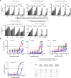
Fig 2. Selection of αReps based on their affinities and neutralization activities.
BLI binding kinetics measurements are shown for F9 (A) and C2 (B). Equilibrium dissociation constants (KD) were determined on the basis of fits, applying 1:1 interaction model; ka, association rate constant; kd, dissociation rate constant. (C) Pseudo-typed SARS-CoV-2 neutralization assay was shown with selected αRep (C2, F9, C7, G1). An αRep specific to influenza polymerase (H7) was chosen as a negative control. To assess αReps specificity, pseudo-typed VSV-G were incubated with the highest concentration of each αRep (3 μM). Pseudo-type particles entry into cells was quantified by measuring luciferase activity (n = 3, mean ± SEM, two-way ANOVA, *P<0.05). (D) Cell viability of infected cells in presence of dilutions of αReps C2, C7, F9, G1 and H7 was monitored using the CellTiter-Glo Luminescent Assay Kit (Promega). Infected cells (triangle) and mock-infected cells (square) were included in the assay as controls (n = 2, mean is presented). (E) Half maximal inhibitory concentration (IC50) were calculated using “log(inhibitor) vs. normalized response” equation from the neutralization potency curves with GraphPad Prism 8 software. ND: Not done, NA: Not available.
Fig 3
Fig 3. Competitive binding assays.
(A and B) BLI experiments showed that C2 and F9 could bind RBD simultaneously. (C) Binding of ACE2 was assessed after a first association phase with αReps C2 and F9, the F9-C2 construct, the VHH72 [19] or with a negative control (NR). F9-C2 and VHH-72 blocked the binding of RBD to ACE2. While F9 inhibited partially ACE2 binding, C2 did not compete with ACE2 binding. (3D-F) Reciprocal competitive binding assays between VHH-72, VHH H11D4, C2 and F9. While C2 and H11D4 competed for binding to the RBD (immobilized on the chip) in a reciprocal manner, F9 and VHH-72 blocked reciprocally their interaction to the RBD.
Fig 4
Fig 4. The F9-C2 and C2-foldon constructs properties.
(A) BLI binding kinetics measurements for F9-C2 to the S1-immobilized biosensor. (B) Pseudo-typed SARS-CoV-2 particles neutralization assay was performed with F9, C2, F9-C2 and C2-foldon constructs (n = 3, mean ± SEM, two-way ANOVA, *P<0.0001). (C) Cell viability of SARS-CoV-2-infected cells in presence of dilutions of F9-C2, C2-foldon, C2, F9 and H7 (an αRep negative control) was monitored using the CellTiter-Glo Luminescent Assay Kit (Promega) (n = 2, mean is presented). Infected cells (triangle) and mock-infected cells (square) were included in the assay. Half maximal inhibitory concentration (IC50) were displayed. (D) SARS-CoV-2 neutralisation by aReps constructs. Virus replication was quantified by qRT-PCR in infected cells treated by C2, F9, F9-C2, C2-foldon (n = 3, mean ± SEM). Half maximal effective concentration (EC50) were shown.
Fig 5
Fig 5. Efficacy of F9-C2 αRep prophylaxis in SARS-CoV-2 infection in a golden Syrian hamster model.
(A) Overview of the experiment design. 6 mg/kg of αReps were delivered intranasally in hamsters 1h prior to infection with 5.103 TCID50 of SARS-CoV-2. (B) Evolution of animal weight (n = 4, mean of the relative weight to 1-day prior infection ± SEM, two-way ANOVA). (C) Evolution of virus titre in nasal swabs (n = 4, mean of TCID50 ± SEM, two-way ANOVA, ****P<0.0001) (D) Quantification of RNA encoding SARS-CoV-2 protein E, IL-6, TNFα, Ncf2 in the olfactory turbinates, relative to viral infection, inflammation and neutrophil respectively (normalized to ß-actin, mean ± SEM, Mann–Whitney *P<0.05). (E) Representative images of the infected olfactory epithelium area treated by G1 or F9-C2 in the rostral zone of the nasal cavity (1 dpi) showing respectively a strong and partial infection. (F) Measurement of the extent area of infection in the dorso-medial part of the hamster nose. Values represent the mean of infected area (Arbitrary Unit ± SEM, Mann–Whitney *P<0.05).
Fig 6
Fig 6. Efficacy of F9-C2 αRep repeated treatments in SARS-CoV-2 infection in a golden Syrian hamster model.
(A) Overview of the experiment design. 6 mg/kg of αReps were delivered intranasally in hamsters 1h prior to infection with 5.103 TCID50 of SARS-CoV-2. The treatment was repeated on 1 dpi and 2 dpi for the group examined at 3 dpi. (B) Evolution of animal weight (n = 4, mean of the relative weight to 1-day prior infection ± SEM, two-way ANOVA). (C) Evolution of virus titre in nasal swabs (n = 4, mean of TCID50 ± SEM, two-way ANOVA, ****P<0.0001) (D) quantification of RNA encoding SARS-CoV-2 protein E, IL-6, TNFα, Ncf2 in the olfactory turbinates, relative to viral infection, inflammation and neutrophil respectively (normalized to ß-actin, mean ± SEM, Mann–Whitney *P<0.05). (E) Representative images of the infected olfactory epithelium area treated by F9-C2 or H7 in the dorso-medial zone of the nasal cavity (1 dpi) showing respectively a partial infection with a low number of Iba1+ immune cell infiltration and a strong infection associated with damage of the olfactory epithelium and Iba1+ cell infiltration as well as desquamated cells in the lumen of the nasal cavity (white asterisk). (F) Measurement of the extent area of infection in the dorso-medial part of the hamster nose. Values represent the mean of infected area (Arbitrary Unit ± SEM, Mann–Whitney *P<0.05).
Fig 7
Fig 7. Neutralization activity of the F9-C2 and C2-foldon constructs against SARS-CoV-2 pseudo-typed and virus variants.
(A) F9, C2, F9-C2 and C2-foldon were tested for their ability to neutralize four SARS-CoV-2 pseudo-typed RBD mutants. Pseudo-typed VSV-G was incubated with the highest concentration of each αRep (500 nM) to validate specificity of αRep neutralization activity (n = 3, mean ± SEM, two-way ANOVA, *P<0.0001). (B) F9, C2, F9-C2 and C2-foldon were tested for their ability to neutralize authentic SARS-CoV-2 virus variants (beta, gamma, delta and omicron) (n = 3, mean ± SEM). (C) Chart listing the EC50 of αReps and derivates towards variant viruses.

References

    1. Hou YJ, Okuda K, Edwards CE, Martinez DR, Asakura T, Dinnon KH, et al.. SARS-CoV-2 Reverse Genetics Reveals a Variable Infection Gradient in the Respiratory Tract. Cell. 2020;182: 429–446.e14. doi: 10.1016/j.cell.2020.05.042 - DOI - PMC - PubMed
    1. Bryche B, St Albin A, Murri S, Lacôte S, Pulido C, Ar Gouilh M, et al.. Massive transient damage of the olfactory epithelium associated with infection of sustentacular cells by SARS-CoV-2 in golden Syrian hamsters. Brain Behav Immun. 2020;89: 579–586. doi: 10.1016/j.bbi.2020.06.032 - DOI - PMC - PubMed
    1. von Bartheld CS, Hagen MM, Butowt R. Prevalence of Chemosensory Dysfunction in COVID-19 Patients: A Systematic Review and Meta-analysis Reveals Significant Ethnic Differences. ACS Chem Neurosci. 2020;11: 2944–2961. doi: 10.1021/acschemneuro.0c00460 - DOI - PMC - PubMed
    1. Valdez-Cruz NA, García-Hernández E, Espitia C, Cobos-Marín L, Altamirano C, Bando-Campos CG, et al.. Integrative overview of antibodies against SARS-CoV-2 and their possible applications in COVID-19 prophylaxis and treatment. Microb Cell Factories. 2021;20: 88. doi: 10.1186/s12934-021-01576-5 - DOI - PMC - PubMed
    1. Chen F, Liu Z, Jiang F. Prospects of Neutralizing Nanobodies Against SARS-CoV-2. Front Immunol. 2021;12: 690742. doi: 10.3389/fimmu.2021.690742 - DOI - PMC - PubMed

MeSH terms

Supplementary concepts