. 2022 Sep 13;119(37):e2121385119.
doi: 10.1073/pnas.2121385119. Epub 2022 Sep 6.

IRF3 inhibits nuclear translocation of NF-κB to prevent viral inflammation

Affiliations

IRF3 inhibits nuclear translocation of NF-κB to prevent viral inflammation

Sonam Popli et al. Proc Natl Acad Sci U S A. .

Abstract

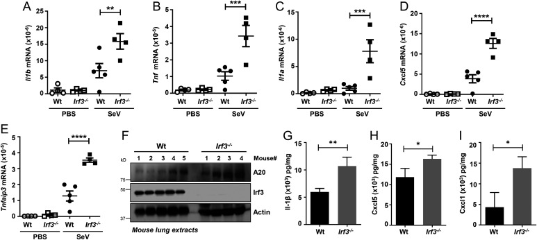
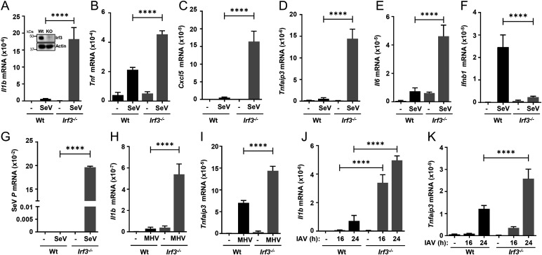
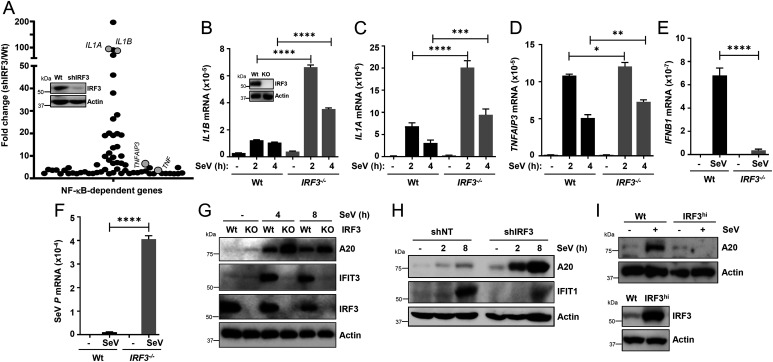
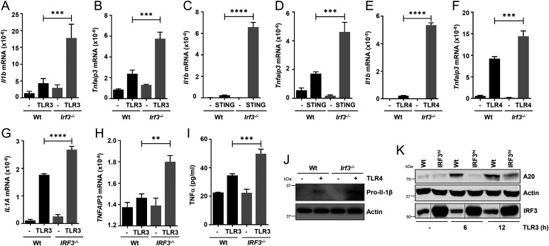
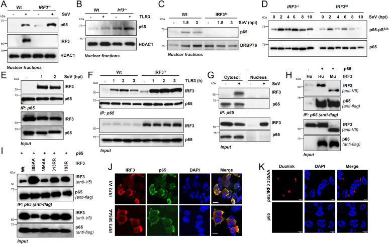
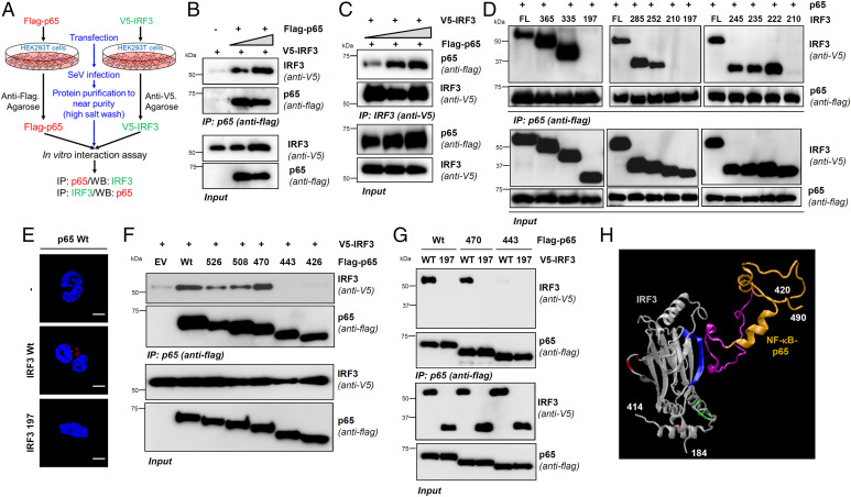
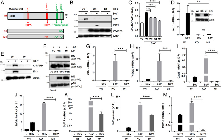
Interferon (IFN) regulatory factor 3 (IRF3) is a transcription factor activated by phosphorylation in the cytoplasm of a virus-infected cell; by translocating to the nucleus, it induces transcription of IFN-β and other antiviral genes. We have previously reported IRF3 can also be activated, as a proapoptotic factor, by its linear polyubiquitination mediated by the RIG-I pathway. Both transcriptional and apoptotic functions of IRF3 contribute to its antiviral effect. Here, we report a nontranscriptional function of IRF3, namely, the repression of IRF3-mediated NF-κB activity (RIKA), which attenuated viral activation of NF-κB and the resultant inflammatory gene induction. In Irf3-/- mice, consequently, Sendai virus infection caused enhanced inflammation in the lungs. Mechanistically, RIKA was mediated by the direct binding of IRF3 to the p65 subunit of NF-κB in the cytoplasm, which prevented its nuclear import. A mutant IRF3 defective in both the transcriptional and the apoptotic activities was active in RIKA and inhibited virus replication. Our results demonstrated IRF3 deployed a three-pronged attack on virus replication and the accompanying inflammation.

Keywords: IRF3; NF-κB; antiviral; innate immunity; viral inflammation.


Conflict of interest statement

The authors declare no competing interest.

Figures

Fig. 1.
Fig. 1.
Increased inflammatory gene expression in Irf3−/− mice upon SeV infection. (AI) Wt or Irf3−/− mice were infected intranasally with SeV (or treated with the vehicle PBS, as indicated). At 5 d postinfection, the lungs were analyzed for the mRNA levels of Il1b, Tnf, Il1a, Cxcl5, and Tnfaip3 by qRT-PCR (AE), the protein expression of Tnfaip3/A20 by immunoblot (F), or the cytokine levels by ELISA (GI). The data represent mean ± SEM; n = 3–5 for each mouse genotype and for each condition, as shown, *P < 0.05, **P < 0.005, ***P < 0.0005, ****P < 0.0001.
Fig. 2.
Fig. 2.
Increased inflammatory gene expression in Irf3−/− mouse macrophages upon virus infection. Wt or Irf3−/− iBMDMs (immunoblot of Irf3 expression; inset in A) were infected with SeV (for 4 h postinfection [hpi]; AG), MHV (for 16 hpi; Hand I), or IAV (for 16 and 24 hpi; J and K), and the mRNA levels of Il1b, Tnf, Cxcl5, Tnfaip3, Il6, Ifnb1 (AF, HK), or viral transcript (G) were analyzed by qRT-PCR. The results are representative of three experiments; the data represent mean ± SEM. KO, knockout, ****P < 0.0001.
Fig. 3.
Fig. 3.
IRF3 inhibits virus-induced inflammatory gene induction in human cells. (A) Graphical presentation of the microarray analyses of the NF-κB–dependent genes in Wt and shIRF3 (immunoblot of IRF3 expression; inset in A) HT1080 cells after SeV infection (2 h postinfection), are as described in Methods; the genes are listed in SI Appendix, Tables S1 and S2. The microarray results are from duplicate samples for each condition. (BF) Wt or IRF3−/− (immunoblot of IRF3 expression; inset in B) HT1080 cells were infected with SeV (BF), and the mRNAs of inflammatory target genes (BE) or viral mRNA (F) were analyzed by qRT-PCR. (G) Wt and IRF3−/− (knockout [KO]) HT1080 cells were infected with SeV for the indicated time, and the cell lysates were analyzed for TNFAIP3/A20, IFIT1, and IRF3 by immunoblot. (H) The IRF3 shIRF3 HT1080 cells were infected with SeV for the indicated time, and the cell lysates were analyzed for TNFAIP3/A20 and IFIT1 by immunoblot. (I) Wt and IRF3hi U4C cells were infected with SeV and analyzed for TNFAIP3/A20 (Upper panel) and IRF3 (Lower panel) by immunoblot. NT, nontargeting. The results are representative of three experiments; the data represent mean ± SEM. SeV P, SeV P gene, *P < 0.05, **P < 0.005, ***P < 0.0005, ****P < 0.0001.
Fig. 4.
Fig. 4.
IRF3 inhibits inflammatory gene expression in response to nonviral stimuli. (AF) Wt or Irf3−/− iBMDMs were stimulated with polyI:C (TLR3; A and B), cGAMP (STING; C and D), or LPS (TLR4; E and F) for 4 h, and the mRNAs of Il1b and Tnfaip3 were analyzed by qRT-PCR. (G and H) Wt or IRF3−/− HT1080 cells were stimulated with polyI:C (TLR3; G and H) for 4 h, and the mRNAs of Il1a and Tnfaip3 were analyzed by qRT-PCR. (I) Wt and IRF3−/− (knockout) HT1080 cells were stimulated with polyI:C (TLR3) for 8 h, and the supernatants were analyzed for TNFα by ELISA. (J) Wt or Irf3−/− iBMDMs were treated with LPS (TLR4) for 4 h and analyzed for pro–IL-1β by immunoblot. (K) Wt and IRF3hi HT1080 cells were infected with SeV and analyzed for TNFAIP3/A20 by immunoblot. The results are representative of three experiments; the data represent mean ± SEM. **P < 0.005, ***P < 0.0005, ****P < 0.0001.
Fig. 5.
Fig. 5.
IRF3 interacts with NF-κB–p65, independent of transcriptional or RIPA functions, and inhibits NF-κB activation. (A) Wt and IRF3−/− HT1080 cells were either mock-infected or infected with SeV for 2 h when the nuclear fractions were analyzed for NF-κB–p65 and IRF3 by immunoblot. HDAC1 is a marker for nuclear fractions. (B) Wt and Irf3−/− iBMDMs were stimulated with polyI:C (TLR3) for 2 h, when the nuclear fractions were analyzed for NF-κB–p65 by immunoblot. (C) Wt or IRF3hi HT1080 cells were infected with SeV for the indicated time, when the nuclear fractions were analyzed for NF-κB–p65 by immunoblot; DRBP76 is a marker for the nuclear fractions. (D) IRF3−/− and IRF3hi HT1080 cells were infected with SeV for the indicated time, and phospho-p65 (on Ser536) was analyzed by immunoblot. (E) HT1080 cells infected with SeV for the indicated time were subjected to co-IP analyses for the endogenous NF-κB–p65 and IRF3 proteins using ExactaCruz. (F) Wt and IRF3hi HT1080 cells stimulated with polyI:C (TLR3) for the indicated time were subjected to co-IP analyses for NF-κB–p65 and IRF3 proteins, as in E. (G) The cytosolic and nuclear fractions, isolated from the SeV-infected Wt and IRF3hi HT1080 cells, were subjected to co-IP analyses for NF-κB–p65 and IRF3, as in E. (H and I) HEK293T cells, cotransfected with Flag–NF-κB–p65 and V5-IRF3 Wt (H) human (Hu), murine (Mu), or IRF3 mutants (I), as indicated, were infected with SeV for 2 h and subjected to co-IP analyses for the Flag–NF-κB–p65 and V5-IRF3, as indicated. (J) HEK293T cells, cotransfected with NF-κB–p65 and V5-IRF3 Wt or IRF3 385AA mutant, were infected with SeV and immunostained with anti-Flag and anti-V5 antibodies and analyzed by confocal microscopy. (K) HEK293T cells, cotransfected with NF-κB–p65 and V5-IRF3 385AA mutant, were infected with SeV and immunostained with anti-Flag and anti-V5 antibodies, and analyzed by proximal ligation assay. The results are representative of three experiments; the data represent mean ± SEM. Scale bar, 10 µm. IP, immunoprecipitation.
Fig. 6.
Fig. 6.
IRF3 and NF-κB interact directly via specific domains. (AC) V5-IRF3 or Flag–NF-κB–p65 were ectopically expressed in HEK293T cells, isolated to near purity, and subjected to cell-free interaction assay, as indicated in A, and the complex was analyzed by co-IP (B and C). (D) HEK293T cells, cotransfected with Wt or the C-terminal deletion mutants of V5-IRF3 and Flag-NF-κB–p65, were infected with SeV for 2 h and subjected to co-IP analyses. (E) HEK293T cells expressing either Flag–NF-κB–p65 or Flag–NF-κB–p65 with V5-IRF3 (Wt or 1–197) were analyzed by proximity ligation assay. (F and G) HEK293T cells, cotransfected with full-length or the deletion mutants of Flag–NF-κB–p65 (F) and V5-IRF3 (Wt in F; Wt and 1–197 in G) were infected with SeV for 2 h and subjected to co-IP analyses. (H) A 3D structural model showing the domains and residues of IRF3 (blue: RIKA; red: RIPA; green: transcription) and NF-κB–p65 (gold and pink: IRF3-binding motif) involved in RIKA. The 3D protein structures of IRF3 and NF-κB–p65 were adapted from Protein Data Bank (PDB) templates 1j2f.2.A and 2lww.1, respectively. EV, empty vector. The results are representative of three experiments; the data represent mean ± SEM. Scale bar, 5 µm. IP, immunoprecipitation.
Fig. 7.
Fig. 7.
IRF3 mutants, active in RIKA but inactive in transcriptional and RIPA, inhibits virus replication and inflammatory gene expression. (A) A cartoon showing mouse Irf3 and its critical residues and domains required for specific functions and the pathway-specific mutants (Irf3-S1 and Irf3-M1). DBD, DNA-binding domain, A, Ala, R, Arg. (B) HT1080/shIRF3 cells, lentivirally expressing Wt or S1 mutant of IRF3, were analyzed for A20 and IFIT1 induction upon SeV infection, by immunoblot 8 h postinfection (hpi). (C) RAW-Blue cells were transfected with Wt or Irf3 mutants (S1 or M1), and the NF-κB–SEAP activity in the culture supernatants was measured upon SeV infection (24 hpi). (D) Wt, Irf3−/−, and Irf3-M1 iBMDMs (Irf3 expression is shown in the inset) were infected with SeV, and Ifnb1 induction was analyzed by qRT-PCR 4 hpi. (E) Wt and Irf3-M1 iBMDMs were transfected with polyI:C for 16 h, when the cell lysates were analyzed for Ifit3 and C-PARP by immunoblot. (F) HEK293T cells, cotransfected with Flag–NF-κB–p65 and V5-Irf3 (Wt or M1), were infected with SeV for 2 h and subjected to co-IP analyses for the Flag–NF-κB–p65 and V5-Irf3. (GJ) Wt, Irf3−/−, and Irf3-M1 iBMDMs were infected with SeV for 4 h (GI) or MHV for 16 h (J), and the inflammatory target genes were analyzed by qRT-PCR. (KM) Wt, Irf3−/−, and Irf3-M1 iBMDMs were infected with SeV for 4 h (K and L) or MHV for 16 h (M), and the viral replication was analyzed by qRT-PCR. EV, empty vector. The results are representative of three experiments; the data represent mean ± SEM. IP, immunoprecipitation; KO, knockout, SeV P, SeV P gene, **P < 0.005, ***P < 0.0005, ****P < 0.0001.

Similar articles

Cited by

References

    1. Samuel C. E., Antiviral actions of interferons. Clin. Microbiol. Rev. 14, 778–809, (2001). - PMC - PubMed
    1. White C. L., Sen G. C., “Interferons and antiviral actions” inCellular Signaling and Innate Immune Responses to RNA Virus Infections, Brasier A. R., Garcia-Sastre A., Lemon S. M., Eds. (ASM Press, Washington, DC, 2008), pp. 91–106.
    1. Biron C. A., Sen G. C., “Innate responses to viral infections” in Fields Virology, Knipe D. M., Howley P. M., Eds. (Lippincott, Williams and Wilkins, Philadelphia, PA, ed. 5, 2006), pp. 249–278.
    1. Fensterl V., Sen G. C., Interferons and viral infections. Biofactors 35, 14–20 (2009). - PubMed
    1. Schoggins J. W., Interferon-stimulated genes: What do they all do? Annu. Rev. Virol. 6, 567–584 (2019). - PubMed

Publication types

LinkOut - more resources