. 2022 Sep 13;119(37):e2202426119.
doi: 10.1073/pnas.2202426119. Epub 2022 Sep 6.

KidA, a multi-PAS domain protein, tunes the period of the cyanobacterial circadian oscillator

Affiliations

KidA, a multi-PAS domain protein, tunes the period of the cyanobacterial circadian oscillator

Soo Ji Kim et al. Proc Natl Acad Sci U S A. .

Abstract

The cyanobacterial clock presents a unique opportunity to understand the biochemical basis of circadian rhythms. The core oscillator, composed of the KaiA, KaiB, and KaiC proteins, has been extensively studied, but a complete picture of its connection to the physiology of the cell is lacking. To identify previously unknown components of the clock, we used KaiB locked in its active fold as bait in an immunoprecipitation/mass spectrometry approach. We found that the most abundant interactor, other than KaiC, was a putative diguanylate cyclase protein predicted to contain multiple Per-Arnt-Sim (PAS) domains, which we propose to name KidA. Here we show that KidA directly binds to the fold-switched active form of KaiB through its N-terminal PAS domains. We found that KidA shortens the period of the circadian clock both in vivo and in vitro and alters the ability of the clock to entrain to light-dark cycles. The dose-dependent effect of KidA on the clock period could be quantitatively recapitulated by a mathematical model in which KidA stabilizes the fold-switched form of KaiB, favoring rebinding to KaiC. Put together, our results show that the period and amplitude of the clock can be modulated by regulating the access of KaiB to the fold-switched form.

Keywords: circadian rhythms; cyanobacteria; diguanylate cyclase.


Conflict of interest statement

The authors declare no competing interest.

Figures
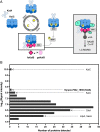
Fig. 1.
Fig. 1.
KidA is identified as a potential clock interactor from fsKaiB co-IP/MS. (A) Experimental scheme to identify potential interactors of the nighttime state of the cyanobacterial oscillator. (Left) the circadian rhythm manifests as an oscillation of protein complexes. Near dusk, fold-switched KaiB molecules bind to KaiC. (Right) Unknown interactors may be detected by coprecipitation with a fold-switched KaiB mutant (G88A;D90R). (B) Histogram of proteins that were detected in the fsKaiB-HA co-IP/MS with 19x or higher signal intensity compared with the negative control (WT strain). Bins containing Kai proteins, known Kai protein interactors, and a previously undescribed candidate (KidA) are marked on the histogram.
Fig. 2.
Fig. 2.
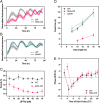
KidA overexpression (KidA-OX) mutants exhibit shorter in vivo clock rhythms and altered entrainment. (A and B) Bioluminescence (PpsbAI::luxAB, PpsbAI::luxCDE) time traces of WT, KidA-OX, and ΔkidA cultures induced with 10 µM IPTG. (A) WT vs. KidA-OX (B) WT vs. ΔkidA. Each trace was normalized by dividing by the maximum value. The solid and dotted lines show the mean normalized signal and the spread shows the SD. Average period obtained from fitting: WT = 23.5 ± 0.2 h, n = 8; KidA-OX = 20.1 ± 0.1 h, n = 7; ΔkidA = 25.3 ± 0.4 h, n = 8. Traces are aligned so that t = 0 occurs at a trough after three cycles in constant light. (C) The mean best-fit periods of the KidA-OX strain induced using different concentrations of IPTG and the WT strain characterized in the same conditions. Average period obtained from fitting at 50 µM IPTG: WT = 24.6 ± 0.6 h; KidA-OX = 18.6 ± 0.1 h. Error bars show the SD (n = 3–4). (D) Clock phases of WT, KidA-OX, and ΔkidA induced with 10 µM IPTG after entrainment to different photoperiods (10L:14D, 12L:12D, 16L:8D). The average time of the first peak of the bioluminescence signal after dawn following four LD cycles is plotted. Error bars show SD (n = 11–23 for each sample). Dotted lines show linear regression to the data: mWT = 0.89 (CI95% = [0.47, 1.31], R2 = 0.98), mKidA-OX = 0.35 (CI95% = [–0.08, 0.78], R2 = 0.99), mΔkidA = 0.74 (CI95% = [–0.54, 2.01], R2 = 0.98). (E) Phase response curves of WT and KidA-OX induced with 10 µM IPTG. Entrained cultures were allowed to free-run in constant light and then were subjected to a 5 h dark pulse at various times. The mean difference between the first peak of the reporter after the dark pulse and the closest peak time in the control (phase shift) is plotted. Error bars show the SD (n = 3–20 for each sample). CT, circadian time.
Fig. 3.
Fig. 3.
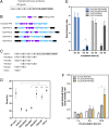
An N-terminal fragment of KidA is sufficient for the clock phenotype and binds to fold-switched KaiB. (A) Proposed KidA domain architecture derived from BLAST alignment and secondary structure prediction. (B) Phyre2 secondary structure prediction suggests four tandem domains with PAS-like folds. Canonical PAS architecture was modified from Möglich et al. (23). In the schematic, the first flanking alpha helix is the same as the last flanking alpha helix of the previous PAS domain. Predicted domain boundaries between the first and last β-sheets: PAS-A (22–114); PAS-B (145– 246); PAS-C (270–350); PAS-D (373–472). (C) Domain truncation mutants of KidA expressed in vivo. (D) Free-running clock periods of S. elongatus strains expressing KidA fragments described in (C). The average periods are plotted, and error bars show the SD (n = 4–16). The PAS-A construct expressed poorly compared with the other constructs. (E) In vitro co-IP of KidA PAS-ABC fragment (3.5 µM) using fsKaiB-FLAG or wtKaiB-FLAG (3.5 µM) as bait in a 30-min or 20-h coincubation; KaiC S431E;T432A (KaiC-EA) served as a positive control for KaiB interaction. Band intensities were determined by densitometry after background subtraction. Mean band intensity ratios are plotted, error bars show the SD (n = 3 for each condition). (F) In vitro co-IP of KaiC-EA using wtKaiB-FLAG (3.5 µM) as bait after preincubating wildtype KaiB-FLAG with varying amounts (0, 0.7, or 3.5 µM) of KidA PAS-ABC for varying lengths of time (1, 5, or 20 h). KaiC-EA was added 5 min prior to IP. Band intensities were determined by densitometry after background subtraction. Mean band intensity ratios are plotted, error bars show the SD (n = 2–3 for each condition). (DF) All significance analyses were performed using Welch’s t test. * = P < 0.05, ** = P < 0.01, *** = P < 0.001, **** = P < 0.0001.
Fig. 4.
Fig. 4.
In vitro and in silico reconstitution of the KidA period-shortening effect. (A) In vitro reconstitution of the period-shortening effect of KidA. The KidA PAS-ABC fragment was added to a standard mixture of KaiA, KaiB, and KaiC. Oscillations were measured by fluorescence polarization of labeled KaiB (averaged traces shown from duplicate experiments). (B) Cartoon overview of a mathematical model based on the hypothesis that KidA stabilizes the fold-switched form of KaiB by direct binding. The Paijmans model was modified to explicitly describe the interconversion between the ground-state and fold-switched state of KaiB.The protein complexes and reaction arrows shown were added to allow KidA to bind fsKaiB (SI Appendix). (C) Time course of oscillations of KaiC-bound KaiB molecules per hexamer in the mathematical model. Model parameters were chosen to have period-dependence close to the experimental value. (D) Plots of the period and amplitude of the fluorescence polarization rhythm (as shown in Fig. 4A) as a function of [KidA] and the predictions from the model with the same parameter set (as shown in Fig. 4C). (Left) Period and (Right) amplitude normalized by dividing by the amplitude value at 0 µM KidA. Mean values are plotted, and the error bars show the SD (Experimental n = 2, Model n = 20).

Similar articles

Cited by

References

    1. Nakajima M., et al. , Reconstitution of circadian oscillation of cyanobacterial KaiC phosphorylation in vitro. Science 308, 414–415 (2005). - PubMed
    1. Rust M. J., Golden S. S., O’Shea E. K., Light-driven changes in energy metabolism directly entrain the cyanobacterial circadian oscillator. Science 331, 220–223 (2011). - PMC - PubMed
    1. Leypunskiy E., et al. , The cyanobacterial circadian clock follows midday in vivo and in vitro. eLife 6, e23539 (2017). - PMC - PubMed
    1. Chavan A. G., et al. , Reconstitution of an intact clock reveals mechanisms of circadian timekeeping. Science 374, eabd4453 (2021). - PMC - PubMed
    1. Chang Y. G., et al. , Circadian rhythms. A protein fold switch joins the circadian oscillator to clock output in cyanobacteria. Science 349, 324–328 (2015). - PMC - PubMed

Publication types

MeSH terms

Substances

Supplementary concepts

LinkOut - more resources