. 2022 Oct;19(10):1185-1195.
doi: 10.1038/s41423-022-00913-x. Epub 2022 Sep 7.

Extracellular sulfatase-2 is overexpressed in rheumatoid arthritis and mediates the TNF-α-induced inflammatory activation of synovial fibroblasts

Affiliations

Extracellular sulfatase-2 is overexpressed in rheumatoid arthritis and mediates the TNF-α-induced inflammatory activation of synovial fibroblasts

Ruby J Siegel et al. Cell Mol Immunol. 2022 Oct.

Abstract

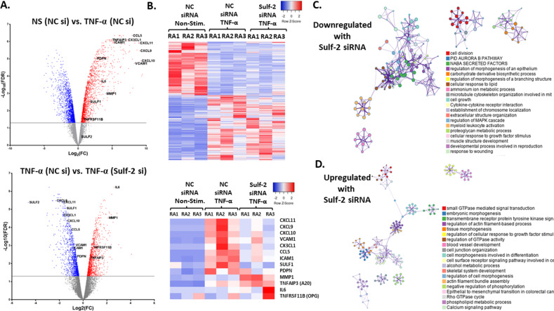
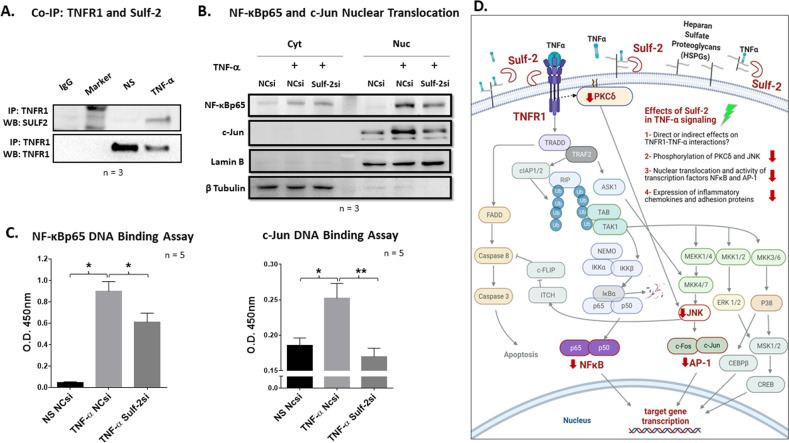
Extracellular sulfatase-2 (Sulf-2) influences receptor-ligand binding and subsequent signaling by chemokines and growth factors, yet Sulf-2 remains unexplored in inflammatory cytokine signaling in the context of rheumatoid arthritis (RA). In the present study, we characterized Sulf-2 expression in RA and investigated its potential role in TNF-α-induced synovial inflammation using primary human RA synovial fibroblasts (RASFs). Sulf-2 expression was significantly higher in serum and synovial tissues from patients with RA and in synovium and serum from hTNFtg mice. RNA sequencing analysis of TNF-α-stimulated RASFs showed that Sulf-2 siRNA modulated ~2500 genes compared to scrambled siRNA. Ingenuity Pathway Analysis of RNA sequencing data identified Sulf-2 as a primary target in fibroblasts and macrophages in RA. Western blot, ELISA, and qRT‒PCR analyses confirmed that Sulf-2 knockdown reduced the TNF-α-induced expression of ICAM1, VCAM1, CAD11, PDPN, CCL5, CX3CL1, CXCL10, and CXCL11. Signaling studies identified the protein kinase C-delta (PKCδ) and c-Jun N-terminal kinase (JNK) pathways as key in the TNF-α-mediated induction of proteins related to cellular adhesion and invasion. Knockdown of Sulf-2 abrogated TNF-α-induced RASF proliferation. Sulf-2 knockdown with siRNA and inhibition by OKN-007 suppressed the TNF-α-induced phosphorylation of PKCδ and JNK, thereby suppressing the nuclear translocation and DNA binding activity of the transcription factors AP-1 and NF-κBp65 in human RASFs. Interestingly, Sulf-2 expression positively correlated with the expression of TNF receptor 1, and coimmunoprecipitation assays demonstrated the binding of these two proteins, suggesting they exhibit crosstalk in TNF-α signaling. This study identified a novel role of Sulf-2 in TNF-α signaling and the activation of RA synoviocytes, providing the rationale for evaluating the therapeutic targeting of Sulf-2 in preclinical models of RA.

Keywords: Rheumatoid arthritis; Signal transduction; Sulfatase-2; Synovial fibroblasts; TNF-α.


Conflict of interest statement

The authors declare no competing interests.

Figures

Fig. 1
Fig. 1
Sulfatase-2 expression is significantly higher in human RA synovial tissues and serum, and SULF2 expression is transiently induced by TNF-α stimulation of RASFs in vitro. A qRT‒PCR analysis revealed significantly higher SULF1 and SULF2 mRNA levels in human RASTs than in NLSTs. Pearson correlation analysis revealed positive coexpression of TNFRSF1A and SULF2. B RASTs have significantly elevated Sulf-2 protein levels compared to NLSTs, as determined by Western blotting followed by densitometry analysis. C Quantitative ELISAs revealed significantly higher soluble Sulf-2 levels in serum from RA patients than in that from normal controls and MS patients. Soluble Sulf-1 was not detected. D Primary human RA synovial fibroblasts stimulated with TNF-α (20 ng/ml) for 0–48 h in vitro showed a transient increase in Sulf-1 and Sulf-2 protein levels in cell lysates. E In a separate experiment, soluble Sulf-2 levels in conditioned medium were quantitated by ELISA. Data are presented as the mean ± SEM. *p < 0.05; **p < 0.01; ***p < 0.001
Fig. 2
Fig. 2
Extracellular sulfatase levels are elevated in inflamed joint tissues and serum of a human TNF-transgenic mouse model of RA. Extracellular sulfatase protein levels were compared in human TNF-transgenic mice of the 3647 line (hTNFtg) and littermate wild-type (WT) C57BL/6 mice. A Murine Sulf-1 and Sulf-2 proteins were at qualitatively higher levels in ankle sections of hTNFtg mice as determined by fluorescence IHC. Images are shown with isotype-matched controls on the same tissue and slide. H&E images illustrate synovial hyperplasia and leukocyte infiltration, which are characteristic of hTNFtg mice. Bone (B) and synovial tissues (S) are labeled. B Murine Sulf-1 and Sulf-2 proteins in serum samples from hTNFtg and littermate WT mice were quantitated by ELISA. Data are presented as the mean ± SEM. *p < 0.05
Fig. 3
Fig. 3
RNA-seq array reveals that the TNF-α-mediated induction of many inflammatory genes in human RASFs in vitro is dependent on Sulf-2. A (Upper volcano plot): Gene expression profiles of TNF-α-stimulated and NS RASFs are compared in a volcano plot. Genes of interest in this study are labeled, including TNF-α-induced inflammatory genes that were highly significant (up) and highly upregulated (right). (Lower volcano plot): TNF-α-stimulated RASFs with or without Sulf-2 are compared in a volcano plot. For GO analysis, DEGs were identified by |log2FC| > 1 and FDR < 0.05. B The upper heatmap shows the relative gene expression of the top 236 DEGs by FDR in the comparison between unstimulated and TNF-α-stimulated RASFs. Modulation of TNF-α-induced gene expression can be seen in RASFs with Sulf-2 knockdown. The lower heatmap shows the relative expression of selected gene targets that are relevant to RA pathogenesis. Blue represents lower expression, and red represents higher expression. CD Diagrams depict networks of enriched GO terms for the 414 downregulated genes (panel C) and 315 upregulated genes (panel D) in TNF-α-stimulated RASFs with Sulf-2 knockdown in vitro; node size represents the number of input genes included in a specific term, colors indicate a shared cluster ID, and line thickness represents the GO similarity score
Fig. 4
Fig. 4
Knockdown of Sulf-2 or pretreatment of human RASFs with the Sulf-2 inhibitor OKN-007 inhibits the TNF-α-mediated induction of adhesion proteins and chemokines. Human RASFs were transfected with scrambled (NC), Sulf-2 or Sulf-1 siRNA (130 pmol) for 48 h, followed by serum starvation and stimulation with TNF-α (20 ng/ml) for 24 h. Knockdown of Sulf-2 significantly inhibited the TNF-α-mediated induction of ICAM-1 and VCAM-1 (A) and of IP-10/CXCL10, and I-TAC/CXCL11 (B). Pretreatment of RASFs for 12 h with the Sulf-2 inhibitor OKN-007 (0–1 mM) followed by TNF-α (20 ng/ml) stimulation for 24 h resulted in the dose-dependent inhibition of the TNF-α-mediated induction of ICAM-1 and VCAM-1 (C) and RANTES/CCL5, ENA-78/CXCL5, IP-10/CXCL10 and I-TAC/CXCL11 (D)
Fig. 5
Fig. 5
TNF-α induces adhesion proteins in human RASFs via the PKCδ (JNK and NF-κB) signaling pathway. Knockdown of Sulf-2 inhibits the TNF-α-induced phosphorylation of PKCδ and JNK. A Human RASFs were preincubated for 2 h with a panel of signaling inhibitors, followed by stimulation with TNF-α for 24 h. Western blots were probed for ICAM-1, VCAM-1, cadherin-11 and podoplanin. Inhibitors of NF-κB, JNK and, most prominently, PKCδ led to significant reductions in TNF-α-induced adhesion proteins, revealing the importance of these pathways. B The central role of PKCδ was confirmed by the dose-dependent decrease in ICAM-1, VCAM-1 and cadherin-11 levels in RASFs after treatment with the PKCδI-specific small-molecule inhibitor NP627
Fig. 6
Fig. 6
Knockdown of Sulf-2 inhibits the TNF-α-induced phosphorylation of PKCδ and JNK and TNF-α-induced RASF proliferation and invasion. A siRNA-mediated knockdown of Sulf-2 in human RASFs significantly inhibited the TNF-α-induced phosphorylation of PKCδ and JNK. Data are presented as the mean ± SEM. *p < 0.05. B Knockdown of Sulf-2 inhibited the TNF-α-induced proliferation of RASFs to almost basal levels. C The Transwell invasion assay showed a trend in decreased (25%) TNF-α-induced RASF invasion in the presence of Sulf-2 siRNA (p < 0.07)
Fig. 7
Fig. 7
Sulf-2 binds TNF receptor 1, and knockdown of Sulf-2 reduces the TNF-α-induced nuclear translocation and DNA binding activity of inflammatory transcription factors. A Immunoprecipitation of TNF receptor 1 revealed binding with the 75 kD subunit of the mature Sulf-2 heterodimeric protein in TNF-α-stimulated human RASFs. B Knockdown of Sulf-2 in RASFs inhibited the TNF-α-induced nuclear translocation of c-Jun and NF-κBp65. C Knockdown of Sulf-2 significantly reduced the TNF-α-induced DNA binding activity of NF-κBp65 and c-Jun. Data are presented as the mean ± SEM. *p < 0.05; **p < 0.01. D Schematic diagram illustrating the effects of Sulf-2 knockdown on TNF-α inflammatory signaling in human RASFs

Comment in

References

    1. Puimège L, Libert C, Van, Hauwermeiren F. Regulation and dysregulation of tumor necrosis factor receptor-1. Cytokine Growth Factor Rev. 2014;25:285–300. - PubMed
    1. Locksley RM, Killeen N, Lenardo MJ. The TNF and TNF receptor superfamilies: integrating mammalian biology. Cell. 2001;104:487–501. - PubMed
    1. Bartok B, Firestein GS. Fibroblast-like synoviocytes: key effector cells in rheumatoid arthritis. Immunol Rev. 2010;233:233–55. - PMC - PubMed
    1. Lowenthal JW, Ballard DW, Bohnlein E, Greene WC. Tumor necrosis factor alpha induces proteins that bind specifically to kappa B-like enhancer elements and regulate interleukin 2 receptor alpha-chain gene expression in primary human T lymphocytes. Proc Natl Acad Sci USA. 1989;86:2331–5. - PMC - PubMed
    1. Roach DR, Bean AG, Demangel C, France MP, Briscoe H, Britton WJ. TNF regulates chemokine induction essential for cell recruitment, granuloma formation, and clearance of mycobacterial infection. J Immunol. 2002;168:4620–7. - PubMed

Publication types

MeSH terms