. 2022 Oct;45(10):1019-1036.
doi: 10.1007/s40264-022-01216-w. Epub 2022 Sep 6.

High-Dose Intravenous Iron with Either Ferric Carboxymaltose or Ferric Derisomaltose: A Benefit-Risk Assessment

Affiliations

High-Dose Intravenous Iron with Either Ferric Carboxymaltose or Ferric Derisomaltose: A Benefit-Risk Assessment

Johannes M M Boots et al. Drug Saf. 2022 Oct.

Abstract

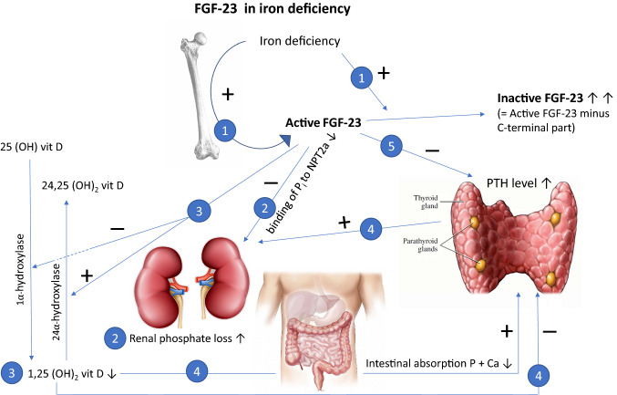
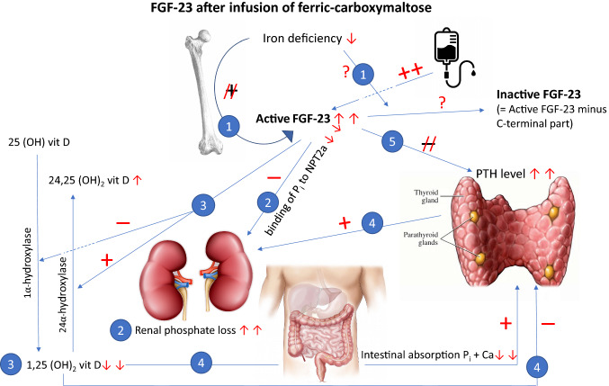
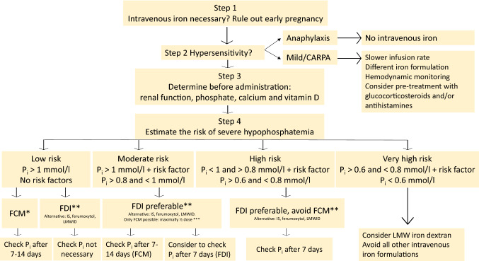
The intravenous iron formulations ferric carboxymaltose (FCM) and ferric derisomaltose (FDI) offer the possibility of administering a large amount of iron in one infusion. This results in faster correction of anemia and the formulations being better tolerated than oral iron formulations. This triad of logistic advantages, improved patient convenience, and fast correction of anemia explains the fact that intravenous iron formulations nowadays are frequently prescribed worldwide in the treatment of iron deficiency anemia. However, these formulations may result in hypophosphatemia by inducing a strong increase in active fibroblast growth factor-23 (FGF-23), a hormone that stimulates renal phosphate excretion. This effect is much more pronounced with FCM than with FDI, and therefore the risk of developing hypophosphatemia is remarkably higher with FCM than with FDI. Repeated use of FCM may result in severe osteomalacia, which is characterized by bone pain, Looser zones (pseudofractures), and low-trauma fractures. Intravenous iron preparations are also associated with other adverse effects, of which hypersensitivity reactions are the most important and are usually the result of a non-allergic complement activation on nanoparticles of free labile iron-Complement Activation-Related Pseudo-Allergy (CARPA). The risk on these hypersensitivity reactions can be reduced by choosing a slow infusion rate. Severe hypersensitivity reactions were reported in < 1% of prospective trials and the incidence seems comparable between the two formulations. A practical guideline has been developed based on baseline serum phosphate concentrations and predisposing risk factors, derived from published cases and risk factor analyses from trials, in order to establish the safe use of these formulations.


Conflict of interest statement

Johannes M.M. Boots received lecture fees and participated in advisory boards from Cablon Medical, The Netherlands; Pharmacosmos, Denmark; and Vifor Pharma, The Netherlands. Rogier A.M. Quax has no disclosures with regard to this manuscript.

Figures

Fig. 1
Fig. 1
The physiological effects of FGF-23 during iron deficiency. During iron deficiency, the production of FGF-23 is increased. Recent experiments in mice showed that bone marrow sinusoidal endothelial cells were a site of FGF-23 upregulation [55]; however, at the same time, the increase in active FGF-23 is counterbalanced by the increase in the disintegration of intact (active) FGF-23 (1). This results in normal physiology of FGF-23 and increased concentrations of inactive FGF-23. FGF-23 results in diminished binding of Pi to NPT2A in the proximal tubule in the kidney, resulting in renal phosphate excretion (2). FGF-23 also interferes with vitamin D metabolism. It stimulates the inactivation of active 1.25 (OH)2 vitamin D and inhibits the activation of inactive 25 (OH)vitamin D (3). Less-active vitamin D results in a decrease in intestinal calcium and phosphate absorption. Lower calcium and phosphate concentrations stimulate the release of PTH, and active vitamin D inhibits the production of PTH. The increased production of PTH further stimulates phosphate excretion in the kidney (4). FGF-23 has also a direct blocking effect on the production of PTH by its co-receptor α-Klotho (5). FGF-23 fibroblast growth factor 23, Pi inorganic phosphate, NPT2A sodium-dependent phosphate cotransporter 2A, PTH parathyroid hormone, Vit D vitamin D
Fig. 2
Fig. 2
The effects of FGF-23 after infusion of ferric carboxymaltose. Although the iron deficiency-driven stimulus for increased FGF-23 production and the simultaneous inactivation is diminished after iron repletion, the active FGF-23 level is remarkably increased following ferric carboxymaltose infusion. The exact mechanism behind this increase is currently unknown (1). This strongly results in diminished binding to NPT2A in the proximal tubule in the kidney and induces renal phosphate losses (2). In addition, vitamin D metabolism is disturbed by blocking the activation of 25 (OH) vitamin D and stimulating the inactivation of active 1.25 (OH)2 vitamin D (3). The markedly reduced active vitamin D concentrations result in lower intestinal calcium and phosphate absorption. By lower calcium and phosphate concentrations, the secretion of PTH is stimulated. In addition, active vitamin D no longer blocks the secretion of PTH. The substantial increase in PTH further stimulates renal phosphate losses (4). The ongoing stimulus of FGF-23 downregulates the blockade of FGF-23 on PTH secretion, which results in a further increase in PTH concentrations and renal phosphate losses (5). FGF-23 fibroblast growth factor 23, Pi inorganic phosphate, NPT2A sodium-dependent phosphate cotransporter 2A, PTH parathyroid hormone, Vit D vitamin D
Fig. 3
Fig. 3
Flowchart for safe use of intravenous iron preparations. The risk of developing hypophosphatemia can be determined by applying the predisposing risk factors from Table 2. When FCM is used, the SmPC advices to measure the serum phosphate concentration before administration and to repeat this after 14 days. However, when only one dose of FCM is administered, the nadir usually occurs after 7 days. A predose serum phosphate concentration of 1 mmol/L is used as a relatively safe border to prevent the development of severe hypophosphatemia, since prospective trials revealed a mean decrease in serum phosphate of 0.4–0.5 mmol/L with the use of FCM. For mg/dL, Pi must be multiplied by 3.1. *Even in low-risk patients, FCM represents a risk of severe hypophosphatemia. The best way to prevent hypophosphatemia is the choice of iron formulation. Local availability, costs, and the risk of other adverse effects will further contribute to the physician’s final decision to choose either FCM or FDI. When iron deficiency anemia cannot be restored by a maximum dose of iron 1000 mg and multiple infusions are likely, FCM should definitely not be used because repeated infusions increase the risk and duration of hypophosphatemia, implying a risk of inducing osteomalacia. An interval of 6 months between dosing may be safe to prevent osteomalacia. **Ferumoxytol and IS have a lower risk of hypophosphatemia than FDI and can also be used, but less iron can be administered in one infusion. Ferumoxytol is only registered in the US. LMWID has not been associated with the development of hypophosphatemia and can therefore also be used. Total correction of the iron deficit is possible using LMWID, but requires a longer infusion rate (4–6 h) and a test dose of 25 mg. ***The use of FCM in this situation is not recommended. In a situation whereby FCM is the only treatment option, use half the maximal dose (500 mg), realizing that iron deficiency cannot be fully restored, and repeated doses are contraindicated. CARPA Complement Activation-Related Pseudo-Allergy, FCM ferric carboxymaltose, SmPC Summary of Product Characteristics, FDI ferric derisomaltose, IS iron sucrose, LMWID low-molecular-weight iron dextran, Pi inorganic phosphate

Similar articles

Cited by

References

    1. Heath CW, Strauss MB, Castle WB. Quantitative aspects of iron deficiency in hypochromic anemia. (The parenteral administration of iron) J Clin Invest. 1932;11:1293–1312. doi: 10.1172/JCI100478. - DOI - PMC - PubMed
    1. Goetsch AT, Moore CV, Minnich V. Observations on the effect of massive doses of iron given intravenously to patients with hypochromic anemia. Blood. 1946;1:129–142. doi: 10.1182/blood.V1.2.129.129. - DOI - PubMed
    1. Nissim JA. Intravenous administration of iron. Lancet. 1947;2(6463):49–51. doi: 10.1016/s0140-6736(47)90053-6. - DOI - PubMed
    1. Nissim JA, Robson JM. Preparation and standardisation of saccharated iron oxide for intravenous administration. Lancet. 1949;1(6556):686–689. doi: 10.1016/S0140-6736(49)91958-3. - DOI - PubMed
    1. Müller A. Classification and properties of iron preparations. Arzneimittelforschung (Drug Res). 1974;24:880–883. - PubMed