. 2022 Sep 6;23(1):232.
doi: 10.1186/s12931-022-02161-z.

Cigarette smoke exposed airway epithelial cell-derived EVs promote pro-inflammatory macrophage activation in alpha-1 antitrypsin deficiency

Affiliations

Cigarette smoke exposed airway epithelial cell-derived EVs promote pro-inflammatory macrophage activation in alpha-1 antitrypsin deficiency

Nazli Khodayari et al. Respir Res. .

Erratum in

Abstract

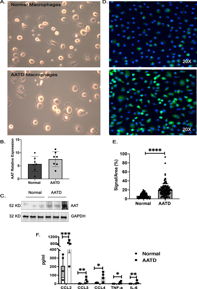
Background: Alpha-1 antitrypsin deficiency (AATD) is a genetic disorder most commonly secondary to a single mutation in the SERPINA1 gene (PI*Z) that causes misfolding and accumulation of alpha-1 antitrypsin (AAT) in hepatocytes and mononuclear phagocytes which reduces plasma AAT and creates a toxic gain of function. This toxic gain of function promotes a pro-inflammatory phenotype in macrophages that contributes to lung inflammation and early-onset COPD, especially in individuals who smoke cigarettes. The aim of this study is to determine the role of cigarette exposed AATD macrophages and bronchial epithelial cells in AATD-mediated lung inflammation.

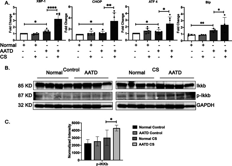
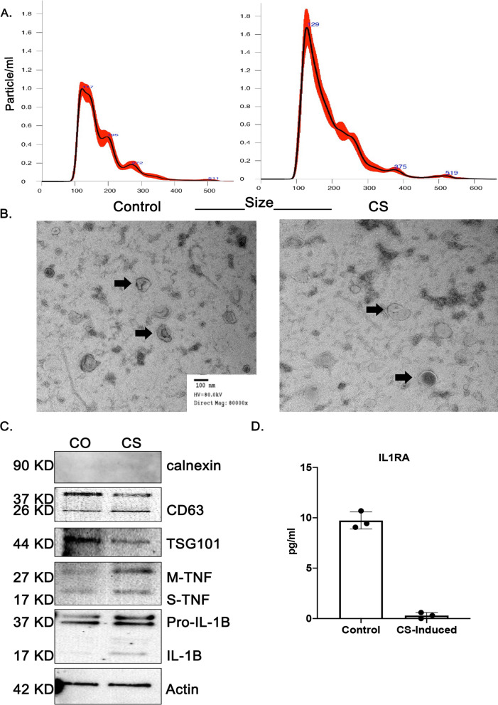
Methods: Peripheral blood mononuclear cells from AATD and healthy individuals were differentiated into alveolar-like macrophages and exposed to air or cigarette smoke while in culture. Macrophage endoplasmic reticulum stress was quantified and secreted cytokines were measured using qPCR and cytokine ELISAs. To determine whether there is "cross talk" between epithelial cells and macrophages, macrophages were exposed to extracellular vesicles released by airway epithelial cells exposed to cigarette smoke and their inflammatory response was determined.

Results: AATD macrophages spontaneously produce several-fold more pro-inflammatory cytokines as compared to normal macrophages. AATD macrophages have an enhanced inflammatory response when exposed to cigarette smoke-induced extracellular vesicles (EVs) released from airway epithelial cells. Cigarette smoke-induced EVs induce expression of GM-CSF and IL-8 in AATD macrophages but have no effect on normal macrophages. Release of AAT polymers, potent neutrophil chemo attractants, were also increased from AATD macrophages after exposure to cigarette smoke-induced EVs.

Conclusions: The expression of mutated AAT confers an inflammatory phenotype in AATD macrophages which disposes them to an exaggerated inflammatory response to cigarette smoke-induced EVs, and thus could contribute to progressive lung inflammation and damage in AATD individuals.

Keywords: Alpha-1 antitrypsin; Cigarette smoke; Extracellular vesicles; Lung disease; Macrophages.


Conflict of interest statement

The authors have no conflict of interest.

Figures

Fig. 1
Fig. 1
Characterization of normal and AATD macrophages. A The morphology of monocyte-derived macrophages from normal and AATD individuals were visualized under the light microscopy. B The relative expression of AAT mRNA form normal and AATD macrophages. C The intracellular protein levels of AAT in representative macrophages. D Immunofluorescent images of normal and AATD macrophages with the magnification of 20X, demonstrate the intracellular AAT levels using FITC- labeled AAT antibody. E Quantification of the intracellular levels of AAT protein. F Comparison of the cytokine levels in the culture media of normal and AATD macrophages, *p < 0.05, **p < 0.005, ***p < 0.0005, ****p < 0.00005
Fig. 2
Fig. 2
Changes in the macrophage’s cytokines profile in response to cigarette smoke. A The cytokine levels in the culture media of normal and AATD macrophages exposed to cigarette smoke (CS) or air. B Heatmap presentation of the cytokines. C The levels of cytokines associated with EVs in the cultured media of normal and AATD macrophages exposed to air or CS. D Heatmap presentation of the EV associated cytokines. *p < 0.05, **p < 0.005
Fig. 3
Fig. 3
Induction of ER stress and NF-κB inflammatory pathway in AATD macrophages in response to cigarette smoke. A Comparison of the mRNA expression levels of XBP-1, CHOP, ATF 4, and BiP in normal and AATD macrophages exposed to air or cigarette. B Western blot analysis of 4 different normal and AATD macrophages incubated with control or CS exposed showing activation of NF-κB pathway in AATD macrophages after CS exposure. C Bar graphs showing the results of quantification and normalization of band intensities, *p < 0.05, **p < 0.005, ****p < 0.00005
Fig. 4
Fig. 4
Characterization of airway epithelial cell-derived EVs. A Nano sight tracking analysis of size and concentration for EVs isolated from conditioned media of control and cigarette smoke-induced EVs. The bold red curves plot the size distribution of EVs. B Morphological characterization of EVs by transmission electron microscopy (black arrows). C Western blotting experiment of the purified EVs showing Calnexin, CD63, TSG101, membrane bound and soluble TNF-α and pro and active IL-1 β. D The levels of cytokines associated with EVs released by small airway epithelial cells
Fig. 5
Fig. 5
Increased secretion levels of pro-inflammatory cytokines and ZAAT polymers by AATD macrophages in response to cigarette smoke—induced EVs. A The levels of secreted cytokines in the conditioned media of normal and AATD macrophages incubated with control or cigarette smoke-induced EVs. B NF-κB pathway was inhibited in AATD macrophages using TPCA-1 and the secretion levels of IL-8 and GM-CSF in response to cigarette smoke -induced EVs were measured. C The levels of secreted cytokines presented in a heatmap graph. D The levels of polymeric AAT in the conditioned media from the normal and AATD macrophages incubated with control or cigarette smoke-induced EVs. E Signal intensity quantification of polymers in AATD macrophages conditioned media. F Total AAT levels were also measured in the conditioned media of normal and AATD macrophages. G The protein expression levels of ER markers were also measured by western blot analysis. *p < 0.05, **p < 0.005, ***p < 0.0005

References

    1. Khodayari N, Oshins R, Holliday LS, Clark V, Xiao Q, Marek G, et al. Alpha-1 antitrypsin deficient individuals have circulating extracellular vesicles with profibrogenic cargo. Cell Commun Signal. 2020;18(1):140. doi: 10.1186/s12964-020-00648-0. - DOI - PMC - PubMed
    1. Krotova K, Marek GW, Wang RL, Aslanidi G, Hoffman BE, Khodayari N, et al. Alpha-1 antitrypsin-deficient macrophages have increased matriptase-mediated proteolytic activity. Am J Respir Cell Mol Biol. 2017;57(2):238–247. doi: 10.1165/rcmb.2016-0366OC. - DOI - PMC - PubMed
    1. Di Stefano A, Caramori G, Oates T, Capelli A, Lusuardi M, Gnemmi I, et al. Increased expression of nuclear factor-kappaB in bronchial biopsies from smokers and patients with COPD. Eur Respir J. 2002;20(3):556–563. doi: 10.1183/09031936.02.00272002. - DOI - PubMed
    1. Schuliga M. NF-kappaB signaling in chronic inflammatory airway disease. Biomolecules. 2015;5(3):1266–1283. doi: 10.3390/biom5031266. - DOI - PMC - PubMed
    1. Lee J, Lu Y, Oshins R, West J, Moneypenny CG, Han K, et al. Alpha 1 antitrypsin-deficient macrophages have impaired efferocytosis of apoptotic neutrophils. Front Immunol. 2020;11:574410. doi: 10.3389/fimmu.2020.574410. - DOI - PMC - PubMed

MeSH terms